

Evaluation of fluoride varnish treatment of postorthodontic white spot lesions by visual inspection and laser fluorescence-A randomized controlled study
- PMID: 35510617
- PMCID: PMC9382033
- DOI: 10.1002/cre2.579
Evaluation of fluoride varnish treatment of postorthodontic white spot lesions by visual inspection and laser fluorescence-A randomized controlled study
Abstract
Objectives: White spot lesions (WSLs), as a side effect of orthodontic therapy, can be treated with fluoride varnish, with the difference in efficiency reported.
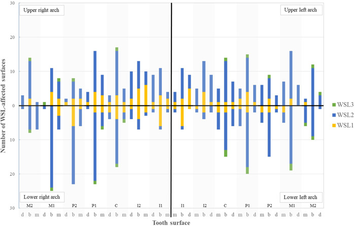
Material and methods: Patients with buccal WSLs were consecutively included in a randomized controlled double-blind study. At first inspection and at three follow-ups over 6 months, 0.1% fluoride varnish and placebo (water) were applied in the test group (N = 21) and control group (N = 21), respectively. The maximum laser fluorescence value (LFV) of WSLs was recorded using DIAGNOdent. Between the groups, differences in the mean numbers of WSLs and the mean LFV of WSLs per patient at different time points were analyzed with mixed-design analysis of variance. Orthodontic therapy duration (OTD) was included in the model as a covariate.
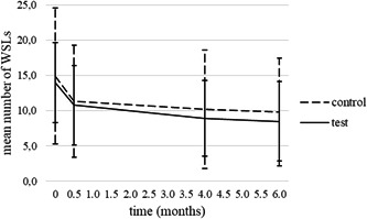
Results: A decrease in the mean WSLs number and LFV was observed; however, there were no significant differences between study groups at any time point. OTD was in interaction only with LFV. Analysis showed a different pattern of mean LFV changes for patients with OTD of >48 months compared to patients with OTD of ≤24.
Conclusion: The changes in numbers of WSLs and LFV over the study period indicated regression of WSLs, but an additional effect of FV was not confirmed.
Keywords: fluoride varnish; postorthodontic; white spot lesion.
© 2022 The Authors. Clinical and Experimental Dental Research published by John Wiley & Sons Ltd.
Conflict of interest statement
The authors declare no conflicts of interest.
Figures


References
-
- Aljehani, A. , Yousif, M. , Angmar‐Mansson, B. , & Shi, X. Q. (2006). Longitudinal quantification of incipient carious lesions in postorthodontic patients using a fluorescence method. European Journal of Oral Sciences, 114, 430–434. - PubMed
-
- Artun, J. , & Thylstrup, A. (1986). Clinical and scanning electron microscopic study of surface changes of incipient caries lesions after debonding. Scandinavian Journal of Dental Research, 94, 193–201. - PubMed
-
- Baeshen, H. A. , Lingstrom, P. , & Birkhed, D. (2011). Effect of fluoridated chewing sticks (Miswaks) on white spot lesions in postorthodontic patients. American Journal of Orthodontics and Dentofacial Orthopedics, 140, 291–297. - PubMed
-
- Beerens, M. W. , Boekitwetan, F. , van der Veen, M. H. , & ten Cate, J. M. (2015). White spot lesions after orthodontic treatment assessed by clinical photographs and by quantitative light‐induced fluorescence imaging; a retrospective study. Acta Odontologica Scandinavica, 73, 441–446. - PubMed
-
- Benson, P. E. , Parkin, N. , Dyer, F. , Millett, D. T. , Furness, S. , & Germain, P. (2013). Fluorides for the prevention of early tooth decay (demineralised white lesions) during fixed brace treatment. Cochrane Database of Systematic Reviews, 12, CD003809. - PubMed
Publication types
MeSH terms
Substances
LinkOut - more resources
Full Text Sources
Medical
