. 2022 Aug;235(3):1287-1301.
doi: 10.1111/nph.18200. Epub 2022 May 26.

Broad-range metalloprotease profiling in plants uncovers immunity provided by defence-related metalloenzyme

Affiliations

Broad-range metalloprotease profiling in plants uncovers immunity provided by defence-related metalloenzyme

Kyoko Morimoto et al. New Phytol. 2022 Aug.

Abstract

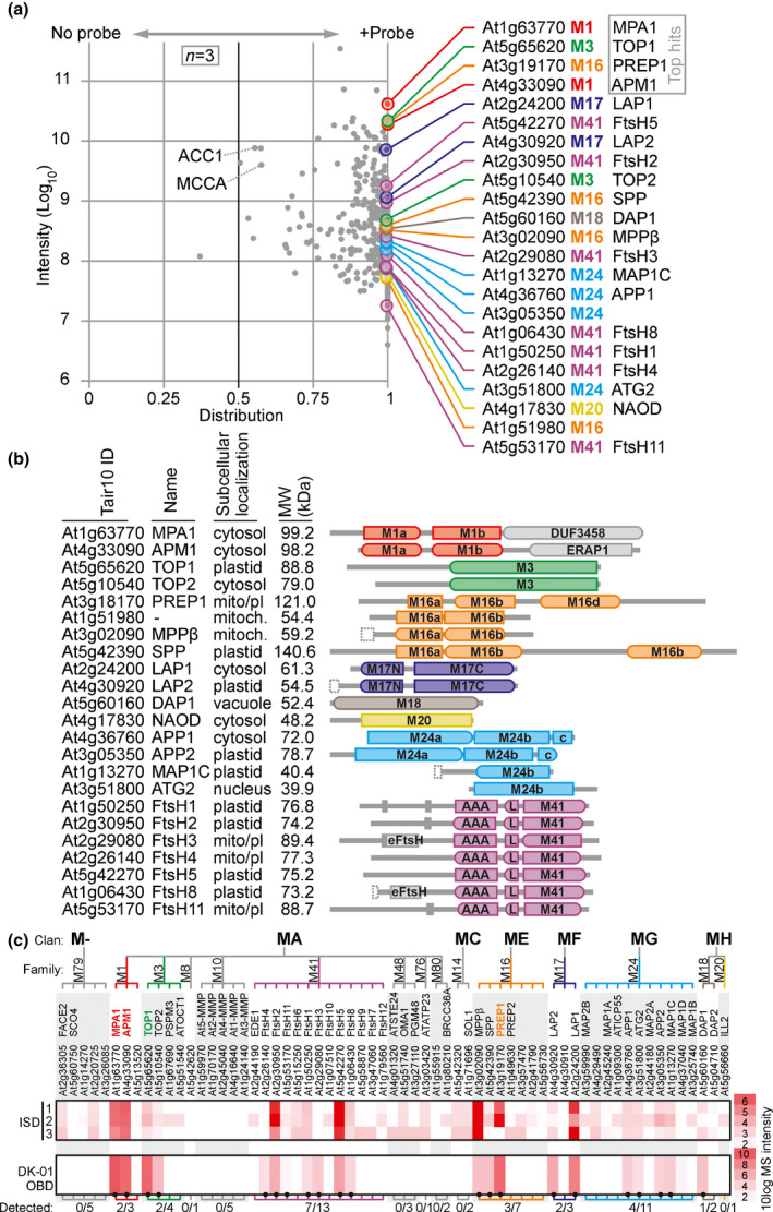
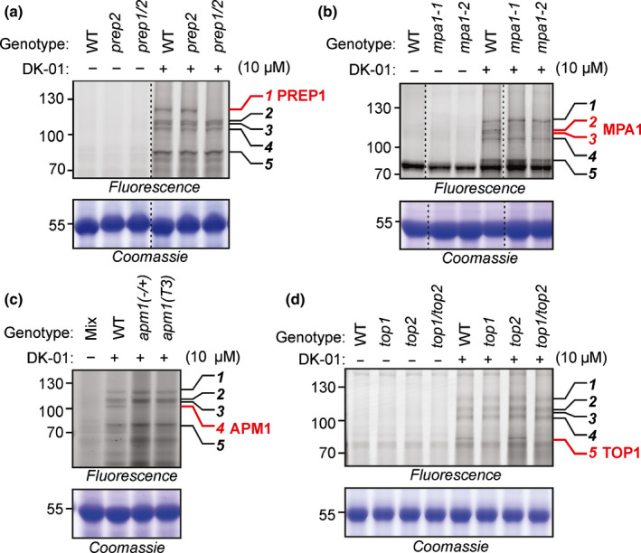
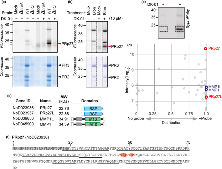
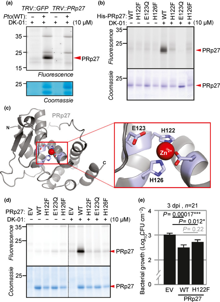
Plants encode > 100 metalloproteases representing > 19 different protein families. Tools to study this large and diverse class of proteases have not yet been introduced into plant research. We describe the use of hydroxamate-based photoaffinity probes to explore plant proteomes for metalloproteases. We detected labelling of 23 metalloproteases in leaf extracts of the model plant Arabidopsis thaliana that belong to nine different metalloprotease families and localize to different subcellular compartments. The probes identified several chloroplastic FtsH proteases, vacuolar aspartyl aminopeptidase DAP1, peroxisomal metalloprotease PMX16, extracellular matrix metalloproteases and many cytosolic metalloproteases. We also identified nonproteolytic metallohydrolases involved in the release of auxin and in the urea cycle. Studies on tobacco plants (Nicotiana benthamiana) infected with the bacterial plant pathogen Pseudomonas syringae uncovered the induced labelling of PRp27, a secreted protein with implicated metalloprotease activity. PRp27 overexpression increases resistance, and PRp27 mutants lacking metal binding site are no longer labelled, but still show increased immunity. Collectively, these studies reveal the power of broad-range metalloprotease profiling in plants using hydroxamate-based probes.

Keywords: Arabidopsis; PRp27; immunity; metalloproteases; photoaffinity labelling.


Figures

Fig. 1
Fig. 1
Hydroxamate‐based probes display five polymorphic signals in Arabidopsis leaf extracts. (a) General structure of the used probes. All compounds carry a hydroxamate zinc‐binding group (blue), an alkyne minitag (green) and a photoreactive benzophenone group (red), connected through a dipeptide with various amino acid residues at P2 (grey). (b) Differential labelling profiles with different hydroxamate probes. Arabidopsis leaf extracts were labelled with and without 1 µM probes by UV irradiation at 366 nm for 30 min on ice. Alkyne‐labelled proteins were coupled to a fluorophore using click‐chemistry with rhodamine‐azide. Proteins were separated on SDS‐PAGE gels and fluorescent proteins were detected by in‐gel fluorescence scanning (ex488/em520). Five specific signals are indicated on the right.
Fig. 2
Fig. 2
Hydroxamate‐based inhibitors cause differential labelling. (a) Structure of DK‐01, containing a hydroxamate (blue), photoreactive benzophenone (red), alkyne minitag (green) and a dipeptide linker with P1 = Leu and P2 = Gly. (b) DK‐01 labels several proteins in Arabidopsis leaf extracts and requires both UV treatment and click‐chemistry. (c) Structures of the hydroxamates used, highlighting the differences with DK‐01 in red. (d) Differential labelling upon preincubation with hydroxamates. Leaf extracts of wild‐type Arabidopsis were preincubated with and without 200 µM inhibitors then end‐labelled with or without 10 µM DK‐01 by UV irradiation at 254 nm for 30 min on ice. Click‐chemistry with and without Cy3‐picolyl‐azide was used to couple the alkyne minitag to a fluorophore. Proteins were separated on SDS‐PAGE gels and fluorescent proteins were detected by in‐gel fluorescence scanning.
Fig. 3
Fig. 3
DK‐01 targets a broad range of Arabidopsis metalloproteases. (a) Arabidopsis leaf extracts were labelled with or without 10 µM DK‐01 by UV irradiation at 254 nm for 30 min on ice. Alkyne‐labelled proteins were biotinylated using click‐chemistry, biotinylated proteins were enriched on streptavidin beads and on‐bead digested with trypsin, and released peptides were analysed by LC‐MS/MS. The sum of the MS signal intensities was plotted against the distribution of the MS signal intensities over the no‐probe‐control and the DK‐01‐labelled sample. Only proteins that were detected in all three DK‐01 samples are shown. Twenty‐six enriched proteins are metalloproteases, identified on the right by accession number, subfamily and name. Nonspecific abundant proteins include endogenously biotinylated proteins MCCA and ACC1. (b) Domain structures and classification of the 23 identified metalloproteases. The protein IDs and names are given along with the predicted subcellular localization based on the Arabidopsis Subcellular Database (SUBA), the predicted molecular weight (MW; kDa) and the protein domain structures (PFAMs) summarized as follows: M1a (PF17900); M1b (PF01433); DUF3458 (PF11940/PF17432); ERAP1‐like domain (PF11838); M3 peptidase (PF01432); M16a (PF00675); M16b (PF05193); M16c (PF16187); M16d (PF08367); M17N (PF02789); M17C catalytic domain (PF00883); M18 peptidase (PF02127); M20 peptidase (PF01546); M24a (PF01321/16189); M24b catalytic M24 peptidase (PF00557); c, C‐terminal M24 domain (PF16188); eFtsH, extracellular FtsH domain (PF06484); AAA, ATPase (PF00004); L, ATPase lid (PF17862); and M41 peptidase (PF01434). (c) Most abundant metalloproteases detected in leaf extracts detected by DK‐01 labelling. The protein intensities of all metalloproteases were extracted from the pep2pro database from three independent MS experiments on leaf extracts and compared to the intensities of the metalloproteases detected in the pull‐down upon DK‐01 labelling. The classification of metalloproteases into families and clans is from the MEROPS database.
Fig. 4
Fig. 4
Knockout mutants deconvolute high‐MW metalloprotease signals. (a) Metalloprotease PREP1 is required for signal 1. (b) MPA1 is required for signals 2 and 3. (c) APM1 is required for signal 4. (d) TOP1 is required for signal 5. Leaf extracts of wild‐type (WT) and mutant plants were labelled with or without 10 µM DK‐01 by UV irradiation at 254 nm for 30 min on ice. Proteins were separated on SDS‐PAGE gels and fluorescent proteins were detected by in‐gel fluorescence scanning at ex488/em520 or ex633/em670. Five specific signals are numbered on the right. Coomassie stains are provided as loading controls.
Fig. 5
Fig. 5
Differential labelling of PRp27 upon infection with WT PtoDC3000. (a) Treatment with WT PtoDC3000 induces a DK‐01‐labelled protein. Nicotiana benthamiana leaves were infiltrated with water (Mock), WT PtoDC3000, and the derived ΔhrpA and ΔhQ mutants. Apoplastic fluids (AFs) were isolated at 2 d postinfiltration (2 dpi) and labelled with or without 10 µM DK‐01 by UV irradiation at 254 nm for 30 min on ice. Alkyne‐labelled proteins were coupled to Cy3‐picolyl‐azide using click‐chemistry. Proteins were separated on SDS‐PAGE gels and fluorescent proteins were detected by in‐gel fluorescence scanning at ex488/em520. The specific signal at 23 kDa and the PR proteins are indicated with red and black arrowheads, respectively. (b) The 23 kDa signal is induced by SA signalling. Nicotiana benthamiana leaves were infiltrated with water or 2 mM Bion. Apoplastic fluids were isolated at 4 dpi and labelled with or without 10 µM DK‐01 by UV irradiation at 254 nm for 30 min on ice. Alkyne‐labelled proteins were coupled to Cy5‐picolyl‐azide using click‐chemistry. Proteins were separated on SDS‐PAGE gels and fluorescent proteins were detected by in‐gel fluorescence scanning at ex633/em670. (c) Analysis of the purified 23 kDa gel region. Apoplastic fluids from WT infiltrated plants were labelled with and without DK‐01 and coupled to biotin. Biotinylated proteins were purified, separated on gels and stained with Sypro Ruby (top). The 20–25 kDa region was excised and incubated with trypsin/LysC, and released peptides were analysed by MS. (d) Distribution graph of proteins identified from 20 to 25 kDa gel slices. The sum of the peptide intensities was plotted against the distribution over the probe and no‐probe control for each protein. Four highly enriched proteins include pathogenesis‐related protein 27 (PRp27), PRp27‐like protein (PRp27L), matrix metalloprotease 1 (MMP1) and MMP‐1‐like protein (MMP1L), highlighted in red or blue, respectively. (e) Domain structures of the four proteins highlighted in (c). The protein IDs, names, predicted MWs (kDa) not including N‐ and C‐terminal signal peptides, and the PFAM protein domains: BSP (PF04450), M10 (PF00413) and PG binding 1 (PF01471) are given. (f) PRp27 contains the consensus zinc‐binding motif HExxH. The predicted signal peptides (grey) and the HExxH motif (red) are highlighted. The identified peptides (underlined) cover c. 61% of the PRp27 protein sequence.
Fig. 6
Fig. 6
Putative active site of PRp27 is required for labelling but not plant immunity. (a) PRp27 depletion by VIGS removes the 23 kDa signal. Apoplastic fluids (AFs) isolated from the TRV::GFP and TRV::PRp27 leaves infected with PtoDC3000 (WT) or water were labelled with or without 10 µM DK‐01 by UV irradiation at 254 nm for 30 min on ice. Alkyne‐labelled proteins were fluorescently coupled to Cy3‐picolyl‐azide using click chemistry. Proteins were separated on SDS‐PAGE gels and fluorescent proteins were detected by in‐gel fluorescence scanning at ex488/em520. (b) Amino acid substitution at the putative zinc‐binding sites abolishes DK‐01 labelling of PRp27 expressed in E. coli. Extracts of E. coli expressing WT and H122F, E123Q and E126F mutant His‐PRp27 proteins were labelled with 10 µM DK‐01 as described in (a). (c) A three‐dimensional model of the PRp27 structure shows the putative architecture of the zinc‐binding domain in PRp27, coordinated by four amino acid residues. The model was generated with swiss model using PDB 1z5h as a template. (d) DK‐01 labels transiently expressed PRp27 (WT), but not the H122F, E123Q or H126F mutants. Apoplastic fluids isolated at 4 dpi from agroinfiltrated leaves coexpressing the empty vector (EV) or PRp27 (WT, H122F, E123Q and E126F) with silencing inhibitor P19 were labelled, with or without 10 µM DK‐01, as described in (a). (e) Both PRp27 WT and the H122F mutant increase immunity to PtoDC3000(WT). Agroinfiltrated leaves expressing the empty vector (EV), PRp27(WT) or PRp27(H122F) were infiltrated with PtoDC3000(WT) at 2 dpi and bacterial growth was measured 3 d later. Error bars represent mean ± SE of n = 21 biological replicates. The experiment shown is representative of three experimental replicates. The P‐value was calculated using the two‐tailed Student t‐test to compare bacterial growth between leaves expressing empty vector, PRp27 WT and PRp27 H122F. *, P < 0.05; ***, P < 0.001.

Similar articles

Cited by

References

    1. Baerenfaller K, Hirsch‐Hoffmann M, Svozil J, Hull R, Russenberger D, Bischof S, Lu Q, Gruissem W, Baginsky S. 2011. pep2pro: a new tool for comprehensive proteome data analysis to reveal information about organ‐specific proteomes in Arabidopsis thaliana . Integrative Biology 3: 225–237. - PubMed
    1. Buscaill P, Sanguankiattichai N, Lee YJ, Kourelis J, Preston G, Van der Hoorn RAL. 2021. Agromonas: a rapid disease assay for Pseudomonas syringae growth in agroinfiltrated leaves. The Plant Journal 105: 831–840. - PMC - PubMed
    1. Cederholm SN, Bäckman HG, Pesaresi P, Leister D, Glaser E. 2009. Deletion of an organellar peptidasome PreP affects early development in Arabidopsis thaliana . Plant Molecular Biology 71: 497–508. - PubMed
    1. Chandrasekar B, Colby T, Emran Khan Emon A, Jiang J, Hong TN, Villamor JG, Harzen A, Overkleeft HS, Van der Hoorn RAL. 2014. Broad‐range glycosidase activity profiling. Molecular & Cellular Proteomics 13: 2787–2800. - PMC - PubMed
    1. Christensen AB, Cho BH, Næsby M, Gregersen PL, Brandt J, Madriz‐Ordeñana K, Collinge DB, Thordal‐Christensen H. 2002. The molecular characterization of two barley proteins establishes the novel PR‐17 family of pathogenesis‐related proteins. Molecular Plant Pathology 3: 135–144. - PubMed

Publication types

MeSH terms