. 2022 Aug;42(6):1111-1129.
doi: 10.1007/s10875-022-01252-2. Epub 2022 May 5.

Early and Rapid Identification of COVID-19 Patients with Neutralizing Type I Interferon Auto-antibodies

Affiliations

Early and Rapid Identification of COVID-19 Patients with Neutralizing Type I Interferon Auto-antibodies

Bengisu Akbil et al. J Clin Immunol. 2022 Aug.

Abstract

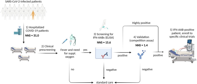
Purpose: Six to 19% of critically ill COVID-19 patients display circulating auto-antibodies against type I interferons (IFN-AABs). Here, we establish a clinically applicable strategy for early identification of IFN-AAB-positive patients for potential subsequent clinical interventions.

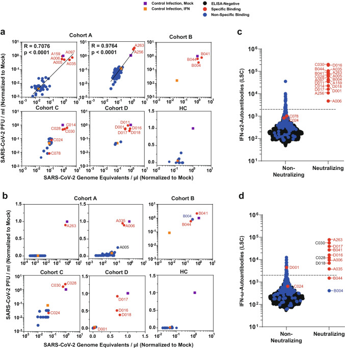
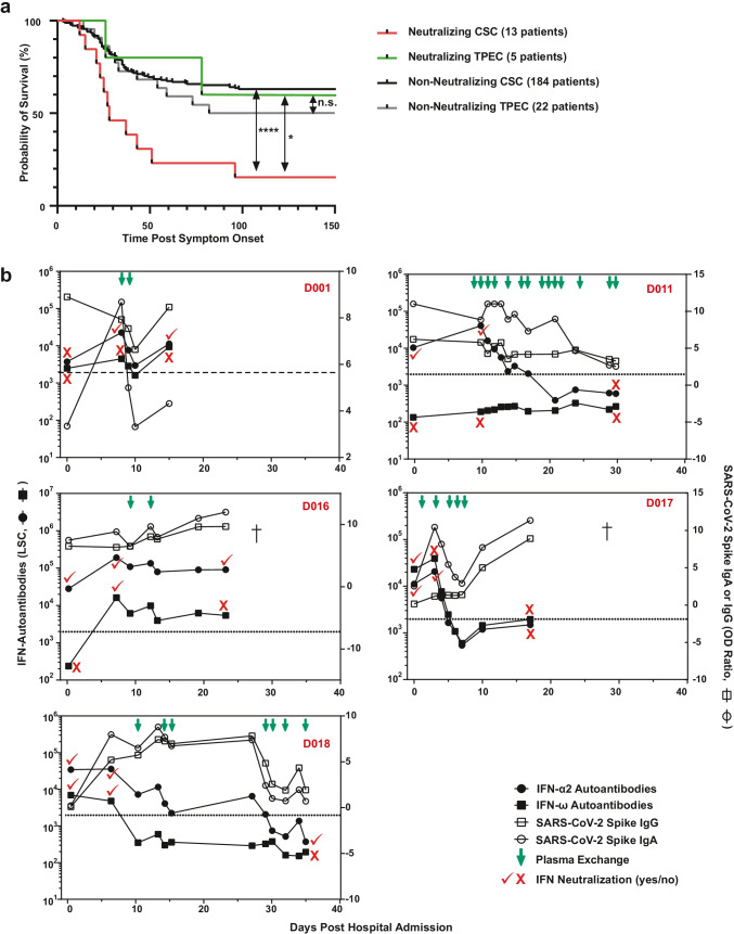
Methods: We analyzed sera of 430 COVID-19 patients from four hospitals for presence of IFN-AABs by ELISA. Binding specificity and neutralizing activity were evaluated via competition assay and virus-infection-based neutralization assay. We defined clinical parameters associated with IFN-AAB positivity. In a subgroup of critically ill patients, we analyzed effects of therapeutic plasma exchange (TPE) on the levels of IFN-AABs, SARS-CoV-2 antibodies and clinical outcome.

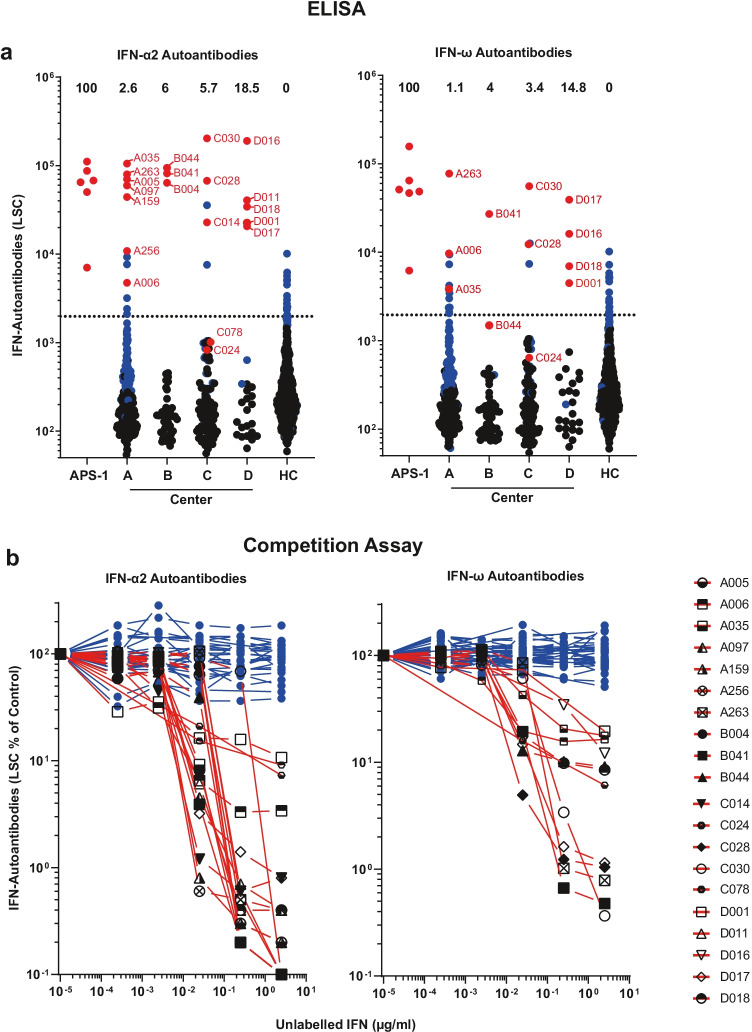
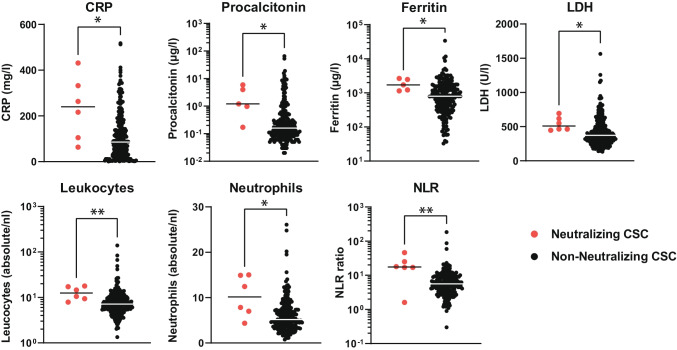
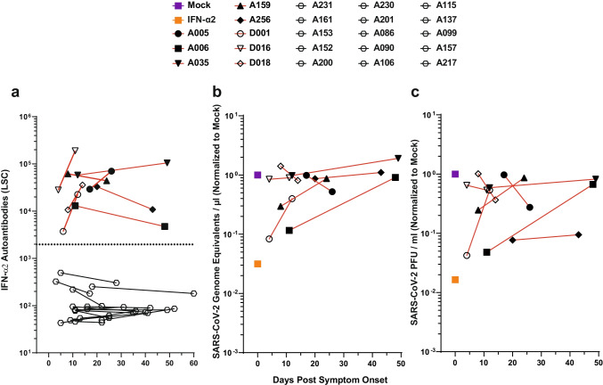
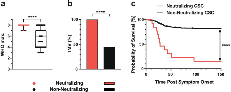
Results: The prevalence of neutralizing AABs to IFN-α and IFN-ω in COVID-19 patients from all cohorts was 4.2% (18/430), while being undetectable in an uninfected control cohort. Neutralizing IFN-AABs were detectable exclusively in critically affected (max. WHO score 6-8), predominantly male (83%) patients (7.6%, 18/237 for IFN-α-AABs and 4.6%, 11/237 for IFN-ω-AABs in 237 patients with critical COVID-19). IFN-AABs were present early post-symptom onset and at the peak of disease. Fever and oxygen requirement at hospital admission co-presented with neutralizing IFN-AAB positivity. IFN-AABs were associated with lower probability of survival (7.7% versus 80.9% in patients without IFN-AABs). TPE reduced levels of IFN-AABs in three of five patients and may increase survival of IFN-AAB-positive patients compared to those not undergoing TPE.

Conclusion: IFN-AABs may serve as early biomarker for the development of severe COVID-19. We propose to implement routine screening of hospitalized COVID-19 patients for rapid identification of patients with IFN-AABs who most likely benefit from specific therapies.

Keywords: Autoantibodies; COVID-19; SARS-CoV-2; Type I interferon.


Conflict of interest statement

V.M.C is named together with Euroimmun GmbH on a patent application filed recently regarding the diagnostic of SARS-CoV-2 by antibody testing. Technische Universität Berlin, Freie Universität Berlin, and Charité—Universitätsmedizin have filed a patent application for siRNAs inhibiting SARS-CoV-2 replication with D.N. as co-author. J.C.S, T.S. (full departmental disclosure): the department of Intensive Care Medicine has/had research and/or development/consulting contracts with (full disclosure) Orion Corporation, Abbott Nutrition International, B. Braun Medical AG, CSEM SA, Edwards Lifesciences Services GmbH/SA, Kenta Biotech Ltd, Maquet Critical Care AB, Omnicare Clinical Research AG, Phagenesis Ltd, Cytel, and Nestlé. No personal financial gains resulted from respective development/consulting contracts and/or educational grants.

Figures

Fig. 1
Fig. 1
Prevalence of AABs against IFN-α2 and IFN-ω in patients with COVID-19. a ECLIA-based assay for detection of IgG AABs against IFN-α2 and IFN-ω in sera from hospitalized patients with COVID-19 from four different university hospital cohorts (Center A, n = 266; Center B, n = 50; Center C, n = 87; Center D, n = 27), in patients with APS-1 (n = 6), and healthy health care workers (HC) without documented SARS-CoV-2 infection (n = 667). Dotted lines indicate the 97.5th percentile of the ECLIA assay LSC in sera from the HC cohort. Dots indicate samples containing AABs scoring specific (red) or unspecific (blue) for IFN-α2 and IFN-ω binding in the competition assay (see b), respectively. Samples depicted as black dots were not tested in the competition assay. The prevalence of sera with specifically binding type I IFN-AABs in each cohort is given in percent. b Specificity of the ECLIA assay signal for IFN-α2- and IFN-ω-AABs was tested in an competition assay by preincubation of sera with increasing concentrations of unlabeled IFN-α2 and IFN-ω protein (0–2.5 µg/ml) before analysis. Samples showing a decrease in assay signal by at least 75% in the presence of the highest competitor concentration were defined as specific for type I IFN antibody reactivity and are indicated with red lines (IFN-α2 n = 20, IFN-ω n = 12). Samples showing no decrease in the presence of excess unlabeled type I IFN protein were regarded as unspecific for type I IFN antibody reactivity and are indicated with blue lines (IFN-α2 n = 62, IFN-ω n = 39)
Fig. 2
Fig. 2
IFN-AABs neutralize exogenous IFN in a virus infection-based assay. a, b Selected sera were analyzed for IFN neutralization activity in a SARS-CoV-2 infection-based assay. The ability of individual sera to neutralize exogenous IFN-α2 (a) and IFN-ω (b) is shown by the rescue of susceptibility to infection as judged by quantification of viral RNA (x-axis) and infectivity (y-axis) in the supernatant. The infection condition in the absence of serum and IFN is set to 1. c, d The LSC value for individual sera, grouped into non-neutralizing and neutralizing sera, for the four COVID-19 cohorts. Dots indicate sera containing AABs scoring specific (red) or unspecific (blue) for IFN-α2 and IFN-ω binding in the competition assay (see b), respectively. Black dots indicate samples that scored below the threshold of the ELISA. Black dotted lines indicate the 97.5th percentile of the ECLIA assay LSC in sera from the healthy health care workers (HC) cohort (see Fig. 1). Neutralization ability of IFN-α and IFN-ω can be predicted at 100% for sera displaying LSCs above the respective red dotted lines (IFN-α: 35,639; IFN-ω: 12,603)
Fig. 3
Fig. 3
Laboratory parameters of COVID-19 patients displaying type I IFN-AABs. Values of C-reactive protein (CRP), procalcitonin, ferritin, lactate dehydrogenase (LDH), absolute leukocyte and neutrophil count, and neutrophil-to-lymphocyte ratio (NLR) of patients with (N = 5–6) and without neutralizing IFN-AABs (N = 200–265) from the cross-sectional cohort (CSC, all WHO scores). For each patient, the first available parameter within 72 h of hospital admission is shown. Statistical testing was performed with Mann–Whitney U test
Fig. 4
Fig. 4
In IFN-AAB-positive patients, high quantities of neutralizing IFN-α2-AABs were present both soon post-symptom onset and at the peak of disease. a Time course of antibody quantities in patient sera that scored IFN-AAB-positive at the peak of disease (N = 8, red lines). Additionally, time course of antibody quantities in patient sera that scored IFN-AAB-negative of the peak of disease is plotted (N = 15, black lines). The dotted line indicates the 97.5th percentile of the ECLIA assay LSC in sera from the HC cohort (see Fig. 1). b, c The ability of the sera to neutralize exogenous IFN-α2 is shown by the rescue of susceptibility to infection as judged by quantification of viral RNA (b) and infectivity (c) in the supernatant. The infection condition in the absence of serum and IFN is set to 1
Fig. 5
Fig. 5
Clinical outcome of COVID-19 patients with neutralizing IFN-AABs. a Median max. WHO score in hospital. Statistical testing was performed using the Mann–Whitney U test. b Proportion of patients requiring invasive mechanical ventilation (IMV) after hospital admission. Statistical testing was performed using the chi-square test. c Probability of survival of patients with and without neutralizing IFN-AABs from the cross-sectional cohort (CSC) from symptom onset until discharge (up to 150 days), death or transferral (p < 0.0001). Statistical testing was performed using a log-rank test. Neutralizing (N = 13), non-neutralizing (panels a and b: N = 372, panel c: N=369)
Fig. 6
Fig. 6
Inter-individual effect of therapeutic plasma exchange on IFN-AABs and SARS-CoV-2 antibodies. a Probability of survival of neutralizing IFN-AAB-positive and -negative patients with critical COVID-19 (max. WHO score 6–8) with and without plasma exchange (CSC and TPEC) from symptom onset until discharge, death or transferral (p = 0.04, neutralizing CSC versus neutralizing TPEC; p < 0.0001, neutralizing CSC versus non-neutralizing CSC). Statistical testing was performed using a log-rank test. Neutralizing CSC (N = 13), non-neutralizing CSC (N = 184), neutralizing TEPC (N = 5), and non-neutralizing TPEC (N = 22). b Antibody profile in serum from individual COVID-19 patients of the TPEC subjected to plasma exchange. The quantity of IFN-α2- and IFN-ω-AABs, SARS-CoV-2-IgG and -IgA, and the IFN-α2 and IFN-ω neutralization status are given for various time points. The patient identifier is given in red. Viral load profiles were only available for patients D011 and D018 and are shown in Supplementary Fig. 7
Fig. 7
Fig. 7
Proposed diagnostic algorithm for rapid identification of neutralizing IFN-AAB-positive patients. The number needed to screen (NNS) is based on results from the cross-sectional cohort (CSC). ELISA for IFN-AAB detection was considered to be positive if it exceeded the 97.5th percentile of the healthy control cohort. (1) NNS of all hospitalized COVID-19 patients without preselection was 31.0 (403 patients in total, 13 patients with neutralizing IFN-AABs). (2) Prescreening of patients using the clinical criteria of fever at admission and need for supplemental oxygen within the first 72 h after hospitalization diminished the NNS in the IFN-AAB ELISA (3) by half, to 15.6 (172/11). For patients identified as positive in the screening ELISA, the NNS in the competition assay to confirm the presence of IFN-specific AABs is reduced to 1.4 (15/11) (4). For patients with high-titer IFN-AABs (light signal count > 35.639), the competition assay can be omitted. Patients highly positive in the IFN-AAB ELISA and those with specific results in the competition assay may be included in clinical studies that aim testing specific therapies, including therapeutic plasma exchange (5). Figure created with BioRender.com

Similar articles

Cited by

References

    1. Pfortmueller CA, Spinetti T, Urman RD, Luedi MM, Schefold JC. COVID-19-associated acute respiratory distress syndrome (CARDS): current knowledge on pathophysiology and ICU treatment–a narrative review. Best Pract Res Clin Anaesthesiol. 2021;35(3):351–368. doi: 10.1016/j.bpa.2020.12.011. - DOI - PMC - PubMed
    1. Sah P, Fitzpatrick MC, Zimmer CF, Abdollahi E, Juden-Kelly L, Moghadas SM, et al. Asymptomatic SARS-CoV-2 infection: A systematic review and meta-analysis. Proceedings of the National Academy of Sciences. 2021;118(34):e2109229118. doi: 10.1073/pnas.2109229118. - DOI - PMC - PubMed
    1. Knight SR, Ho A, Pius R, Buchan I, Carson G, Drake TM, et al. Risk stratification of patients admitted to hospital with Covid-19 using the ISARIC WHO Clinical Characterisation Protocol: development and validation of the 4C Mortality Score. BMJ. 2020;370:m3339. doi: 10.1136/bmj.m3339. - DOI - PMC - PubMed
    1. Williamson EJ, Walker AJ, Bhaskaran K, Bacon S, Bates C, Morton CE, et al. Factors associated with COVID-19-related death using OpenSAFELY. Nature. 2020;584(7821):430–436. doi: 10.1038/s41586-020-2521-4. - DOI - PMC - PubMed
    1. COVID-19 Host Genetics Initiative. Mapping the human genetic architecture of COVID-19. Nature. 2021;600(7889):472–7. 10.1038/s41586-021-03767-x. - PMC - PubMed

Publication types