

ISCEV Standard for full-field clinical electroretinography (2022 update)
- PMID: 35511377
- PMCID: PMC9192408
- DOI: 10.1007/s10633-022-09872-0
ISCEV Standard for full-field clinical electroretinography (2022 update)
Abstract
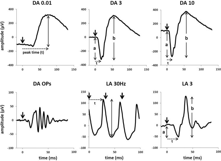
The full-field electroretinogram (ERG) is a mass electrophysiological response to diffuse flashes of light and is used widely to assess generalized retinal function. This document, from the International Society for Clinical Electrophysiology of Vision (ISCEV), presents an updated and revised ISCEV Standard for clinical ERG testing. Minimum protocols for basic ERG stimuli, recording methods and reporting are specified, to promote consistency of methods for diagnosis, monitoring and inter-laboratory comparisons, while also responding to evolving clinical practices and technology. The main changes in this updated ISCEV Standard for clinical ERGs include specifying that ERGs may meet the Standard without mydriasis, providing stimuli adequately compensate for non-dilated pupils. There is more detail about analysis of dark-adapted oscillatory potentials (OPs) and the document format has been updated and supplementary content reduced. There is a more detailed review of the origins of the major ERG components. Several tests previously tabulated as additional ERG protocols are now cited as published ISCEV extended protocols. A non-standard abbreviated ERG protocol is described, for use when patient age, compliance or other circumstances preclude ISCEV Standard ERG testing.
Keywords: Clinical standards; Electroretinogram (ERG); Full-field ERG; International Society of Clinical Electrophysiology of Vision (ISCEV).
© 2022. The Author(s).
Conflict of interest statement
The authors declare that they have no Conflict of interest.
Figures
References
MeSH terms
LinkOut - more resources
Full Text Sources
Other Literature Sources
Medical
