

Combinations of anti-GITR antibody and CD28 superagonist ameliorated dextran sodium sulfate-induced mouse colitis
- PMID: 35511600
- PMCID: PMC9226153
- DOI: 10.1093/cei/uxac039
Combinations of anti-GITR antibody and CD28 superagonist ameliorated dextran sodium sulfate-induced mouse colitis
Abstract
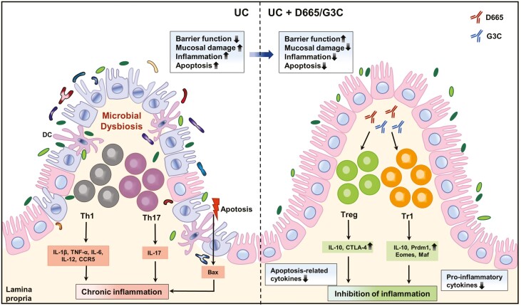
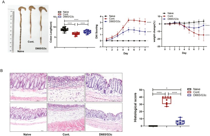
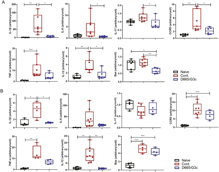
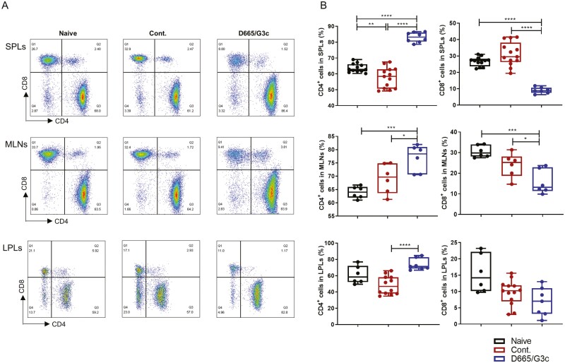
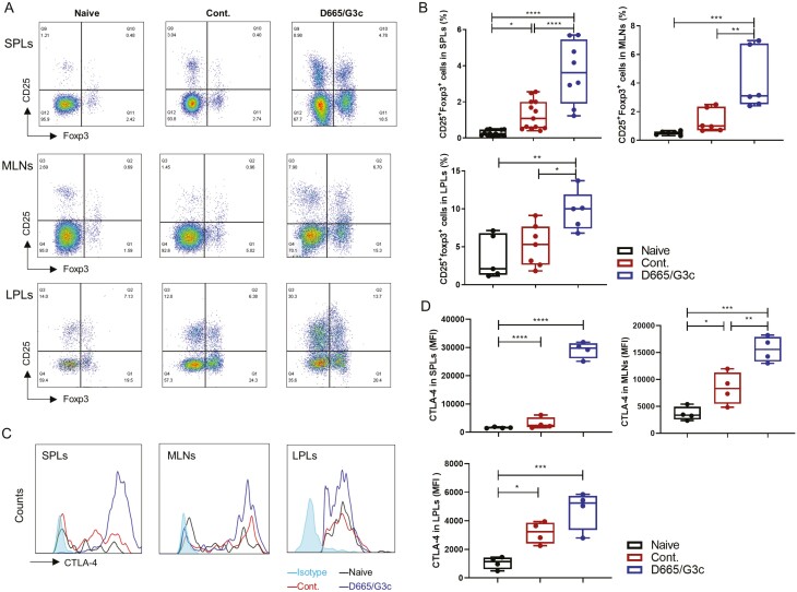
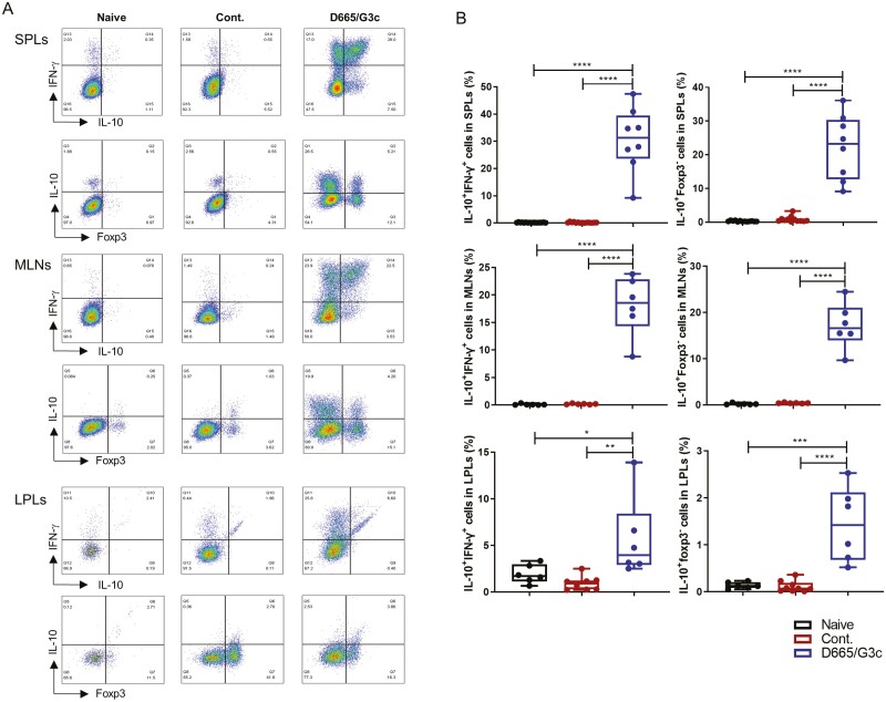
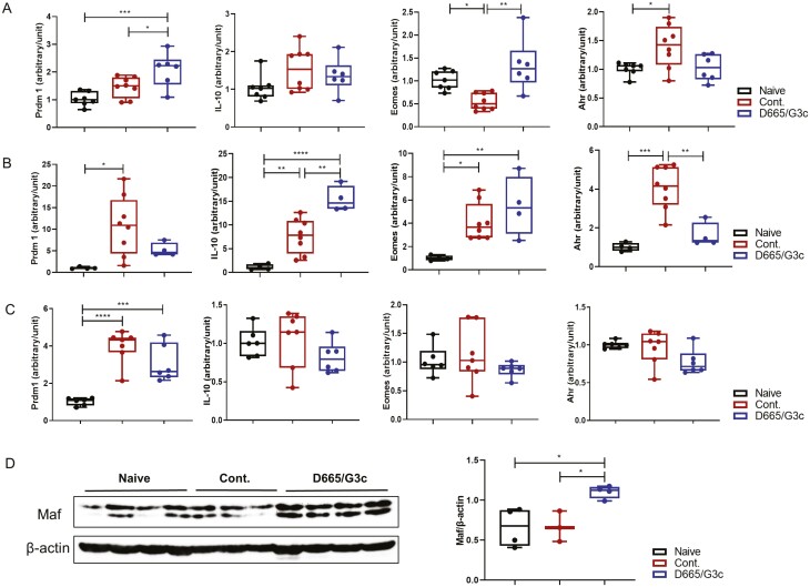
Ulcerative colitis (UC) is one of the two main forms of inflammatory bowel disease (IBD) and is an idiopathic, chronic inflammatory disease of the colonic mucosa with an unclear etiology. Interleukin (IL)-10 has been reported to play a crucial role in the maintenance of immune homeostasis in the intestinal environment. Type 1 regulatory T (Tr1) cells are a subset of CD4+Foxp3- T cells able to secrete high amounts of IL-10 with potent immunosuppressive properties. In this study, we found that the combination of anti-GITR antibody (G3c) and CD28 superagonist (D665) treatment stimulated the generation of a large amount of Tr1 cells. Furthermore, G3c/D665 treatment not only significantly relieved severe mucosal damage but also reduced the incidence of colonic shortening, weight loss, and hematochezia. Dextran sodium sulfate (DSS) upregulated the mRNA levels of IL-6, IL-1β, IL-17, IL-12, tumor necrosis factor-alpha, C-C chemokine receptor type 5, and Bax in splenic lymphocytes (SPLs) and colon tissues, while G3c/D665 treatment conversely inhibited the increase in mRNA levels of these genes. In addition, G3c/D665 treatment altered the proportion of CD4+ and CD8+ T cells and increased CD4+CD25+Foxp3+ regulatory T cells in SPLs, mesenteric lymph nodes (MLNs), and lamina propria lymphocytes (LPLs). Thus, the combination of G3c and D665 treatment showed efficacy against DSS-induced UC in mice by inducing a large amount of Tr1 cell generation via the musculoaponeurotic fibrosarcoma pathways in vivo and relieving inflammatory responses both systematically and locally.
Keywords: CD28 superagonist; anti-GITR antibody; dextran sulfate sodium; inflammatory bowel disease; type 1 regulatory T cells.
© The Author(s) 2022. Published by Oxford University Press on behalf of the British Society for Immunology. All rights reserved. For permissions, please e-mail: journals.permissions@oup.com.
Figures

Similar articles
-
The effects of Foxp3-expressing regulatory T cells expanded with CD28 superagonist antibody in DSS-induced mice colitis.Int Immunopharmacol. 2011 May;11(5):610-7. doi: 10.1016/j.intimp.2010.11.034. Epub 2010 Dec 14. Int Immunopharmacol. 2011. PMID: 21163250
-
Combinations of anti-GITR antibody and CD28 superagonist induce permanent allograft acceptance by generating type 1 regulatory T cells.Sci Adv. 2022 Aug 5;8(31):eabo4413. doi: 10.1126/sciadv.abo4413. Epub 2022 Aug 3. Sci Adv. 2022. PMID: 35921418 Free PMC article.
-
Berberine ameliorates chronic relapsing dextran sulfate sodium-induced colitis in C57BL/6 mice by suppressing Th17 responses.Pharmacol Res. 2016 Aug;110:227-239. doi: 10.1016/j.phrs.2016.02.010. Epub 2016 Mar 9. Pharmacol Res. 2016. PMID: 26969793
-
Indirubin ameliorates dextran sulfate sodium-induced ulcerative colitis in mice through the inhibition of inflammation and the induction of Foxp3-expressing regulatory T cells.Acta Histochem. 2016 Jul;118(6):606-614. doi: 10.1016/j.acthis.2016.06.004. Epub 2016 Jul 7. Acta Histochem. 2016. PMID: 27396532
-
Yogurt starter cultures of Streptococcus thermophilus and Lactobacillus bulgaricus ameliorate symptoms and modulate the immune response in a mouse model of dextran sulfate sodium-induced colitis.J Dairy Sci. 2019 Jan;102(1):37-53. doi: 10.3168/jds.2018-14520. Epub 2018 Oct 19. J Dairy Sci. 2019. PMID: 30343915
Cited by
-
Targeted spleen modulation: a novel strategy for next-generation disease immunotherapy.Theranostics. 2025 Mar 18;15(10):4416-4445. doi: 10.7150/thno.111116. eCollection 2025. Theranostics. 2025. PMID: 40225564 Free PMC article. Review.
References
-
- Ordas I, Eckmann L, Talamini M, et al. . Ulcerative colitis. Lancet 2012, 380, 1606–19. - PubMed
-
- Geremia A, Biancheri P, Allan P, et al. . Innate and adaptive immunity in inflammatory bowel disease. Autoimmun Rev 2014, 13, 3–10. - PubMed
-
- Cao H, Liu J, Shen P, et al. . Protective effect of naringin on DSS-induced ulcerative colitis in mice. J Agric Food Chem 2018, 66, 13133–40. - PubMed
-
- Cassinotti A, Passamonti F, Segato S.. Cell therapy in inflammatory bowel disease. Pharmacol Res 2021, 163, 105247. - PubMed
Publication types
MeSH terms
Substances
LinkOut - more resources
Full Text Sources
Medical
Research Materials
