. 2022 Dec 1;24(12):2121-2132.
doi: 10.1093/neuonc/noac111.

HIP1R and vimentin immunohistochemistry predict 1p/19q status in IDH-mutant glioma

Affiliations

HIP1R and vimentin immunohistochemistry predict 1p/19q status in IDH-mutant glioma

Marius Felix et al. Neuro Oncol. .

Abstract

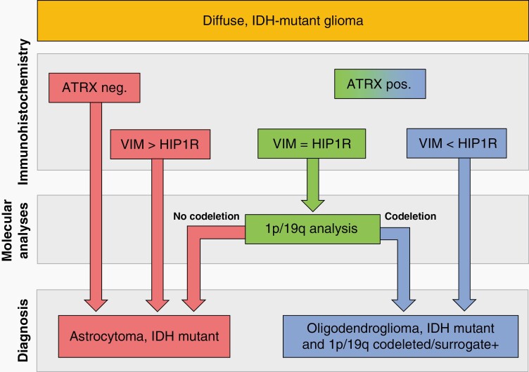
Background: IDH-mutant gliomas are separate based on the codeletion of the chromosomal arms 1p and 19q into oligodendrogliomas IDH-mutant 1p/19q-codeleted and astrocytomas IDH-mutant. While nuclear loss of ATRX expression excludes 1p/19q codeletion, its limited sensitivity prohibits to conclude on 1p/19q status in tumors with retained nuclear ATRX expression.

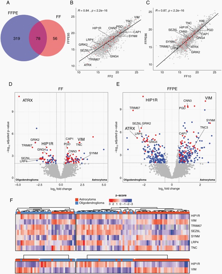
Methods: Employing mass spectrometry based proteomic analysis in a discovery series containing 35 fresh frozen and 72 formalin fixed and paraffin embedded tumors with established IDH and 1p/19q status, potential biomarkers were discovered. Subsequent validation immunohistochemistry was conducted on two independent series (together 77 oligodendrogliomas IDH-mutant 1p/19q-codeleted and 92 astrocytomas IDH-mutant).

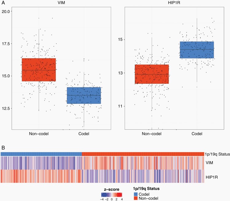
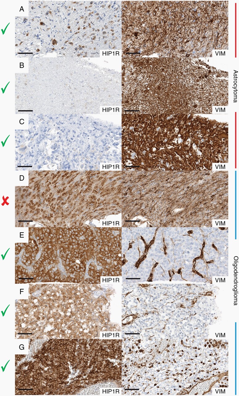
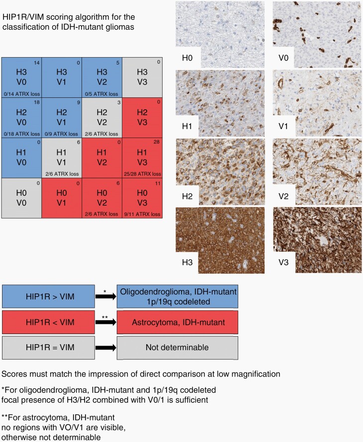
Results: We detected highly specific protein patterns distinguishing oligodendroglioma and astrocytoma. In these patterns, high HIP1R and low vimentin levels were observed in oligodendroglioma while low HIP1R and high vimentin levels occurred in astrocytoma. Immunohistochemistry for HIP1R and vimentin expression in 35 cases from the FFPE discovery series confirmed these findings. Blinded evaluation of the validation cohorts predicted the 1p/19q status with a positive and negative predictive value as well as an accuracy of 100% in the first cohort and with a positive predictive value of 83%; negative predictive value of 100% and an accuracy of 92% in the second cohort. Nuclear ATRX loss as marker for astrocytoma increased the sensitivity to 96% and the specificity to 100%.

Conclusions: We demonstrate that immunohistochemistry for HIP1R, vimentin, and ATRX predict 1p/19q status with 100% specificity and 95% sensitivity and therefore, constitutes a simple and inexpensive approach to the classification of IDH-mutant glioma.

Keywords: 1p/19q; ATRX; HIP1R; IDH; Vimentin; proteomics.


Figures

Fig. 1.
Fig. 1.
Differential protein abundance analysis of astrocytoma and oligodendroglioma. (A) Venn diagramm depicting number of significantly DAP in FF and FFPE tissue. Overlap shows the number of proteins which were significantly different in both cohorts. Pearson correlation analyses between a matched pair of FF and FFPE of an astrocytoma (B) and an oligodendroglioma (C). Volcano plot of significantly differentially expressed proteins (DAP) between astrocytoma and oligodendroglioma in FF (D) and FFPE (E) tissue. Blue dots represent proteins which were found either in the FF or FFPE tissues exclusively, red dots represent proteins which were found consistently in both cohorts. Dashed lines represent threshold for significance (P-value <.01) and log2 fold change (1,5). (F): Hierarchical clustering of the FF (left) and FFPE (right) cohort using a subset of DAP (top) and HIP1R and VIM (bottom) between astrocytoma and oligodendroglioma.
Fig. 2.
Fig. 2.
mRNA expression levels of IDH-mutant glioma. Top: Boxplots log2 normalized expression values of VIM (left) and HIP1R (right) originating from the TCGA database. High levels of VIM coding mRNA in IDH-mutant glioma without 1p/19 codeletion and low levels of VIM in glioma with 1p/19q codeletion are observed. Vice versa high levels of HIP1R coding mR NA are found in 1p/19q codeleted glioma and low levels of HIP1R in glioma without 1p/19 codeletion. Bottom: Heatmap showing Z-scored expression values, samples were ordered by their 1p/19q status. Pairwise comparison of z-scored expression values demonstrates that 1p/19q codeleted glioma exhibiting low levels of VIM and high levels of HIP1R. In contrast glioma without 1p/19q codeletion exhibit high levels of VIM and lower levels of HIP1R.
Fig. 3.
Fig. 3.
Examples of HIP1R/VIM immunohistochemistry for the classification of IDH-mutant glioma. Pairwise comparison of HIP1R (left side) and VIM (right side) in astrocytomas (top 3) and oligodendrogliomas (bottom 3). (A–C) Astrocytomas with low-level HIP1R expression and high-level VIM expression. (D) A nondeterminable oligodendroglioma with similar HIP1R and VIM staining intensities. (E–G) Oligodendrogliomas with high-level HIP1R expression and low-level VIM expression. Check marks indicate determinable cases, cross indicates not-determinability. Black line indicates a length of 60 µm.
Fig. 4.
Fig. 4.
Graphical algorithm for the classification of IDH-mutant glioma according to HIP1R/VIM immunohistochemistry. The HIP1R (H) level should be evaluated first. If the H score is higher than the VIM (V) score the tumor is identified as oligodendroglioma. Note that focal presence of a moderate to strong HIP1R level (H3/H2) in combination with low-level VIM (V0/V1) in the same area is sufficient for making this conclusion. If the VIM score is higher than the HIP1R score the tumor is identified as astrocytoma, as long as there are no areas in the tumor with sparse VIM expression (V0, V1) as well. If the HIP1R and the VIM levels are ambigous, the stains should be compared directly at low magnification. If HIP1R is clearly stronger, the tumor is identified as oligodendroglioma. If VIM is clearly stronger, the tumor is identified as astrocytoma. Similar expression levels of HIP1R and VIM in the tumor are interpreted as not determinable. The distribution of HIP1R and VIM expression from the HD validation cohort (n = 100; observer 1) is shown in the top right corner and the fraction of samples with ATRX loss is noted on the bottom of every combination box.
Fig. 5.
Fig. 5.
Flowchart for the efficient use of ATRX and HIP1R/VIM for the classification of IDH-mutant gliomas. Tumors with nuclear loss of ATRX can be classified as astrocytoma. Tumors with retained or nondeterminable ATRX status are evaluated with HIP1R/VIM immunohistochemistry. VIM > HIP1R tumors are classified as astrocytomas, VIM < HIP1R are classified as oligodendrogliomas. VIM = HIP1R tumors need molecular 1p/19q testing for classification.

Similar articles

Cited by

References

    1. Louis DN, Ohgaki H, Wiestler OD, et al. . WHO Classification and Grading of Tumours of the Central Nervous System. Lyon: IARC Press; International Agency for Research on Cancer; 2016.
    1. Weller M, van den Bent M, Preusser M, et al. . EANO guidelines on the diagnosis and treatment of diffuse gliomas of adulthood. Nat Rev Clin Oncol. 2021; 18(3):170–186. - PMC - PubMed
    1. Balss J, Meyer J, Mueller W, et al. . Analysis of the IDH1 codon 132 mutation in brain tumors. Acta Neuropathol. 2008; 116(6):597–602. - PubMed
    1. Capper D, Zentgraf H, Balss J, Hartmann C, von Deimling A. Monoclonal antibody specific for IDH1 R132H mutation. Acta Neuropathol. 2009; 118(5):599–601. - PubMed
    1. Woehrer A, Hainfellner JA. Molecular diagnostics: techniques and recommendations for 1p/19q assessment. CNS Oncol. 2015; 4(5):295–306. - PMC - PubMed

Publication types

MeSH terms