. 2022 Jul 30;34(8):2969-2988.
doi: 10.1093/plcell/koac133.

A coiled-coil protein associates Polycomb Repressive Complex 2 with KNOX/BELL transcription factors to maintain silencing of cell differentiation-promoting genes in the shoot apex

Affiliations

A coiled-coil protein associates Polycomb Repressive Complex 2 with KNOX/BELL transcription factors to maintain silencing of cell differentiation-promoting genes in the shoot apex

Feng-Quan Tan et al. Plant Cell. .

Abstract

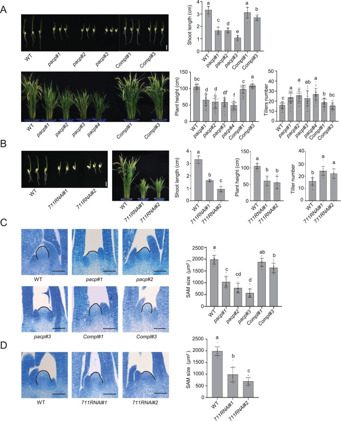
Polycomb repressive complex 2 (PRC2), which mediates the deposition of H3K27me3 histone marks, is important for developmental decisions in animals and plants. In the shoot apical meristem (SAM), Three Amino acid Loop Extension family KNOTTED-LIKE HOMEOBOX /BEL-like (KNOX/BELL) transcription factors are key regulators of meristem cell pluripotency and differentiation. Here, we identified a PRC2-associated coiled-coil protein (PACP) that interacts with KNOX/BELL transcription factors in rice (Oryza sativa) shoot apex cells. A loss-of-function mutation of PACP resulted in differential gene expression similar to that observed in PRC2 gene knockdown plants, reduced H3K27me3 levels, and reduced genome-wide binding of the PRC2 core component EMF2b. The genomic binding of PACP displayed a similar distribution pattern to EMF2b, and genomic regions with high PACP- and EMF2b-binding signals were marked by high levels of H3K27me3. We show that PACP is required for the repression of cell differentiation-promoting genes targeted by a rice KNOX1 protein in the SAM. PACP is involved in the recruitment or stabilization of PRC2 to genes targeted by KNOX/BELL transcription factors to maintain H3K27me3 and gene repression in dividing cells of the shoot apex. Our results provide insight into PRC2-mediated maintenance of H3K27me3 and the mechanism by which KNOX/BELL proteins regulate SAM development.


Figures

Figure 1
Figure 1
Identification of PACP as a PRC2-associated protein. A, Mass spectrometric identification of FLAG antibody affinity-purified proteins from the extracts of FIE2FLAG SAM tissues (FIE2-FLAG). Extract from WT SAM tissues was used as a control (CK). Two biological repeats (separate experiments) were performed. The total numbers of identified unique peptides for each protein are indicated. B, Anti-FLAG Co-IP assays of proteins isolated from SAM tissues of FIE2-FLAG and WT plants. The precipitated proteins were analyzed by immunoblotting with the indicated antibodies. Anti-OsbZIP23 was used as a negative control. C, Y2H assay of the interactions of PACP with PRC2 proteins. The empty vector pGADT7 was used as a negative control. D, Split-luciferase complementation assay. Nicotiana benthamiana leaf cells were co-transformed with cLUC-tagged PRC2 proteins (SDG711, SDG718, EMF2b, FIE2, and LHP1) or cLUC alone with nLUC-tagged PACP or nLUC alone as shown. Diagrams of the constructs are shown on the right. E, Y2H assay of the truncated regions of PACP and PRC2 proteins. Full-length SDG711 (aa 1–896), SDG711-1 (aa 121–896), SDG711-2 (aa 249–896), SDG711-3 (aa 324–896), SDG711-4 (aa 523–896), SDG711-5 (aa 1–248), SDG711-6 (aa 121–323), SDG711-7 (aa 121–248), SDG711-8 (aa 249–522), EMF2b-FL (aa 1–604), EMF2b-1 (aa 1–207), EMF2b-2 (aa 208–452), EMF2b-3 (aa 453–604), EMF2b-4 (aa 208–604), PACP (aa 1–170), PACP-N (aa 1–76), and PACP-C (aa 77–170) were fused to the GAL4 BD or activation domain as indicated. Yeast cells were spotted onto stringent selection medium −WLHA with 40 �g mL−1 X-Gal or a nonselective medium −WL as a control. Right: Structures of SDG711, EMF2b, and PACP proteins. F, In vitro pull-down assay of the interaction between PACP and SDG711 or EMF2b. PACP–SUMO–6xHis was incubated with SDG711-8–GST, EMF2b-4–GST, or GST alone. Following pull down with GST beads, the eluates were analyzed by immunoblotting using the indicated antibodies.
Figure 2
Figure 2
PACP is required for shoot development. A, Phenotypes of WT, pacp mutant, and complementation lines at 7 days after germination (DAG) (upper, Bar = 1 cm) and the mature (heading) stage (lower). Shoot lengths of seedlings (10 plants per line), mature plant height, and tiller number (>25 plants/line) of the different genotypes were measured (right). B, Phenotypes of WT and SDG711 RNAi plants at 7 DAG (left, Bar = 1 cm) and the mature (heading) stage. The plant lengths and tiller number were measured as in (A). C, Shoot apex sections of WT, pacp, and complementation lines at 5 DAG. Bars = 50 μm. SAM size was determined by measuring the dome area delimited by drawing a straight line between the basal edges of the two opposing youngest leaf primordia. Ten plants per line were measured. D, Shoot apex sections of WT and SDG711 RNAi lines at 5 DAG. Bar = 50 μm. SAM size was measured as in (C). Data represent the means � sd (10 plants/line). Significance of differences (denoted by different letters) was tested by ANOVA.
Figure 3
Figure 3
The pacp mutation has a similar effect as SDG711 RNAi on the SAM transcriptome. A, Average read count plots of gene expression changes in pacp versus WT SAM. The x axis represents average gene expression level (FPKM), and the y axis represents log2 FPKM FC (pacp/WT). Significant [FDR < 0.05 and 1.5-FC or 2-FC (in parentheses)] upregulated and downregulated DEGs are indicated. B, Relative enrichment of transcription factor genes in the pacp DEGs. The frequency of transcription factor genes among upregulated or downregulated DEGs was compared with the expected genomic frequency. P-values were calculated using Fisher’s test. C, Relative enrichments of transcription factor families among pacp DEGs. Detected gene number over the total gene number is indicated for each family. D, Average count plots of gene expression changes in 711RNAi versus WT. Significant (FDR < 0.05 and 1.5-FC or 2 FC [in parentheses]) upregulated and downregulated DEGs are indicated. E, pacp DEGs positively correlated with 711RNAi DEGs. The x-axis shows pacp DEGs, and the y-axis shows 711RNAi DEGs. F, Venn diagrams showing the overlap between pacp and up and downregulated DEGs with FC > 1.5 and 2.0. P-values were calculated by hypergeometric test.
Figure 4
Figure 4
Loss of PACP leads to decreased EMF2b binding. A, Violin plots (with boxplots inside) of normalized EMF2b ChIP-seq read enrichment scores in WT and pacp SAM. The P-value for log2 normalized read counts between WT and pacp was calculated based on a two-sided Wilcoxon rank-sum test with Bonferroni correction. B, Metaplots of EMF2b-binding peaks in WT and pacp. C, Scatter plots of changes in EMF2b-binding in pacp versus WT. All EMF2b peaks (n = 9,937) were plotted. Significantly (FC > 1.5, FDR < 0.01) increased and reduced EMF2b binding peaks and numbers are indicated. D, Metaplots and violin plots of H3K27me3 levels of peaks with (left) and without (right) EMF2b binding in WT and pacp. The P-values were calculated based on a two-sided Wilcoxon rank-sum test with Bonferroni correction. E, Venn diagram showing the overlap between H3K27me3-marked genes with decreased EMF2b binding and genes with decreased H3K27me3 in pacp compared to the WT. The P-value was calculated by hypergeometric test.
Figure 5
Figure 5
Identification of PACP-binding targets. A, Heatmaps of PACP and EMF2b binding and H3K27me3 signals at the PACP-binding peaks (n = 10,769). The top tier (3,239/10,769) PACP-binding peaks were bound by EMF2b and marked by H3K27me3. B, PACP-binding genes are highly enriched for H3K27me3 relative to the genomic average. C, Metaplots and violin plots of H3K27me3 levels of the 3,239 peaks commonly bound by EMF2b and PACP (upper) compared to those without PACP binding (lower) in WT and pacp. The P-value was calculated based on a two-sided Wilcoxon rank-sum test. D, Metaplots and violin plots of EMF2b-binding signals of the 3,239 commonly marked peaks (upper) compared to those without PACP binding (lower) in WT and pacp. The P-value was calculated based on a two-sided Wilcoxon rank-sum test. E, Correlation of changes between H3K27me3 and EMF2b binding levels of the 1,289 genes commonly marked by H3K27me3 and PACP and EMF2b binding (from 3,239 peaks) in pacp versus WT. F, Heatmaps of H3K27me3 and EMF2b-binding signals of the 1,289 genes in WT and pacp. Examples of target genes are shown on the right. G, Venn diagram showing the overlap between genes of cluster 2 of the heatmap (F) and upregulated DEGs in pacp.
Figure 6
Figure 6
Integrative Genomics Viewer image of 12 transcription factor genes. PACP and EMF2b binding, H3K27me3, and RNA-seq data of 12 transcription factor genes selected from Table�1 in WT and pacp. The significant binding regions are indicated in black dotted boxes.
Figure 7
Figure 7
PACP interacts with KNOX and BELL proteins in vitro and in vivo. A, PACP interacts with KNOX and BELL proteins in an Y2H assay. WOX11 was used as a control. B, Split-luciferase complementation assay of the interactions of PACP with the indicated KNOX and BELL proteins. cLUC-tagged PACP or cLUC alone was co-transformed into N. benthamiana leaf cells with nLUC-tagged KNOX/BELL or nLUC alone. C, Y2H assay examining the interaction between the truncated regions of PACP and OSH1. The KN1 and KN2 domains of OSH1 and the C-terminus of PACP are required for this interaction. D, In vitro pull-down assay of the interaction of PACP with OSH1. Arrows mark the protein band as indicated. E, PACP interacts with OSH1 in N. benthamiana cells. The 35S:PACPGFP or 35S:GFP vector was transiently co-transfected into N. benthamiana leaf cells with the 35S:OSH13xFLAG vector. Total proteins were precipitated with anti-GFP beads. Anti-FLAG was used to detect OSH1 by immunoblotting. Arrows mark the protein band as indicated. F, PACP interacts with OSH1 in rice cells. Nuclei isolated from SAM tissue of 5-day-old WT and Compl#1 (PACP–2xFLAG) seedlings were precipitated with anti-FLAG and analyzed by immunoblotting with anti-FLAG to detect PACP and with anti-OSH1 antibody to detect OSH1.
Figure 8
Figure 8
PACP represses the expression of lateral organ development-related genes in the SAM. A, RT-qPCR validation of the expression of the six genes in the SAM of WT, pacp mutants, and the complementation line. Data represent the means � sd (three separate experiments per sample). Significance of differences (denoted by different letters) was tested by one-way ANOVA. B, In situ hybridization of DL and YAB4 transcripts in the shoot apex of WT, pacp and OSH1 RNAi plants. Arrows indicate signals in the SAM and leaf primordia. Bar = 50 μm.

Comment in

  • PACP recruits PRC2 to TALE targets.
    Vadde BVL. Vadde BVL. Plant Cell. 2022 Jul 30;34(8):2821-2822. doi: 10.1093/plcell/koac159. Plant Cell. 2022. PMID: 35666578 Free PMC article. No abstract available.

Similar articles

Cited by

References

    1. Belles-Boix E, Hamant O, Witiak SM, Morin H, Traas J, Pautot V (2006) KNAT6: an Arabidopsis homeobox gene involved in meristem activity and organ separation. Plant Cell 18: 1900–1907 - PMC - PubMed
    1. Berger N, Dubreucq B, Roudier F, Dubos C, Lepiniec L (2011) Transcriptional regulation of Arabidopsis LEAFY COTYLEDON2 involves RLE, a cis-element that regulates trimethylation of histone H3 at lysine-27. Plant Cell 23: 4065–4078 - PMC - PubMed
    1. Blackledge NP, Rose NR, Klose RJ (2015) Targeting Polycomb systems to regulate gene expression: modifications to a complex story. Nat Rev Mol Cell Biol 16: 643–649 - PMC - PubMed
    1. Bolger AM, Lohse M, Usadel B (2014) Trimmomatic: a flexible trimmer for Illumina sequence data. Bioinformatics 30: 2114–2120 - PMC - PubMed
    1. Bouyer D, Roudier F, Heese M, Andersen ED, Gey D, Nowack MK, Goodrich J, Renou JP, Grini PE, Colot V, et al (2011) Polycomb repressive complex 2 controls the embryo-to-seedling phase transition. PLoS Genet 7: e1002014. - PMC - PubMed

Publication types