. 2022 Sep 14;145(9):3108-3130.
doi: 10.1093/brain/awac165.

Altered SOD1 maturation and post-translational modification in amyotrophic lateral sclerosis spinal cord

Affiliations

Altered SOD1 maturation and post-translational modification in amyotrophic lateral sclerosis spinal cord

Benjamin G Trist et al. Brain. .

Abstract

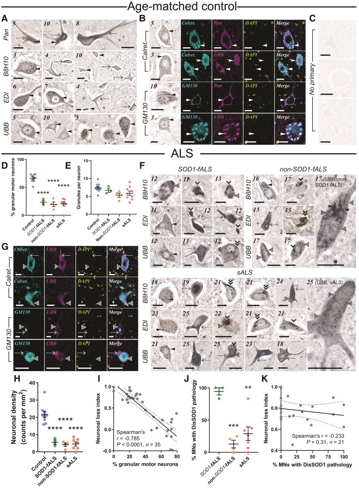
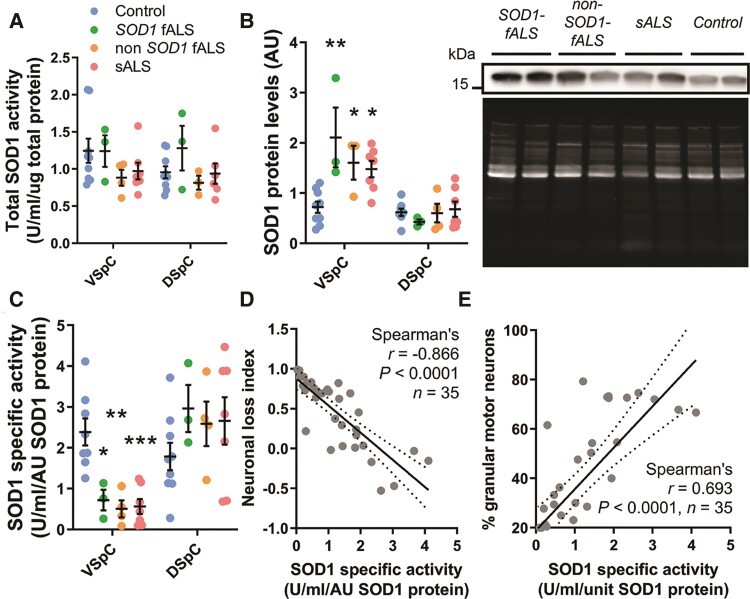
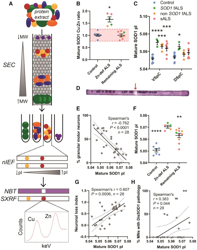
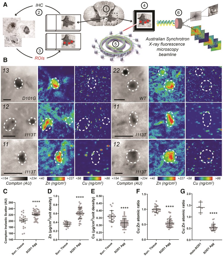
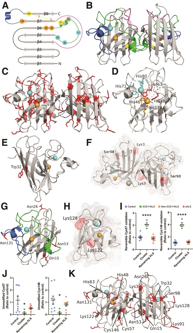
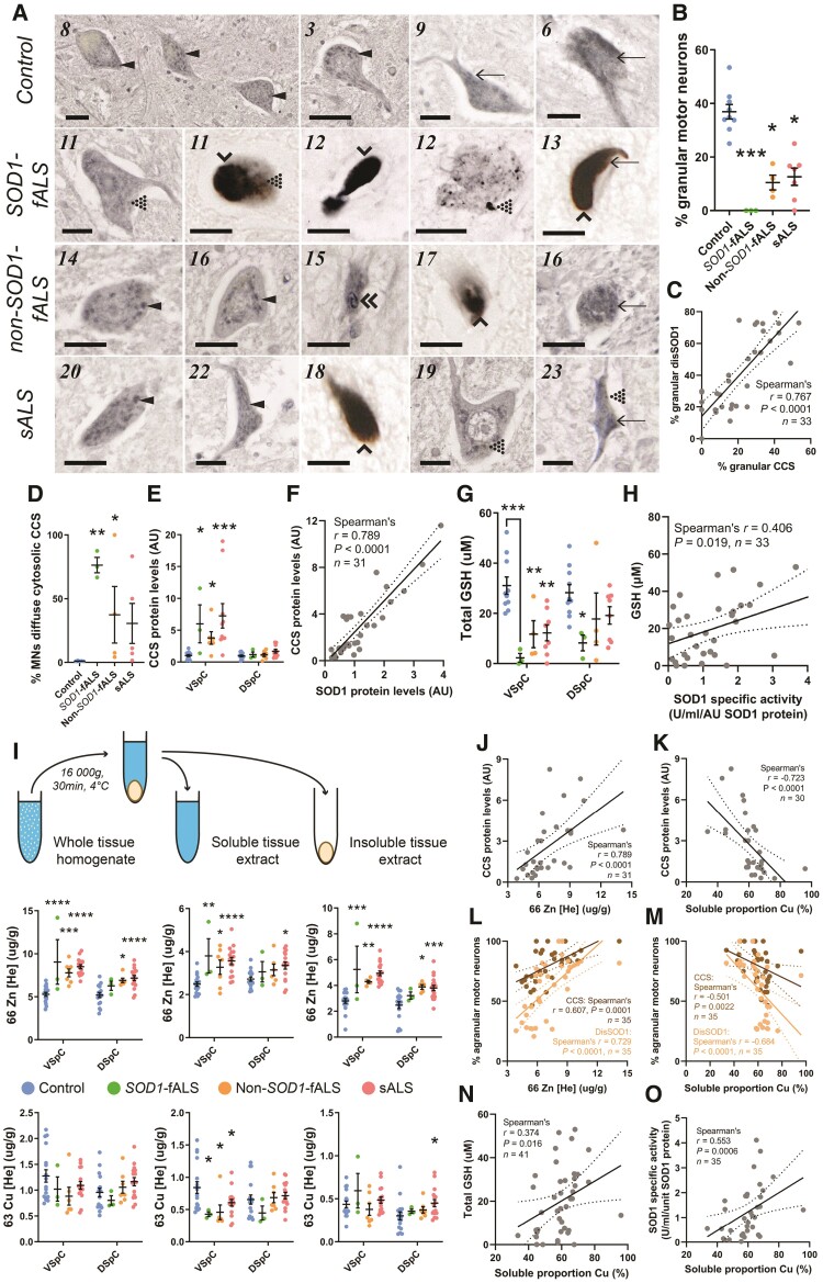
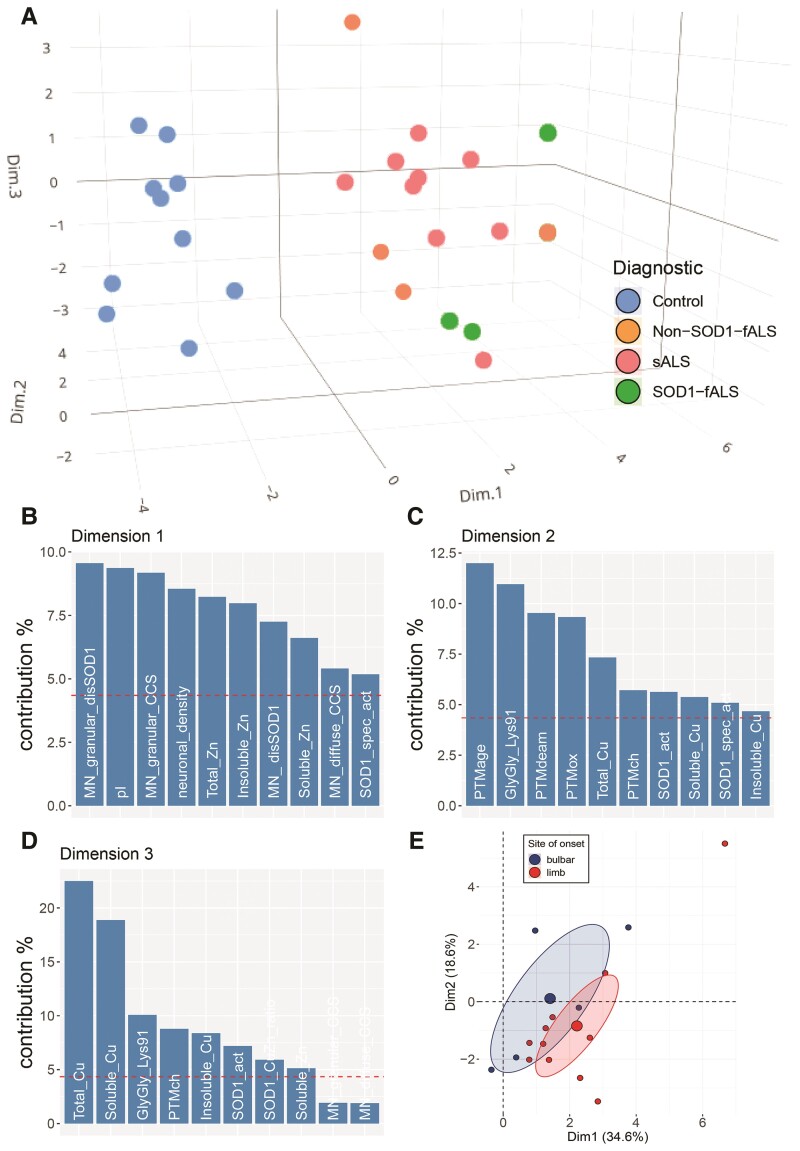
Aberrant self-assembly and toxicity of wild-type and mutant superoxide dismutase 1 (SOD1) has been widely examined in silico, in vitro and in transgenic animal models of amyotrophic lateral sclerosis. Detailed examination of the protein in disease-affected tissues from amyotrophic lateral sclerosis patients, however, remains scarce. We used histological, biochemical and analytical techniques to profile alterations to SOD1 protein deposition, subcellular localization, maturation and post-translational modification in post-mortem spinal cord tissues from amyotrophic lateral sclerosis cases and controls. Tissues were dissected into ventral and dorsal spinal cord grey matter to assess the specificity of alterations within regions of motor neuron degeneration. We provide evidence of the mislocalization and accumulation of structurally disordered, immature SOD1 protein conformers in spinal cord motor neurons of SOD1-linked and non-SOD1-linked familial amyotrophic lateral sclerosis cases, and sporadic amyotrophic lateral sclerosis cases, compared with control motor neurons. These changes were collectively associated with instability and mismetallation of enzymatically active SOD1 dimers, as well as alterations to SOD1 post-translational modifications and molecular chaperones governing SOD1 maturation. Atypical changes to SOD1 protein were largely restricted to regions of neurodegeneration in amyotrophic lateral sclerosis cases, and clearly differentiated all forms of amyotrophic lateral sclerosis from controls. Substantial heterogeneity in the presence of these changes was also observed between amyotrophic lateral sclerosis cases. Our data demonstrate that varying forms of SOD1 proteinopathy are a common feature of all forms of amyotrophic lateral sclerosis, and support the presence of one or more convergent biochemical pathways leading to SOD1 proteinopathy in amyotrophic lateral sclerosis. Most of these alterations are specific to regions of neurodegeneration, and may therefore constitute valid targets for therapeutic development.

Keywords: amyotrophic lateral sclerosis; mislocalization; neurodegeneration; post-translational modifications; superoxide dismutase-1.


Figures

Figure 1
Figure 1
DisSOD1 accumulation, aggregation and mislocalization in the vulnerable ventral spinal cord of ALS cases. (A) Pan-SOD1 antibodies, as well as conformation-specific antibodies raised against disordered mutant SOD1 (B8H10), disSOD1 with an exposed dimer interface and disSOD1 with an unfolded β-barrel (UBB) were used to profile the distribution of mature SOD1 and disSOD1 conformers within control (n = 10) ventral spinal cord tissues. These SOD1 antibodies (Supplementary Table 3) detected granular cytoplasmic staining (arrowheads) and diffuse cytosolic staining (arrows) within control motor neurons. (B) Visualization of granular SOD1 immunostaining within control spinal cord motor neurons using the UBB conformation-specific SOD1 antibody and DAB chromogen. Granular SOD1 immunostaining strongly colocalized with the ER–Golgi markers Calreticulin and GM130 in control motor neurons. DAPI was also used to visualize cell nuclei. (C) No immunostaining was observed in spinal cord tissue sections processed as before in the absence of primary antibodies. (D) Motor neurons possessing granular SOD1 immunostaining were less abundant in all ALS subgroups compared with controls (one-way ANOVA: P < 0.0001, F = 55.82; Dunnett’s multiple comparisons post hoc tests: P < 0.0001 for all comparisons). (E) Granule numbers within surviving ALS motor neurons exhibiting granular SOD1 immunostaining were equivalent to those within control motor neurons (Kruskal–Wallis H-test, P = 0.073, H = 6.975). (F) In addition to granular and diffuse cytosolic immunostaining, conformation-specific antibodies labelled a variety of disSOD1 inclusions in ventral spinal cord motor neurons of SOD1-fALS (n = 3), non-SOD1-fALS (n = 4) and sALS (n = 9) cases; punctate inclusions (dotted arrowheads), globular inclusions (single downwards arrowheads) and fibrillar skein-like inclusions (double downwards arrowheads). Case numbers (Supplementary Table 1) are listed in the top left corner of each panel in A and F. (G) DisSOD1 conformers immunolabelled by the UBB SOD1 antibody were not colocalized with the ER–Golgi markers Calreticulin and GM130 in motor neurons of ALS cases. DAPI was also used to visualize cell nuclei. Scale bars = 25 µm in AC, F and G. (H) Significant reductions in motor neuron density were observed in all ALS subgroups compared with controls (one-way ANOVA: P < 0.0001, F = 30.65; Dunnett’s multiple comparisons post hoc tests: P < 0.0001 for all comparisons). (I) The proportion of granular spinal cord motor neurons was correlated with great indices of motor neuronal loss in the ventral spinal cord. (J) The proportion of motor neurons (MNs) with punctate, globular and skein-like disSOD1 inclusions was higher in SOD1-fALS cases compared with non-SOD1-fALS and sALS cases (one-way ANOVA: P = 0.0011, F = 13.39; Dunnett’s multiple comparisons post hoc tests: non-SOD1-fALS: P = 0.0008; sALS: P = 0.002). Data in D, E, H and J represent mean ± SEM. *P < 0.05, ***P < 0.001, ****P < 0.0001. (K) The proportion of spinal cord motor neurons with disSOD1 inclusion pathology was not correlated with neuronal loss in the ventral spinal cord. Spearman’s r coefficient, the P-value and the number of XY pairs analysed (n) are stated within I and K. A correlation is strong if Spearman’s r = 0.5 or higher.
Figure 2
Figure 2
Alterations to SOD1 protein levels and SOD1 specific activity, but not total SOD1 activity, in the vulnerable ventral spinal cord of all ALS cases. (A) Total SOD1 antioxidant activity was unchanged in the ventral and dorsal spinal cord of ALS cases compared with controls [two-way ANOVA; F(3,36) = 1.888, P = 0.1491]. (B) SOD1 protein levels were significantly increased in the ventral spinal cord (VSpC) (two-way ANOVA with Sidak’s multiple comparisons post hoc test; SOD1-fALS: P = 0.0035; non-SOD1-fALS: P = 0.03; sALS: P = 0.0065), but not DSpC (SOD1-fALS: P = 0.677; non-SOD1-fALS: P = 0.974; sALS: P = 0.999), of all ALS subgroups compared with controls. Representative SOD1 immunoblots (SOD12 antibody, Supplementary Table 3) and Sypro Ruby blot staining for total protein in the VSpC are shown. (C) SOD1 specific activity (antioxidant activity per unit of SOD1 protein) was significantly decreased in the VSpC (two-way ANOVA with Sidak’s multiple comparisons post hoc test; SOD1-fALS: P = 0.0488; non-SOD1-fALS: P = 0.0011; sALS: P = 0.0002), but not DSpC (two-way ANOVA with Sidak’s multiple comparisons post hoc test; SOD1-fALS: P = 0.495; non-SOD1-fALS: P = 0.661; sALS: P = 0.724), of all ALS subgroups compared with controls. Data in AC represent mean ± SEM. *P < 0.05, **P < 0.01, ***P < 0.001. (D and E) Reductions in SOD1 specific activity were strongly correlated with higher indices of neuron loss (D) and greater proportions of motor neurons lacking granular staining with UBB conformation-specific SOD1 antibody (E). Spearman’s r coefficient, P-values and the number of XY pairs analysed (n) are stated within D and E. A correlation is strong if Spearman’s r = 0.5 or higher.
Figure 3
Figure 3
Mature SOD1 metallation and surface charge are altered in the vulnerable ventral spinal cord of ALS cases. (A) Experimental workflow for measuring mature SOD1 metal content after purification of SOD1 from post-mortem tissue protein extracts. SEC and nIEF yielded over 99-fold enrichment of enzymatically active, SOD1 protein dimers by separating protein extracts according to molecular weight (MW) and pI, respectively. The absence of purple nitroblue-tetrazolium (NBT) staining identified enzymatically active SOD1 in the first of two duplicate IPG gels, which informed on the location of mature SOD1 in the second unstained IPG gel for SXRF analysis of metal content. (B) Mature SOD1 exhibited an increased Cu:Zn atomic ratio in 5 of 12 ALS cases compared with controls (Kruskal–Wallis H-test: P = 0.0014, H = 10.59; Dunn’s multiple comparisons post hoc tests: Zn-def ALS: P = 0.0118; remaining ALS: P = 0.9999). Control n = 7, remaining ALS n = 7. For ALS cases to be included in the Zn-deficient (Zn-def) group, their soluble mature SOD1 Cu:Zn ratio had to lie outside of 2 SD from the mean control Cu:Zn ratio. This range is commonly used to statistically delineate outliers and is represented by the red shaded band in this panel. (C) Mature SOD1 pI was significantly elevated in the ventral spinal cord (VSpC) of all ALS subgroups (two-way ANOVA with Sidak’s multiple comparisons post hoc test; SOD1-fALS: P < 0.0001; non-SOD1-fALS: P = 0.0002; sALS: P = 0.0002), as well as in the dorsal spinal cord (DSpC) of SOD1-fALS cases (two-way ANOVA with Sidak’s multiple comparisons post hoc test; SOD1-fALS: P = 0.0036; non-SOD1-fALS: P = 0.65; sALS: P = 0.163), compared with controls. (D) Representative nitroblue-tetrazolium-stained IEF gel, with enzymatically active SOD1 clearly visible by an achromatic gel band (red arrow). (E) The pI of mature SOD1 in cases exhibiting an increased Cu:Zn ratio of enzymatically active SOD1 dimers was significantly elevated compared with both controls, and ALS cases in which alterations to mature SOD1 metal content were absent (one-way ANOVA: P < 0.0001, F = 29.13; Holm–Sidak’s multiple comparisons post hoc test: control: P < 0.0001; remaining ALS: P = 0.009). Data in B, C and E represent mean ± SEM. *P < 0.05, **P < 0.01, ***P < 0.001, ****P < 0.0001. (F) Increases in mature SOD1 pI were significantly correlated with lower proportions of spinal cord motor neuron possessing granular SOD1 staining. (G and H) Increases in mature SOD1 pI were significantly correlated with greater loss of spinal cord motor neurons (G) and more disSOD1 deposition (H) in these same post-mortem tissue cases. Spearman’s r coefficient, the P-value and the number of XY pairs analysed (n) is stated within FH. A correlation is strong if Spearman’s r = 0.5 or higher.
Figure 4
Figure 4
Cu:Zn ratios within individual SOD1 aggregates are distinct from purified soluble mature SOD1. (A) Experimental workflow for identifying and analysing SOD1 aggregate metal content in post-mortem spinal cord tissues from ALS patients. Following serial sectioning of frozen post-mortem spinal cord tissues onto alternating Superfrost slides and Si3N4 synchrotron grids (1), SOD1 aggregates were immunostained on Superfrost slides using B8H10 conformation-specific SOD1 antibody (2) and their locations mapped as regions of interest (ROI) using an Olympus Slide Scanner (3). Tissue maps containing regions of interest were superimposed over differential interference contrast images of facing, serial, unstained frozen tissue sections mounted on Si3N4 grids (4). Synchrotron radiation was directed at regions of interest within unstained tissues mounted on Si3N4 grids using the XRF microscopy (XFM) beamline of the Australian Synchrotron (5) and XRF emission spectra and Compton scatter spectra collected by the Maia detector (6). (B and C) Compton scatter and XFM images of representative SOD1 aggregates in the ventral spinal cord (VSpC) of three SOD1-fALS and one sALS case (B). Scale bars = 10 µm, regions of interest are marked by white dashed circles. Case numbers and SOD1 mutations are listed in the top left corner of each panel. Higher Compton scatter values, represented by darker pixels, signify greater tissue matter densities in SOD1 aggregates compared with surrounding ventral horn grey matter tissue (Surr. Tissue; C; Unpaired t-test; P < 0.0001, t = 4.731, df = 66). (DG) Zinc was increased by 70% (D; Unpaired t-test; P < 0.0001, t = 9.339, df = 67) and copper reduced by 12% (E; Unpaired t-test; P < 0.0001, t = 5.012, dfCu:Zn = 62), within SOD1 aggregates compared with surrounding grey matter tissues, resulting in a significant decrease in the atomic ratio of within SOD1 aggregates compared with surrounding tissues (F; Mann–Whitney U-test; P < 0.0001, U = 8.5) and the Cu:Zn ratio of soluble mature SOD1 measured in these cases (G; Mann–Whitney U-test; P < 0.0001, U = 0). Data in CG represent mean ± SEM. ****P < 0.0001.
Figure 5
Figure 5
SOD1 protein structure and locations of PTMs which are significantly altered in the ventral spinal cord of fALS and sALS cases. (A and B) Mature SOD1 is dimeric, with each monomer comprising an eight-stranded β-barrel (grey) with one bound Cu (orange) and Zn (cyan) ion. The electrostatic loop (blue) contains charged and polar residues important for guiding anionic superoxide towards Cu in the active site. Three histidine residues and one aspartic acid residue (cyan) within the metal-binding loop (green) facilitate Zn coordination, while four histidine residues (orange) mediate Cu coordination. The disulphide loop (yellow) is a substructure within the metal-binding loop, containing one of two cysteine residues that form an intramolecular disulphide bond within SOD1 protein (yellow). The Greek key loop (pink) forms a plug at one pole of the β-barrel and contributes to dimer interface stability. (C) Distribution of all residues identified as sites of PTMs in SOD1 protein isolated from the ventral spinal cord (VSpC) of ALS cases and controls. Significant differences in the oxidation of His48 and His63 (D) oxidation and nitration of Trp32 (E) acetylation of Lys3 (F) phosphorylation of Ser98 (F) deamidation of Gln15, Asn26, Asn53 and Asn131 (G) and in the levels of carboxyethyllysine at Lys122 and Lys128 (H) were identified between a proportion of ALS cases and controls. Residues are labelled in wild-type SOD1 using three letter amino acid codes and the side chains (DH) of altered residues are highlighted in red. Details of specific ALS cases exhibiting alterations to each PTM are presented in Supplementary Table 9. Reversible PTMs to Cys57 and Cys146 (I) were significantly increased in SOD1 protein isolated from three SOD1-fALS cases and one sALS case (cluster), compared with controls and remaining ALS cases (one-way ANOVA: Cys 57, P < 0.0001, F = 36.72; Cys146, P < 0.0001, F = 120.2; Dunnett’s multiple comparisons post hoc tests: P < 0.0001 when comparing both Cys57 and Cys146 to controls and remaining ALS cases). Corresponding significant reductions in unmodified Cys57 and Cys146 (J) residues were also identified in these cases compared with controls and remaining ALS cases (Kruskal–Wallis H-test with Dunn’s multiple comparisons post hoc tests for both residues; Cys 57—cluster versus control: P = 0.03; Cys146—cluster versus control: P = 0.04). Complete details of statistical analyses presented in I and J are presented in Supplementary Table 8. Data in I and J represent mean ± SEM. *P < 0.05, ****P < 0.0001. (K) Distribution of all residues whose post-translational modification is significantly altered in ALS cases.
Figure 6
Figure 6
Alterations to the subcellular localization and levels of CCS protein, as well as in the levels of reduced GSH, copper and zinc, in the vulnerable ventral spinal cord of ALS cases. (A) Pan-CCS antibodies were used to profile the distribution of CCS within ventral spinal cord (VSpC) motor neurons from SOD1-fALS (n = 3), non-SOD1-fALS (n = 4) and sALS (n = 9) cases, and controls (n = 10). CCS antibodies (Supplementary Table 3) detected granular cytoplasmic staining (arrowheads) and diffuse cytosolic staining (arrows) within ALS and control motor neurons. In addition to granular and diffuse cytosolic immunostaining, CCS antibodies labelled motor neuron inclusions of punctate (dotted arrowheads), globular (single downwards arrowheads) and fibrillar skein-like morphologies (double downward arrowheads). Case numbers (Supplementary Table 1) are listed in the top left corner of each panel. Scale bars = 25 µm. (B) Motor neurons lacking granular CCS immunostaining were more abundant in all ALS cases compared with controls (Kruskal–Wallis H-test: P = 0.0005, H = 17.71; Dunn’s post hoc tests: SOD1-fALS: P = 0.0008; non-SOD1-fALS: P = 0.036; sALS: P = 0.011 compared with controls). (C) The proportion of spinal cord motor neurons lacking granular CCS and SOD1 immunostaining were strongly correlated. (D) The proportion of motor neurons possessing diffuse cytosolic CCS immunoreactivity was significantly higher in SOD1-fALS and non-SOD1-fALS cases compared with controls (Kruskal–Wallis H-test: P = 0.0014, H = 11.33; Dunn’s post hoc tests: SOD1-fALS: P = 0.006; non-SOD1-fALS: P = 0.05), with a strong trend for a similar increase in sALS cases compared with controls (sALS: P = 0.07). (E) CCS protein levels were significantly increased in the ventral, but not dorsal, spinal cord of all ALS subgroups [two-way ANOVA: F(3,58) = 11.89, P < 0.0001; Sidak’s multiple comparisons post hoc tests: VSpC—SOD1-fALS: P = 0.002; non-SOD1-fALS: P = 0.0011; sALS: P < 0.0001; DSpC—SOD1-fALS: P = 0.931; non-SOD1-fALS: P = 0.995; sALS: P = 0.163]. (F) CCS and SOD1 protein levels were significantly correlated within the VSpC. (G) GSH concentrations were significantly reduced in the ventral and DSpC of SOD1-fALS cases, and in the ventral spinal cord of non-SOD1­-fALS and sALS cases (Two-way ANOVA: F (3, 42) = 10.52, P < 0.0001; Sidak’s multiple comparisons post hoc tests: VSpC—SOD1-fALS: P = 0.0007; non-SOD1-fALS: P = 0.0127; sALS: P = 0.002; DSpC—SOD1-fALS: P = 0.022; non-SOD1-fALS: P = 0.291; sALS: P = 0.231). (H) Reduced cellular GSH concentrations are correlated with reduced SOD1 specific activity in the VSpC. (I) Levels of zinc and copper were measured in whole tissue homogenates, as well as in Tris buffer-soluble and Tris buffer-insoluble tissue extracts, prepared from fresh frozen ventral and DSpC tissues of ALS cases and controls. Tissues were homogenized in Tris buffer and homogenates centrifuged at 16 000g for 30 min at 4°C to separate soluble and insoluble tissue fractions. Metal data are given as micrograms Cu or Zn per gram wet weight tissue. Complete details of statistical tests performed and test statistics are reported in Supplementary Table 11. Data in B, D, E, G and I represent mean ± SEM. *P < 0.05, **P < 0.01, ***P < 0.001, ****P < 0.0001. Strong correlations were identified between biometal level changes and alterations to CCS protein levels (J and K) as well as increases in the proportions of motor neurons lacking granular disSOD1 or CCS immunostaining (L and M) in the VSpC. Reductions in the proportion of copper in the soluble tissue fraction were also associated with reduced GSH concentrations (N) and SOD1 specific activity (O) in the VSpC. Spearman’s r coefficient, the P-value and the number of XY pairs analysed (n) are stated within C, F, H and JO. A correlation is strong if Spearman’s r = 0.5 or higher.
Figure 7
Figure 7
PCA of variables measured in this study. Active variables for this PCA were: percentage of motor neurons containing granular SOD1 (MN_granular_disSOD1), SOD1 inclusions (MN_disSOD1), granular CCS (MN_granular_CCS) and diffuse cytoplasmic CCS (MN_diffuse_CCS), motor neuron density (neuronal_density), total (SOD1_act) and specific SOD1 activity (SOD1_spec_act), SOD1 (SOD1 level) and CCS protein levels (CCS level), Cu:Zn atomic ratio within enzymatically active soluble mature SOD1 (SOD1_CuZn_ratio), active SOD1 pI (pI), GSH levels (GSH) and copper and zinc levels in whole tissues (Total_Cu, Total_Zn), as well as soluble (Soluble_Cu, Soluble_Zn) and insoluble tissue extracts (Insoluble_Cu, Insoluble_Zn). SOD1 PTM data were also included, although these were grouped on the basis of similarities in expected functional consequences; PTMox—oxidation His, Trp, Cys, nitration Trp; PTMch—acetylation Lys, Phosphorylation Ser; PTMdeam—deamidation Asn; PTMage—CEL Lys; GlyGly_Lys91. Each point represents a sample of ventral spinal cord (VSpC) sample in this study—that is, each dot expresses every measurement obtained for that particular sample as a single value in two-dimensional space. Large dots with coloured ellipses represent the centroid for that category or grouping and its 95% confidence interval. (A) PCA plot of SOD1-fALS (green), non-SOD1-fALS (orange), sALS (red) and control (blue) cases across dimension 1 (x-axis), dimension 2 (z-axis) and dimension 3 (y-axis). (BD) The percentage contribution of the top 10 most influential variables comprising dimensions 1–3. The mean percentage contribution of all variables to each dimension is indicated by a red dotted line. (E) PCA plot of ALS cases grouped according to site of onset; bulbar (blue) or limb (red).

References

    1. Mejzini R, Flynn LL, Pitout IL, Fletcher S, Wilton SD, Akkari PA.. ALS genetics, mechanisms, and therapeutics: Where are we now? Front Neurosci. 2019;13:1310. - PMC - PubMed
    1. Rosen DR, Siddique T, Patterson D, et al. . Mutations in Cu/Zn superoxide-dismutase gene are associated with familial amyotrophic lateral sclerosis. Nature.1993;362:59–62. - PubMed
    1. Trist BG, Hilton JB, Hare DJ, Crouch PJ, Double KL. Superoxide dismutase 1 in health and disease: How a frontline antioxidant becomes neurotoxic. Angew Chem Int Ed Engl. 2021;60:9215–9246. - PMC - PubMed
    1. Guareschi S, Cova E, Cereda C, et al. . An over-oxidized form of superoxide dismutase found in sporadic amyotrophic lateral sclerosis with bulbar onset shares a toxic mechanism with mutant SOD1. Proc Natl Acad Sci USA. 2012;109:5074–5079. - PMC - PubMed
    1. Tokuda E, Takei YI, Ohara S, Fujiwara N, Hozumi I, Furukawa Y. Wild-type Cu/Zn-superoxide dismutase is misfolded in cerebrospinal fluid of sporadic amyotrophic lateral sclerosis. Mol Neurodegener. 2019;14:42. - PMC - PubMed

Publication types

MeSH terms

Supplementary concepts