. 2022 May 5;5(9):e202101215.
doi: 10.26508/lsa.202101215. Print 2022 Sep.

miR-486 is essential for muscle function and suppresses a dystrophic transcriptome

Affiliations

miR-486 is essential for muscle function and suppresses a dystrophic transcriptome

Adrienne Samani et al. Life Sci Alliance. .

Abstract

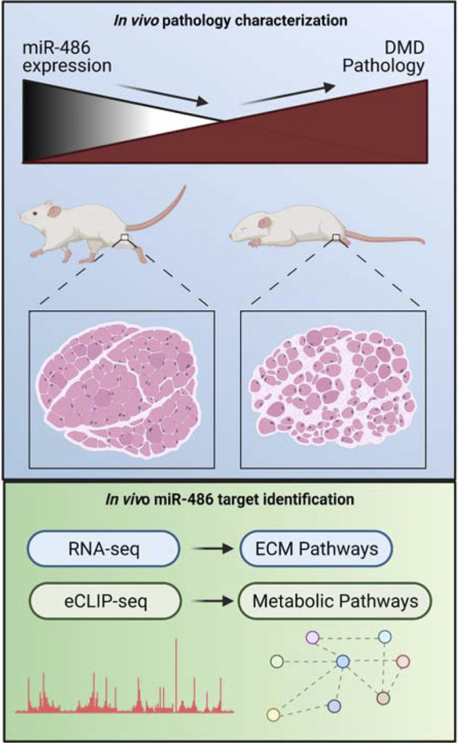
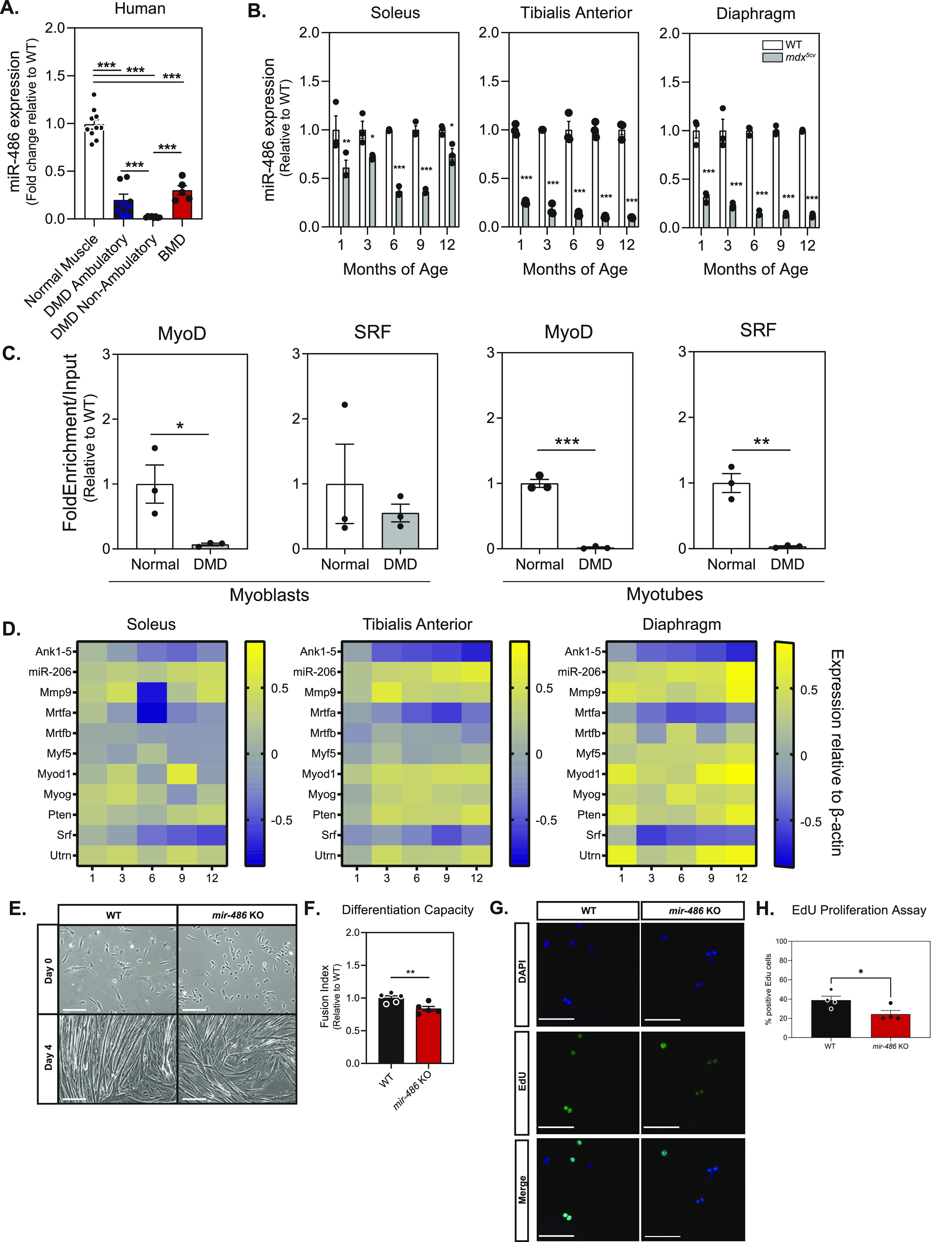
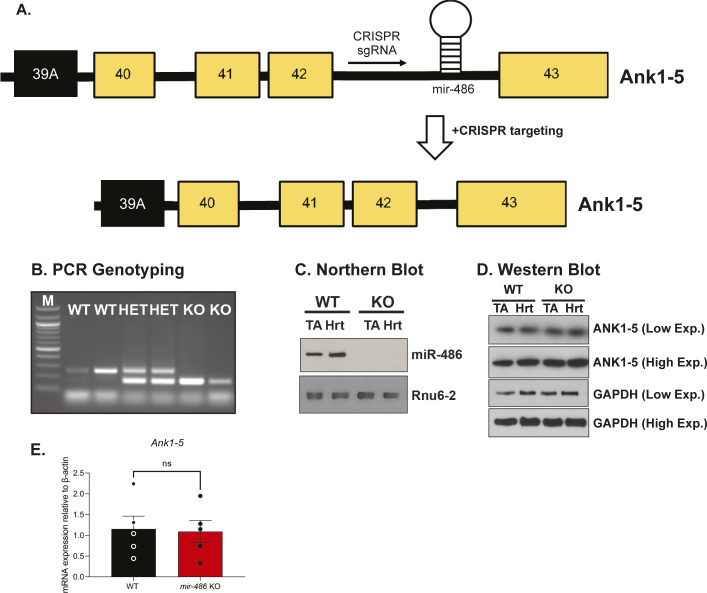
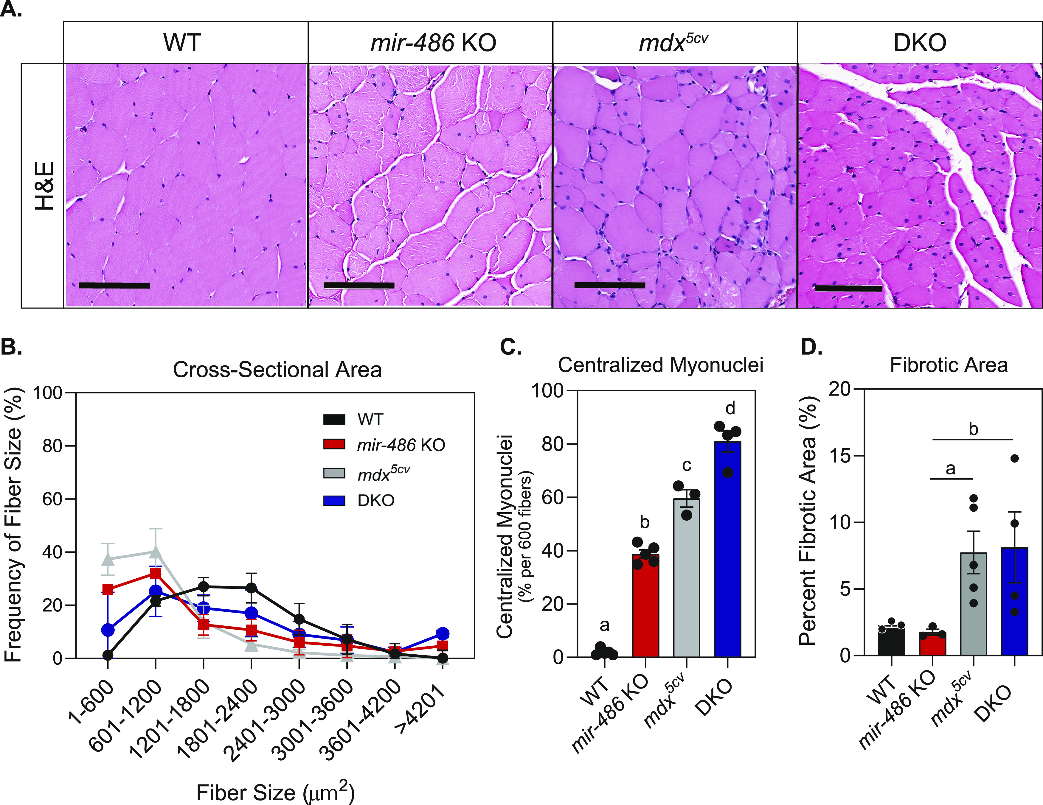
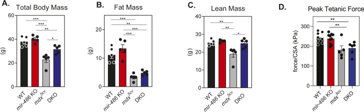
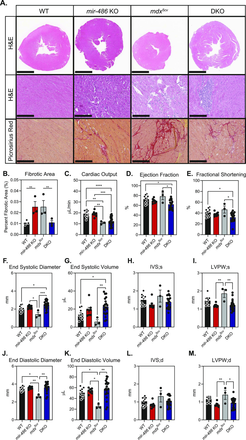
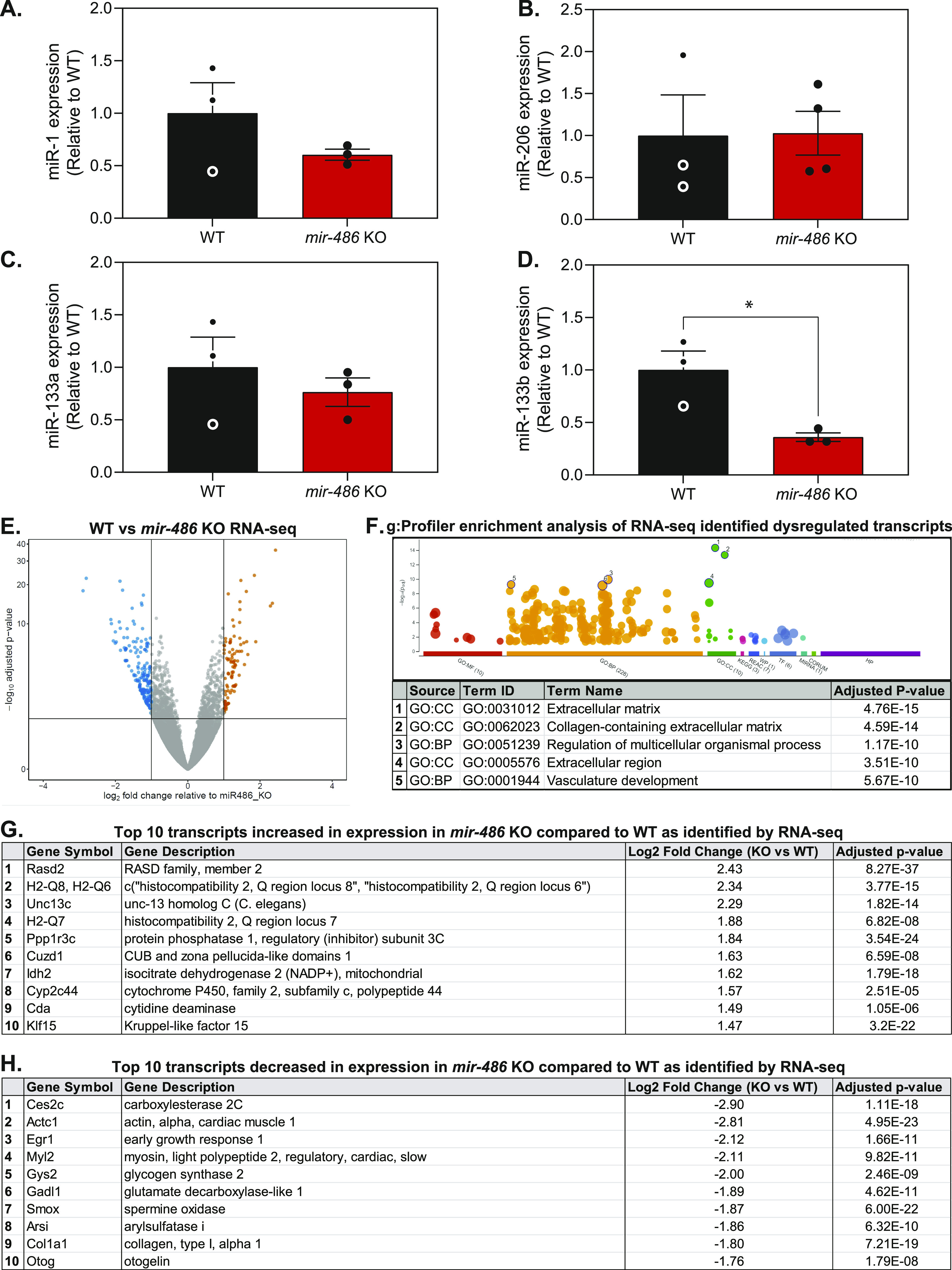
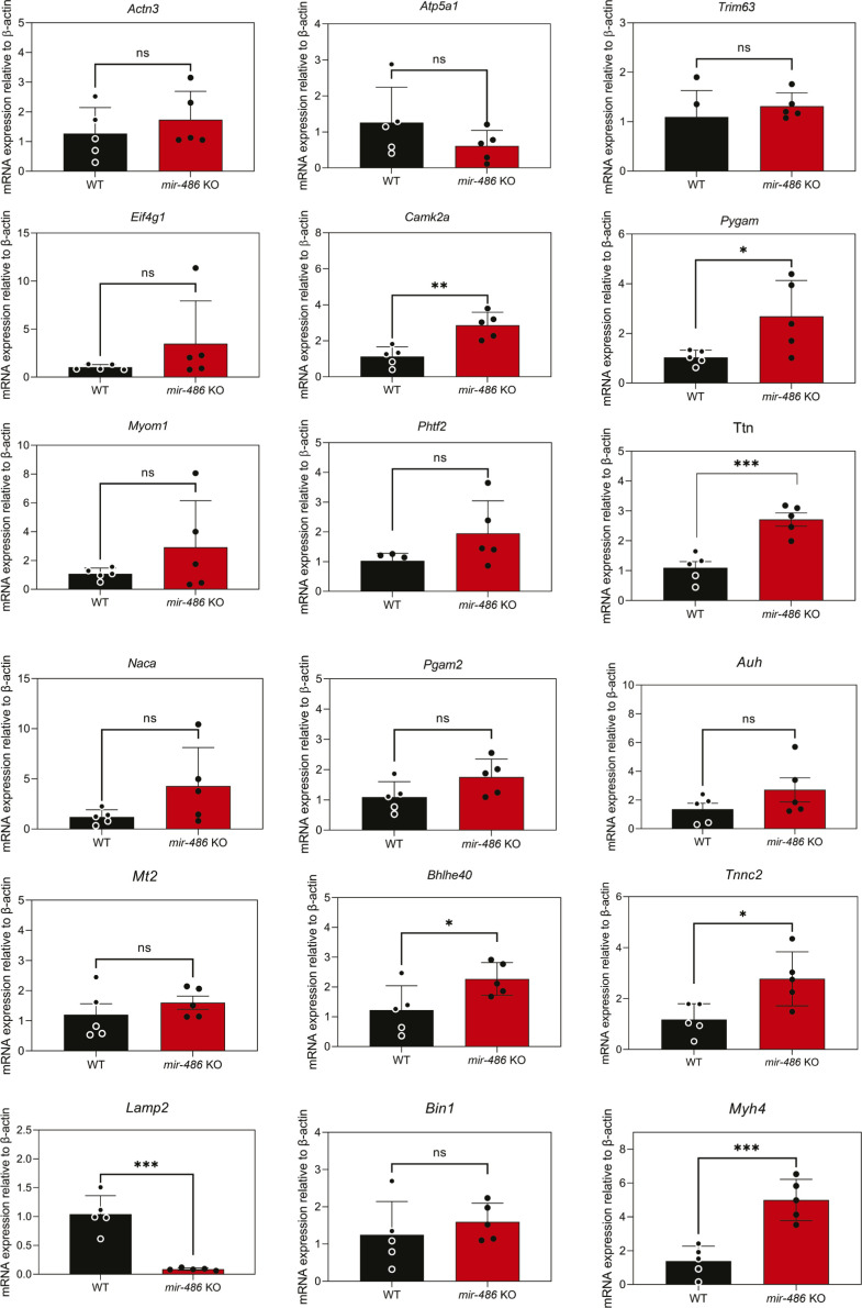
miR-486 is a muscle-enriched microRNA, or "myomiR," that has reduced expression correlated with Duchenne muscular dystrophy (DMD). To determine the function of miR-486 in normal and dystrophin-deficient muscles and elucidate miR-486 target transcripts in skeletal muscle, we characterized mir-486 knockout mice (mir-486 KO). mir-486 KO mice developed disrupted myofiber architecture, decreased myofiber size, decreased locomotor activity, increased cardiac fibrosis, and metabolic defects were exacerbated in mir-486 KO:mdx 5cv (DKO) mice. To identify direct in vivo miR-486 muscle target transcripts, we integrated RNA sequencing and chimeric miRNA eCLIP sequencing to identify key transcripts and pathways that contribute towards mir-486 KO and dystrophic disease pathologies. These targets included known and novel muscle metabolic and dystrophic structural remodeling factors of muscle and skeletal muscle contractile transcript targets. Together, our studies identify miR-486 as essential for normal muscle function, a driver of pathological remodeling in dystrophin-deficient muscle, a useful biomarker for dystrophic disease progression, and highlight the use of multiple omic platforms to identify in vivo microRNA target transcripts.


Conflict of interest statement

The authors declare that they have no conflict of interest.

Figures

None
Graphical abstract
Figure 1.
Figure 1.. miR-486 skeletal muscle expression decreases in DMD.
(A) Quantitative PCR reveals decreased expression of miR-486 in dystrophic human skeletal muscle compared with control human skeletal muscle and Becker muscular dystrophy muscle (N = 8 individual samples: normal muscle, DMD ambulatory, and DMD nonambulatory; N = 5 for Becker muscular dystrophy samples). (B) Quantitative PCR reveals decreasing expression of miR-486 in the mdx5cv dystrophic mouse model skeletal muscle at 1, 3, 6, 9, and 12 mo of age compared with WT control muscle (N = 3 samples per genotype and time point). (C) ChIP revealed myogenic factors MyoD and SRF demonstrate decreased binding at the promoter of ANK1-5 in isolated mdx5cv myoblasts and differentiated myotubes compared with WT controls (n = 3 replicates). (D) Heat maps demonstrate changes in expression of myogenic factors in mdx5cv tibialis anterior, soleus, and diaphragm muscles over 1, 3, 6, 9, and 12 mo of age compared with WT control muscle. Yellow indicates an increase in expression and blue indicates a decrease in expression relative to WT control muscle (N = 3 replicates). (E) Phase contrast reveals disrupted myoblast differentiation capacity. Photomicrographs show differentiated myotubes after 4 d of culturing from primary isolated satellite cells. WT and mir-486 KO satellite cells were isolated from < p10 pups and cultured for 4 d. 10×, scale bar = 200 μm. (F) Myogenic fusion indices calculated from day 4 myotube culture images depicted as calculated by dividing the number of nuclei within multinucleated myofibers by the total number of nuclei (N = 5 replicates per genotype). (G) Images assessing myoblast proliferation in mir-486 KO mice versus WT. Cells were stained with DAPI and EdU and quantified. (H) Quantification of the EdU proliferation assay, N = 4 separate fields of 100 cells per genotype cohort (mean ± SEM).
Figure S1.
Figure S1.. Identification and selection of mir-486 KO mice.
(A) Schematic highlighting the CRISPR sgRNA targeting sites at the mouse Ank1 locus. Black boxes indicate alternative exon 39a, which is the starting exon for Ank1-5 transcript. (B) PCR genotyping reveals an 85-bp deletion in intron 42 of the larger, non-muscle–enriched Ankyrin1 transcripts. Deletion contains the entire stem loop of the mature miR-486 transcript. (C) Northern blot confirms no miR-486 detected in mir-486 KO tibialis anterior or heart muscle. (D) Western blot confirms no change in expression of host gene Ank1, which is critical to evaluating phenotypes as a direct result of mir-486 disruption. (E) Real time qRT-PCR shows no change in expression level in Ank1-5 transcript between WT and mir-486 KO TA muscles. n = 5 samples per cohort. ns, non-significant.
Figure 2.
Figure 2.. mir-486 knockout mice demonstrate histological defects in skeletal muscle.
(A) H&E staining of transverse sections of tibialis anterior (TA) muscles at 6 mo of age. Scale bars = 200 μM. (B) Cross-sectional area of myofibers in TA muscles were measured using ImageJ based on H&E staining. Six hundred fibers from five mice of each genotype were counted. (C) Centralized myonuclei in WT and mir-486 KO TA muscle at 6 mo of age were counted using ImageJ. Six hundred fibers from five mice of each genotype were counted. Means with different letters are significantly different (Tukey’s HSD, P < 0.05). Sections cut at a thickness between 7 and 15 μm. (D) Fibrotic area was quantified as a percentage of total area using ImageJ. Five mice of each genotype were counted. *P ≤ 0.05. N = 5 mice per genotype cohort.
Figure S2.
Figure S2.. mir-486 KO mice demonstrate differences in mass distribution and muscle physiology.
(A) Total body mass shows overall weight of mice, measured in grams. *P ≤ 0.05, **P ≤ 0.01, ***P ≤ 0.001. (B, C) Fat and lean masses measured using quantitative magnetic resonance (QMR) demonstrating differences in mass distribution between genotype cohorts. **P ≤ 0.01, ***P ≤ 0.001. (D) Ex-vivo isolated muscle physiology reveals differences in peak tetanic force relative to myofiber cross-sectional area between genotype cohorts. **P ≤ 0.01. **P ≤ 0.01, ***P ≤ 0.001.
Figure 3.
Figure 3.. mir-486 KO mice develop functional and histological cardiac dysfunction.
(A) Top row: representative hematoxylin and eosin-stained transverse sections of isolated left ventricle (LV). Scale bar = 2 mm. Middle row: representative hematoxylin and eosin-stained left ventricular myocardial sections. Scale bar = 300 μM. Bottom row: representative picrosirius red–stained images of left ventricular myocardial sections. Scale bar = 200 μM. Dark red areas indicate fibrotic tissue in picorosirus-stained sections. Sections cut at a thickness between 50 and 200 μm. (B) Fibrotic area calculated from picrosirius red–stained sections using ImageJ (five images from three animals per genotype). **P ≤ 0.01. N = 3 mice per genotype cohort. (C) Cardiac output (μL/min). (D) Percent ejection fraction. (E) Percent fractional shortening. (F) End systolic diameter (mm). (G) End systolic volume (μL). (H) IVS; s = intraventricular septum thickness during systole (mm). (I) LVPW; s = left ventricular posterior wall thickness during systole (mm). (J) End diastolic diameter (mm). (K) End diastolic volume. (L) IVS; d = intraventricular septum thickness during diastole (mm). (M) LVPW; d = left ventricular posterior wall thickness during diastole (mm). Approximately 8–20 mice per genotype were analyzed. Data are represented as mean ± SEM. *P ≤ 0.05, **P ≤ 0.01. Cardiac function parameters were obtained using VisualSonics small animal echocardiogram equipment and analyzed using VevoLab software.
Figure 4.
Figure 4.. RNA-seq reveals extracellular matrix pathways in miR-486 KO muscle compared with WT.
(A, B, C, D) Quantitative PCR reveals differential expression of myomiRs in 6-mo-old mir-486 KO tibialis anterior (TA) muscle compared with WT. n = 3 mice per genotype, replicated in triplicate. (E) Volcano plot demonstrating the fold change and significance of differential gene expression in 6-mo-old mir-486 KO TA muscle compared with WT controls. N = 5 mice/cohort were used for comparative analysis. N = 4 mice per genotype. (F) g:Profiler enrichment analysis of 85 transcripts increased in expression, and 159 transcripts decreased in expression based on ≥ 2.0 log2 fold change of WT versus mir-486 KO RNA-seq analysis. The top 5 pathway hits are listed below the graph. (G) Table of top 10 transcripts increased in expression in 6-mo-old mir-486 KO mouse TA muscle compared with WT as identified by RNA-seq. (H) Table of top 10 transcripts decreased in expression in 6-mo-old mir-486 KO mouse TA muscle compared with WT as identified by RNA-seq.
Figure 5.
Figure 5.. Targeted chimeric miR-486 eCLIP-seq in WT and mir-486 KO muscles.
(A) Schematic demonstrating the workflow for the chimeric eCLIP sequencing platform to identify miR-486 in vivo skeletal muscle regulated transcripts. Tibialis anterior muscles were harvested from 6-mo-old WT and mir-486 KO male mice, and total RNA isolation was completed. The Ago2-miR-486 complex bound to target RNA transcripts was isolated, and then sequencing was performed to map the miR-486–associated peak tracks. The sequence logo (AUGUACAG) represents the consensus sequence for the top 18 miR-486–associated peak reads based on the miRNA:mRNA target sequencing alignments. The miR-486 seed sequence is shown in the 5′–3′ direction. (B) Chromosomal location of a single top hit peak transcript, Mt2, as identified by eCLIP-seq as a direct target of miR-486. Peaks generated using Integrative Genome Viewer. (C) Metagene plot demonstrating overall miR-486 binding location by a relative position on the target gene. (D) Pie chart demonstrating the proportion of miR-486 gene targets and the respective intragenic binding location of miR-486. (E) Table outlining the top 10 transcripts that were identified as direct targets of miR-486 via CLIP-seq. (F) g:Profiler enrichment analysis graph demonstrates the most significant cellular pathways associated with the 18 direct miR-486 targets identified via chimeric eCLIP-seq. The pathway ID number in the table correlates with the numbered dots in the accompanying graph above. (G) Heat map expression of quantitative PCR of 18 miR-486 eCLIP-seq targets in mir-486 KO and mdx5cv tibialis anterior muscles expression levels compared with WT controls in separate cohort analyses. Data points are individual biological replicates, N = 4/cohort, and logarithmic fold change normalized to both wild type and β-actin is shown. **P ≤ 0.01 N = 3 replicates per cohort. Transcript levels are normalized to β-actin, and mir-486 KO levels are shown as relative to WT.
Figure S3.
Figure S3.. Quantitative real-time PCR validation of all 18 miR-486 targets transcripts identified through chimeric eCLIP sequencing.
All 18 transcript targets validated by TaqMan qRT-PCR assays with expression levels normalized to the β-actin housekeeping control. Tissues used for analyses were tibialis anterior muscles from mir-486 KO and WT mice (n = 5 mice per cohort). Statistical analyses were conducted using unpaired t tests. *P ≤ 0.05, **P ≤ 0.01, ***P ≤ 0.0001.
Figure S4.
Figure S4.. shRNAi knockdown of Dp427 transcript in primary myotubes reduces miR-486 expression.
(A, B, C) Real-time qPCR expression fold changes of miR-486, Ank1-5, and Dp427 transcripts. AAV-mediated shRNAi knockdown in primary mouse myotubes using AAV shRNAi constructs targeting either shLuc (control, nontargeting), shDp427, or mock (1xDPBS) controls. Multiplicity of infections of 1, 100, and 200 were used across 2 × 10⁵ myoblasts originally seeded. Fold changes normalized to either RNU6-2 (miR-486) or Actb housekeeping controls (Ank1-5 and Dp427). One-way ANOVA test with the Tukey HSD test performed *P-value < 0.005. (D) Representative Western blot demonstrating a strong knockdown of the mouse dystrophin (Dp427) protein isoform using shRNAi Dp427 at MOI’s of 100 and 200. β-tubulin was used as a housekeeping loading control.

Similar articles

Cited by

References

    1. Alexander MS, Casar JC, Motohashi N, Myers JA, Eisenberg I, Gonzalez RT, Estrella EA, Kang PB, Kawahara G, Kunkel LM (2011) Regulation of DMD pathology by an ankyrin-encoded miRNA. Skelet Muscle 1: 27. 10.1186/2044-5040-1-27 - DOI - PMC - PubMed
    1. Alexander MS, Casar JC, Motohashi N, Vieira NM, Eisenberg I, Marshall JL, Gasperini MJ, Lek A, Myers JA, Estrella EA, et al. (2014) MicroRNA-486-dependent modulation of DOCK3/PTEN/AKT signaling pathways improves muscular dystrophy-associated symptoms. J Clin Invest 124: 2651–2667. 10.1172/JCI73579 - DOI - PMC - PubMed
    1. Alexander MS, Kawahara G, Motohashi N, Casar JC, Eisenberg I, Myers JA, Gasperini MJ, Estrella EA, Kho AT, Mitsuhashi S, et al. (2013) MicroRNA-199a is induced in dystrophic muscle and affects WNT signaling, cell proliferation, and myogenic differentiation. Cell Death Differ 20: 1194–1208. 10.1038/cdd.2013.62 - DOI - PMC - PubMed
    1. Aránega AE, Lozano-Velasco E, Rodriguez-Outeiriño L, Ramírez de Acuña F, Franco D, Hernández-Torres F (2021) MiRNAs and muscle regeneration: Therapeutic targets in Duchenne muscular dystrophy. Int J Mol Sci 22: 4236. 10.3390/ijms22084236 - DOI - PMC - PubMed
    1. Archacka K, Ciemerych MA, Florkowska A, Romanczuk K (2021) Non-coding RNAs as regulators of myogenesis and postexercise muscle regeneration. Int J Mol Sci 22: 11568. 10.3390/ijms222111568 - DOI - PMC - PubMed

Publication types