. 2022 May 5;5(8):e202101358.
doi: 10.26508/lsa.202101358. Print 2022 Aug.

Molecular analysis and essentiality of Aro1 shikimate biosynthesis multi-enzyme in Candida albicans

Affiliations

Molecular analysis and essentiality of Aro1 shikimate biosynthesis multi-enzyme in Candida albicans

Peter J Stogios et al. Life Sci Alliance. .

Abstract

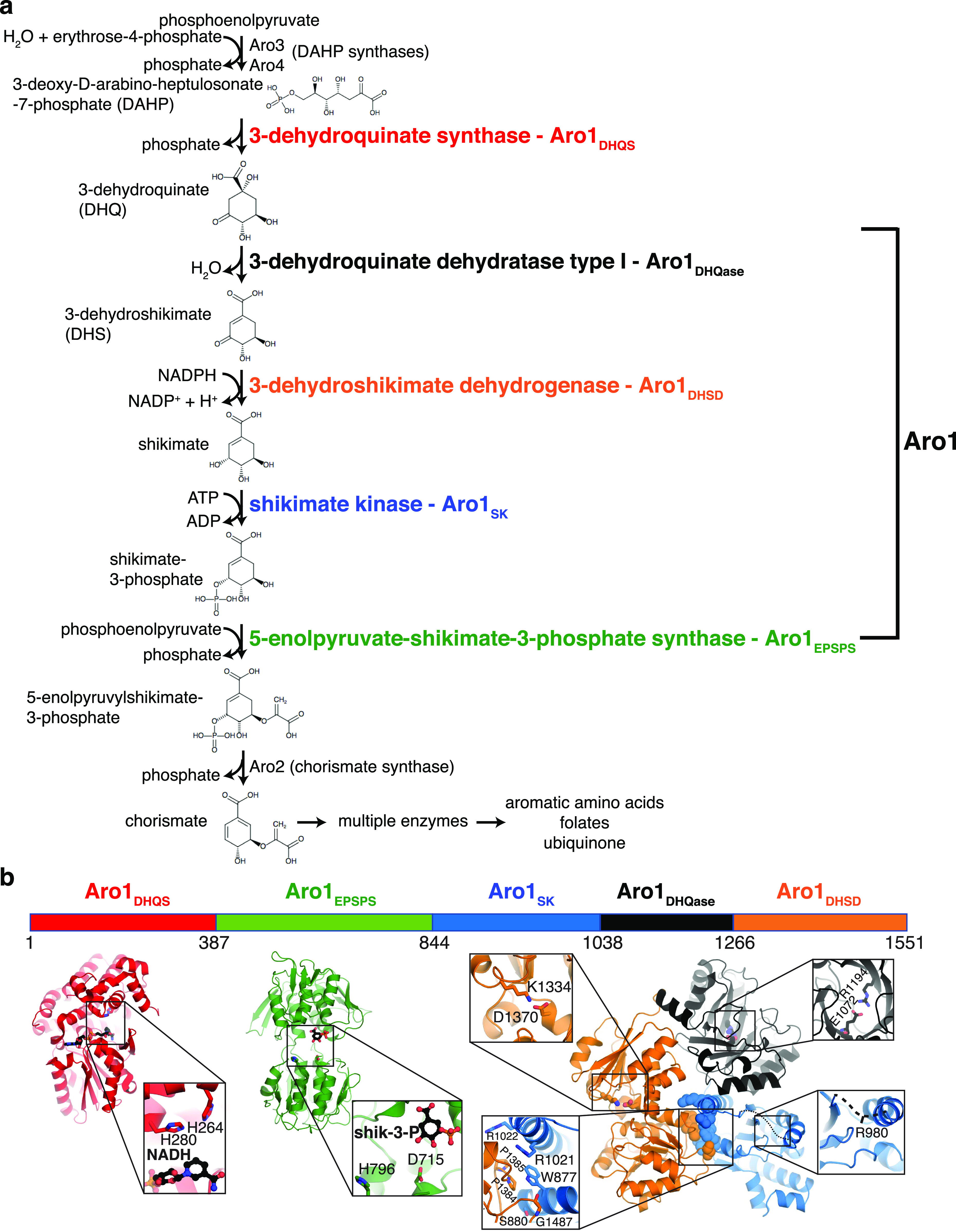
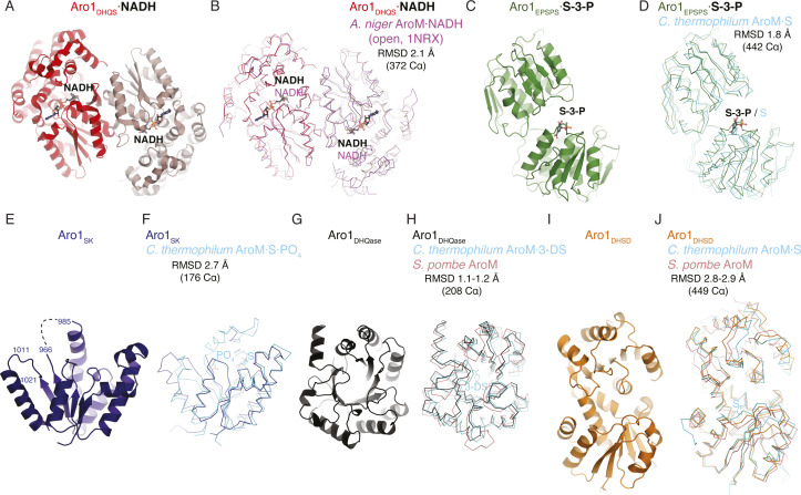
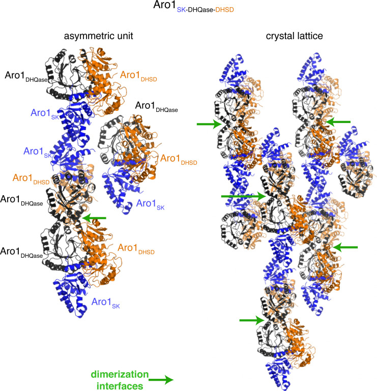
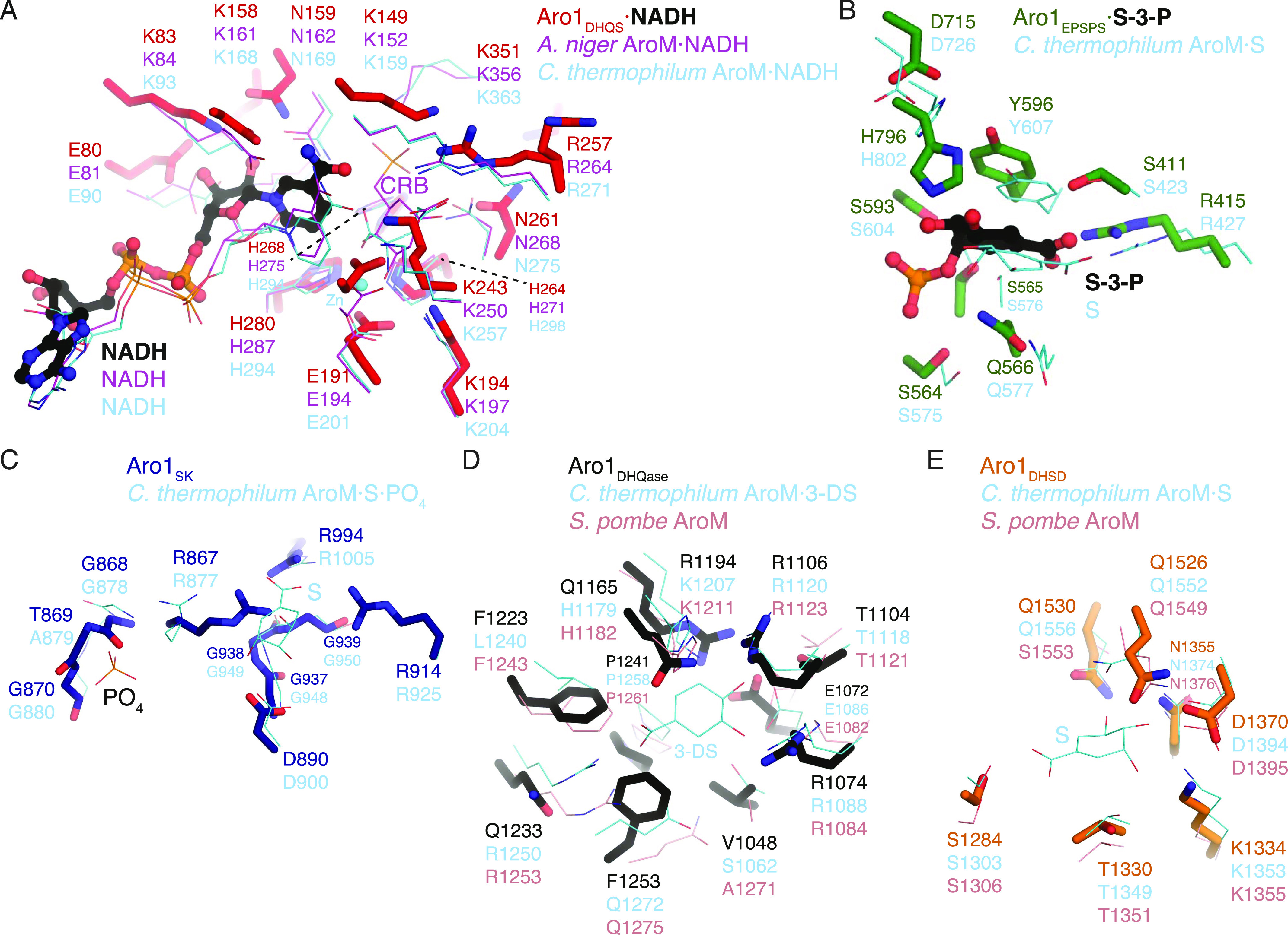
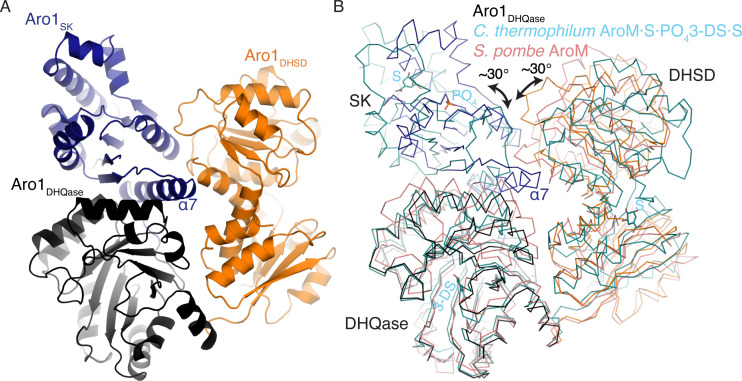
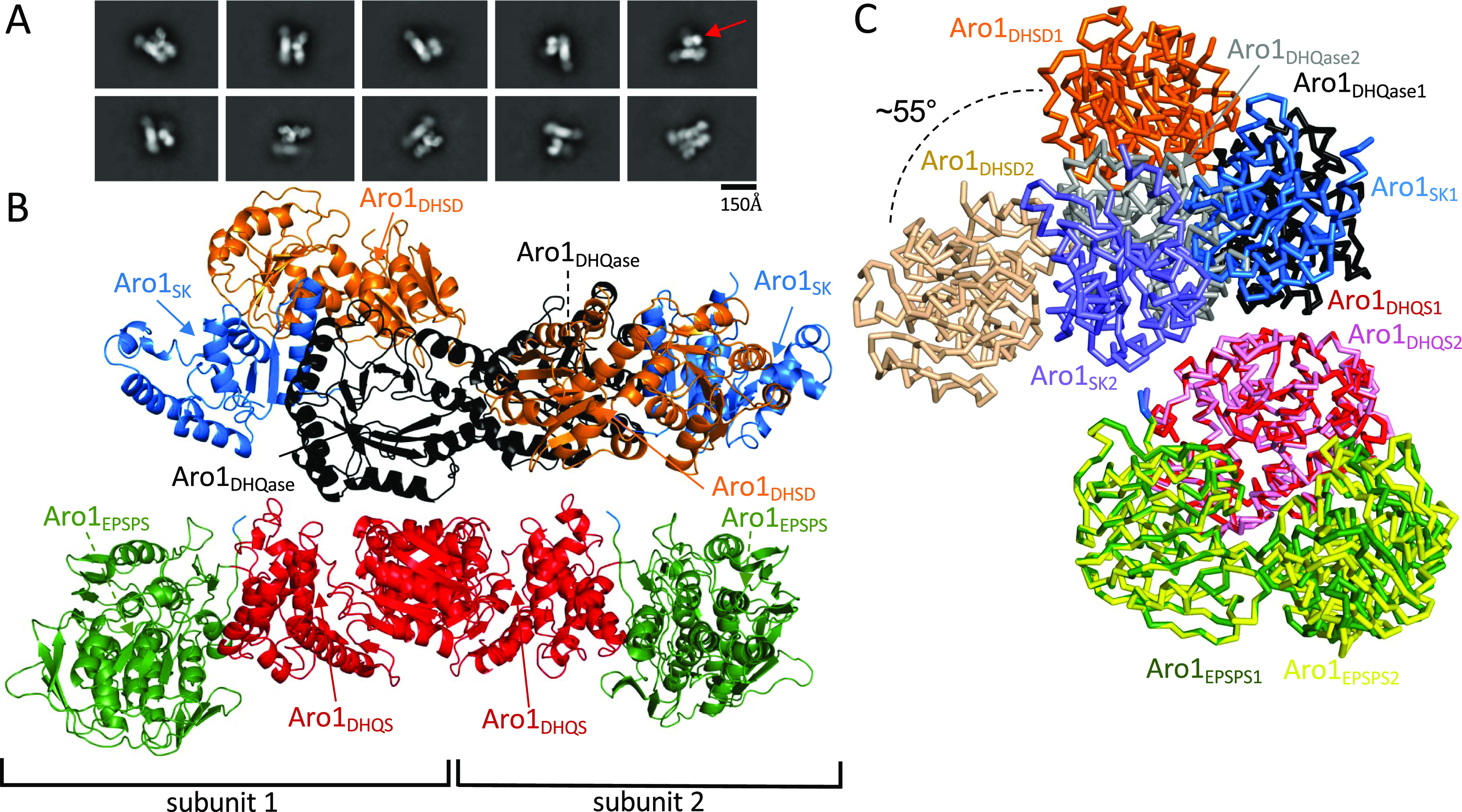
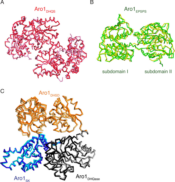
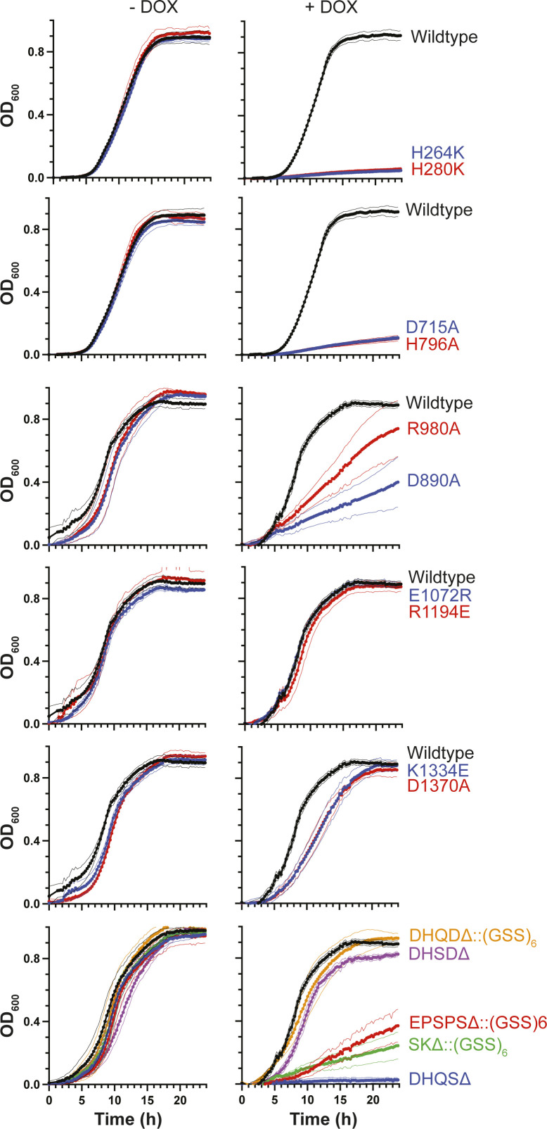
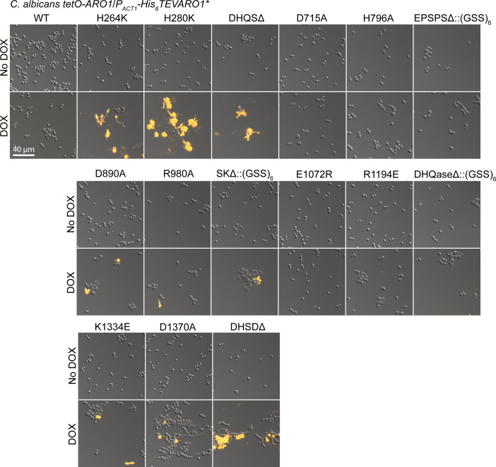
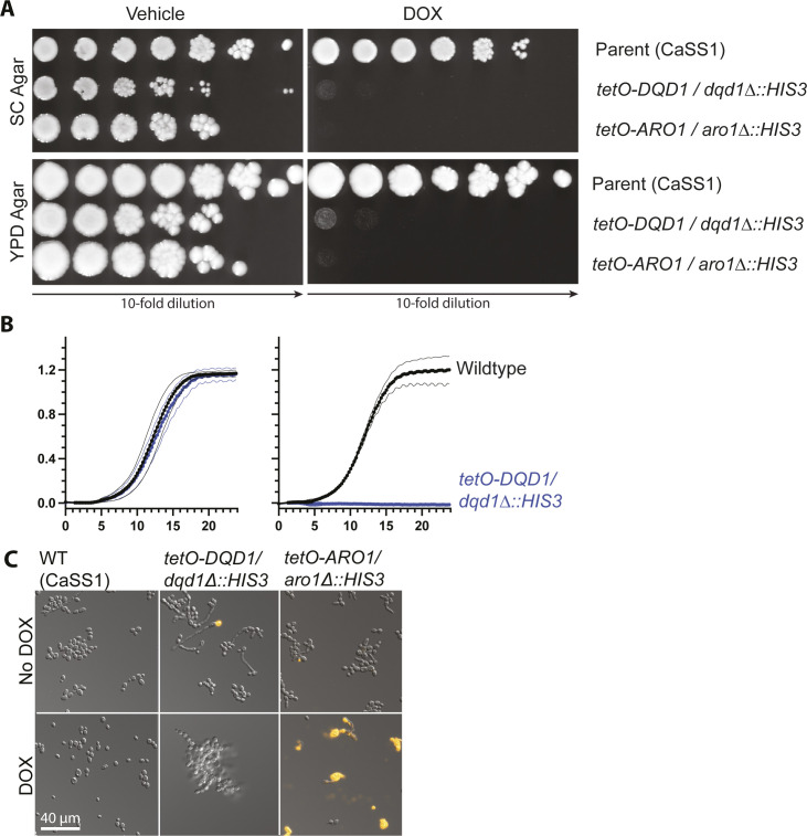
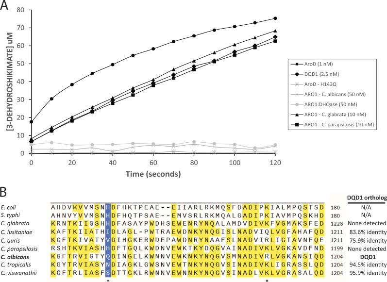
In the human fungal pathogen Candida albicans, ARO1 encodes an essential multi-enzyme that catalyses consecutive steps in the shikimate pathway for biosynthesis of chorismate, a precursor to folate and the aromatic amino acids. We obtained the first molecular image of C. albicans Aro1 that reveals the architecture of all five enzymatic domains and their arrangement in the context of the full-length protein. Aro1 forms a flexible dimer allowing relative autonomy of enzymatic function of the individual domains. Our activity and in cellulo data suggest that only four of Aro1's enzymatic domains are functional and essential for viability of C. albicans, whereas the 3-dehydroquinate dehydratase (DHQase) domain is inactive because of active site substitutions. We further demonstrate that in C. albicans, the type II DHQase Dqd1 can compensate for the inactive DHQase domain of Aro1, suggesting an unrecognized essential role for this enzyme in shikimate biosynthesis. In contrast, in Candida glabrata and Candida parapsilosis, which do not encode a Dqd1 homolog, Aro1 DHQase domains are enzymatically active, highlighting diversity across Candida species.


Conflict of interest statement

LE Cowen is a co-founder and shareholder in Bright Angel Therapeutics, a platform company for development of novel antifungal therapeutics. LE Cowen is a consultant for Boragen, a small-molecule development company focused on leveraging the unique chemical properties of boron chemistry for crop protection and animal health. SD Liston was supported by a Mitacs postdoctoral fellowship funded in partnership with Amplyx Pharmaceuticals Inc., now a subsidiary of Pfizer. D Borek and Z Otwinowski are co-founders of Ligo Analytics, a company that develops a software for cryogenic electron microscopy. Z Otwinowski is a co-founder of HKL Research, a company that develops software for X-ray diffraction data analysis.

Figures

Figure 1.
Figure 1.. C. albicans shikimate pathway.
(A) Shikimate pathway, with enzyme names indicated to the right of each reaction. Aro1 individual domains are highlighted in colours. (B) Crystal structures of Aro1 fragments solved in this work, including Aro1DHQS, Aro1EPSPS, Aro1SK+DHQase+DHSD. Zooms show active sites, key catalytic residues, or domain–domain interactions in the Aro1SK+DHQase+DHSD structure.
Figure S1.
Figure S1.. GRACE library mutants for ARO1 and DQD1 do not grow in the presence of DOX.
(A) Dilutions of C. albicans CaSS1 and derived conditional expression strains for DQD1 and ARO1 were prepared as described in Fig 4. SC medium lacking arginine and supplemented with 150 μl NAT was inoculated with single colonies of C. albicans and incubated overnight at 30°C. Subcultures were prepared at 1:1,000 in SC medium supplemented with 50 ng/ml DOX and incubated overnight at 30°C. Cultures were normalized to 10 OD600 units/ml then 10-fold serial diluted in water. Diluted cultures were spotted onto SC Agar and SC Agar supplemented with 5 μg/ml DOX, then photographed after 48 h incubation at 30°C. (B) Growth curves were performed as described in Fig S12. (C) Cultures were prepared and imaged as described in Fig S10.
Figure S2.
Figure S2.. Structural analysis of domains of Aro1.
(A) Structure of dimeric Aro1DHQS, coloured by chain. NADH show as ball-and-stick. (B) Superposition of Aro1DHQS with DHQS domain from A. niger (PDB 1NRX) in the open conformation. (C) Structure of Aro1EPSPS. Shikimate-3-phosphate (S3P) shown in ball-and-stick. (D) Superposition of Aro1EPSPS and C. thermophilum AroM (PDB 6QHV). Shikimate (S) and S3P bound to C. thermophilum are shown in ball-and-stick. (E) Structure of Aro1SK from Aro1SK+DHQase+DHSD structure. Region with missing electron density indicated in dashed line (986-967). (F) Superposition of Aro1SK and C. thermophilum AroM (PDB 6QHV). Shikimate (S) and phosphate ions bound to AroM shown in ball-and-stick. (G) Structure of Aro1DHQase from Aro1SK+DHQase+DHSD structure. (H) Superposition of Aro1DHQase, C. thermophilum AroM (PDB 6QHV), and S. pombe AroM (PDB 5SVW). 3-dehydroshikimate (3-DS) bound to C. thermophilum AroM shown in ball-and-stick. (I) Structure of Aro1DHSD from Aro1SK+DHQase+DHSD structure. (J) Superposition of Aro1DHSD, C. thermophilum AroM (PDB 6QHV), and S. pombe AroM (PDB 5SVW). Shikimate (S) bound to C. thermophilum AroM shown in ball-and-stick.
Figure 2.
Figure 2.. Molecular architecture of active sites in C. albicans Aro1 domains.
In all panels, Aro1 residues are shown as sticks and the comparative structure’s residues are shown as thin lines. (A) Aro1DHQS superposed with A. niger AroM DHQS domain (PDB 1NVB). NAD molecules bound to the respective structures are shown in sticks. Carbaphosphonate bound to AroM DHQS shown in sticks. Zn2+ ion bound to Aro1DHQS shown as a sphere. (B) Aro1EPSPS superposed with C. thermophilum AroM EPSPS domain (PDB 6HQV). Shikimate-3-phosphate and shikimate bound to Aro1DHQS and AroM, respectively, shown as sticks. (C) Aro1SK from Aro1SK+DHQase+DHSD structure superposed with C. thermophilum AroM SK domain (PDB 6HQV). Shikimate bound to AroM shown in sticks. (D) Aro1DHQase from Aro1SK+DHQase+DHSD structure superposed with C. thermophilum AroM DHQase domain (PDB 6HQV) and S. pombe AroM DHQase domain (PDB 5SWV). 3-dehydroshikimate bound to C. thermophilum AroM shown in sticks. (E) Aro1DHSD from Aro1SK+DHQase+DHSD structure superposed with C. thermophilum AroM DHSD (PDB 6HQV) and S. pombe AroM DHSD domain (PDB 5SWV).
Figure S3.
Figure S3.. Crystal packing of Aro1SK+DHQase+DHSD structure showing dimerization via Aro1DHQase.
left, asymmetric unit; right, crystal lattice. Green arrows highlight dimerization via Aro1DHQase.
Figure S4.
Figure S4.. Comparison of C. albicans Aro1SK+DHQase+DHSD and AroM SK+DHQase+DHSD regions.
S and 3-DS bound to AroM shown in ball-and-stick. Rotational differences between SK and DHSD domains differ by ∼30°.
Figure S5.
Figure S5.. Purification and size-exclusion profiles of Aro1.
(A) Size-exclusion chromatography profile of Aro1 purified from C. albicans. Fractions collected are demarcated in red. (B) SDS–PAGE of Aro1 SEC fractions.
Figure 3.
Figure 3.. Cryo-EM structure of the unliganded full-length Aro1 from C. albicans.
(A) Selected 2D class averages of the full-length Aro1 dimer. The red arrow points to the Aro1SK-DHQase-DHSD region, which is highly heterogeneous and found tilted at various angles relative to the Aro1DHQS-EPSPS region. (B) The structure of the full-length Aro1 from C. albicans. The final resolution for the Aro1SK-DHQase-DHSD domain dimer is 3.16 Å and the Aro1DHQS-EPSPS domain dimer is 3.43 Å. Coloured arrows point towards the ligand binding sites of each domain, which are unoccupied in our structure. The Aro1SK-DHQase-DHSD region of subunit 1 is tilted away from the Aro1DHQS-EPSPS region and is referred to as the “loose” monomer, whereas the Aro1SK-DHQase-DHSD region of subunit 2 is nearly parallel to the Aro1DHQS-EPSPS region and is referred to as the “tight” monomer. (C) Comparison of “tight” and “loose” monomers from the full-length Aro1 dimer superposed on the Aro1DHQS-EPSPS region. Although the backbones of each domain overlays with low RMSD, the relative orientations of the Aro1SK-DHQase-DHSD and Aro1DHQS-EPSPS regions change dramatically.
Figure S6.
Figure S6.. Cryo-EM image processing flow chart.
(A) Example high quality micrograph of Aro1 and selected 2D class averages showing broken particles consisting of Aro1DHQS and Aro1EPSPS as well as full-length Aro1. (B) FSC curves for homogeneous refinement of the full-length Aro1 dimer (red), local refinement of Aro1SK-DHQase-DHSD dimer (orange), local refinement of Aro1DHQS-EPSPS dimer (green), local refinement of Aro1DHQS-EPSPS subunit 1 after EPSPS domain 1 focused classification (blue), and local refinement of Aro1DHQS-EPSPS subunit 2 after EPSPS domain 1 focused classification (purple). (C) Image processing flow chart for structure determination of full-length Aro1.
Figure S7.
Figure S7.. Superposition of individual domains and regions of the full-length monomers and crystal structures.
(A) Superposition of the Aro1DHQS dimer from the crystal structure (pink) and from the full-length dimer (red). (B) Superposition of Aro1EPSPS from the crystal structure (yellow) and from a monomer of the full-length dimer (green). (C) Superposition of the Aro1SK-DHQase-DHSD region from the crystal structure (cyan, light orange, and grey) and the Aro1SK-DHQase-DHSD region (blue, orange, and black) from a monomer of the full-length dimer.
Figure S8.
Figure S8.. Validation of tetO-driven overexpression and DOX-mediated repression of ARO1 and DQD1 transcripts by RT-qPCR.
(A) Cultures were grown in SC medium overnight at 30°C, subcultured into SC medium at 1:1,000 supplemented with 0.05 μg/ml DOX overnight, then subcultured 1:100 into SC medium supplemented with 5 μg/ml DOX for 4 h. RNA was extracted, DNAse-treated, and used to synthesize cDNA. Transcript abundance was quantified by real-time PCR. Data are mean relative ARO1 transcript abundance normalized to ACT1 ± SEM for three independent cultures, each assayed in technical triplicate. Left: conditional expression strains constructed for this study in C. albicans SN95. Right: conditional expression strain for ARO1 in C. albicans CaSS1, from the GRACE library. (B) Data are mean relative DQD1 transcript abundance normalized to ACT1 ± SEM for three independent cultures assayed in technical triplicate. Conditional expression strain for DQD1 in C. albicans CaSS1, from the GRACE library.
Figure S9.
Figure S9.. Aro1 and mutant derivatives are expressed in C. albicans.
(A) The tetO promoter drives DOX-repressible overexpression of Aro1. Panel depicts SDS–PAGE of whole-cell lysates prepared from C. albicans, stained with Coomassie Blue R-250. Cultures were grown in SC medium at 30°C overnight, then subcultured into SC medium and SC medium supplemented with 5 μg/ml DOX for 4 h at 30°C. (B) The tetO promoter drives DOX-dependent overexpression of N-terminally His6TEV tagged Aro1. The Western immunoblot (upper) was probed with anti-His5 monoclonal antibody. Equivalent loading was confirmed by Coomassie Blue R-250 stained SDS–PAGE of identical samples (lower). (A) Lysates were prepared as in (A). (C) His6TEV-tagged truncation/domain deletion mutants of Aro1 expressed under the ACT1 promoter are produced at equal or more abundant levels than the full-length protein in C. albicans. Cultures were grown in SC medium overnight at 30°C, subcultured into SC medium supplemented with 0.050 μg/ml DOX overnight, then subcultured into SC medium supplemented with 5 μg/ml DOX for 4 h. (D) Amino acid substitutions in PACT1-His6TEVAro1 do not alter abundance of Aro1 in C. albicans. (C) Lysates were prepared as in (C).
Figure 4.
Figure 4.. Effect of Aro1 active site mutations on C. albicans viability.
(A) ARO1 activities are required for growth of C. albicans. SC medium lacking arginine and supplemented with 150 μl NAT was inoculated with single colonies of C. albicans and incubated overnight at 30°C. Subcultures were prepared at 1:1,000 in SC medium supplemented with 50 ng/ml DOX and incubated overnight at 30°C. Cultures were normalized to 10 OD600 units/ml then 10-fold serial diluted in water. Diluted cultures were spotted onto SC Agar and SC Agar supplemented with 5 μg/ml DOX, then photographed after 48 h incubation at 30°C. Asterisk indicates substitutions to amino acids identified to mediate interdomain contacts. (B) ARO1 is required for growth of C. albicans. SC medium lacking arginine and supplemented with 150 μl NAT was inoculated with single colonies of C. albicans and incubated overnight at 30°C. 80 μl SC cultures were prepared at 0.001 OD600 unit/ml, in the presence and absence of 5 μg/ml DOX, then incubated at 30°C for 24 h. Data are mean optical densities for quadruplicate cultures ± SD for a representative experiment. (B, C) ARO1 is required for viability of C. albicans. Representative fluorescence micrographs of cultures from (B), stained with 1 μg/ml propidium iodide to label inviable cells.
Figure S10.
Figure S10.. Catalytically inactive derivatives of ARO1 have domain-specific effects on viability of C. albicans in liquid culture.
C. albicans CaLC6598 and derivative strains encoding amino acid substitutions, deletions, and truncations of His6TEV-Aro1 were examined by fluorescence microscopy to assess morphology and viability. Cultures were grown in SC medium overnight at 30°C, subcultured into SC medium and SC medium supplemented with 0.05 μg/ml DOX overnight. Cultures were normalized to an OD600 of 1, then diluted at 1:1,000 into 96-well plates containing 80 μl of SC medium (top) or SC medium supplemented with 5 μg/ml DOX (bottom). After 24-h incubation at 30°C, propidium iodide was added to 1 μg/ml (final). Cells were spotted on glass slides after 10 min incubation at room temperature and then examined by fluorescence microscopy. Panels depict representative fields of view for wells from three independent experiments. Scale bar is 40 μm.
Figure S11.
Figure S11.. Comparison of enzymatic activity between ARO1 purified from C. albicans and recombinant ARO1 purified from E. coli.
Enzymatic activity, expressed as percent relative to wild type, is shown. The data depicted for the EPSPS, SK, and DHSD domains is from recombinant full-length enzyme purified from E. coli. The wild-type data are from C. albicans Aro1 and normalized to 100%.
Figure S12.
Figure S12.. Catalytically inactive derivatives of ARO1 have domain-specific effects on growth of C. albicans in liquid culture.
Cultures were grown in SC medium overnight at 30°C, subcultured into SC medium and SC medium supplemented with 0.05 μg/ml DOX overnight. Cultures were normalized to an OD600 of 1, then diluted at 1:1,000 into 96-well plates containing 80 μl of SC medium (left) or SC medium supplemented with 5 μg/ml DOX (right). Growth at 30°C was monitored by optical density at 15-min intervals for 24 h. Data are mean OD600 ± SD for quadruplicate cultures in a representative experiment of three performed.
Figure 5.
Figure 5.. Diversity of type I DHQase active site composition in Candida species.
(A) C. albicans Aro1 is catalytically inactive in a DHQase activity assay. Candida glabrata and C. parapsilosis Aro1 were active, along with C. glabrata Dqd1. E. coli AroD was included as a positive control. (B) Multiple sequence alignment of the region of DHQase containing residues that comprise the catalytic dyad. Positions where residues involved in catalysis are expected are denoted with a (*), and the position where the catalytic His is expected is highlighted in blue. If a DQD1 ortholog was found in the genome, it is listed on the right and given as percent shared amino acid sequence identity.

Similar articles

Cited by

References

    1. Afonine PV, Grosse-Kunstleve RW, Echols N, Headd JJ, Moriarty NW, Mustyakimov M, Terwilliger TC, Urzhumtsev A, Zwart PH, Adams PD (2012) Towards automated crystallographic structure refinement with phenix.refine. Acta Crystallogr D Biol Crystallogr 68: 352–367. 10.1107/S0907444912001308 - DOI - PMC - PubMed
    1. Bagautdinov B, Kunishima N (2007) Crystal structures of shikimate dehydrogenase AroE from thermus thermophilus HB8 and its cofactor and substrate complexes: Insights into the enzymatic mechanism. J Mol Biol 373: 424–438. 10.1016/j.jmb.2007.08.017 - DOI - PubMed
    1. Becker JM, Kauffman SJ, Hauser M, Huang L, Lin M, Sillaots S, Jiang B, Xu D, Roemer T (2010) Pathway analysis of Candida albicans survival and virulence determinants in a murine infection model. Proc Natl Acad Sci U S A 107: 22044–22049. 10.1073/pnas.1009845107 - DOI - PMC - PubMed
    1. Blanco B, Prado V, Lence E, Otero JM, Garcia-Doval C, van Raaij MJ, Llamas-Saiz AL, Lamb H, Hawkins AR, González-Bello C (2013) Mycobacterium tuberculosis shikimate kinase inhibitors: Design and simulation studies of the catalytic turnover. J Am Chem Soc 135: 12366–12376. 10.1021/ja405853p - DOI - PubMed
    1. Bongomin F, Gago S, Oladele RO, Denning DW (2017) Global and multi-national prevalence of fungal diseases-estimate precision. J Fungi (Basel) 3: 57. 10.3390/jof3040057 - DOI - PMC - PubMed

Publication types

LinkOut - more resources