. 2022 Jun 15:445:116043.
doi: 10.1016/j.taap.2022.116043. Epub 2022 May 2.

The role of Iron in lipid peroxidation and protein nitration during acetaminophen-induced liver injury in mice

Affiliations

The role of Iron in lipid peroxidation and protein nitration during acetaminophen-induced liver injury in mice

Olamide B Adelusi et al. Toxicol Appl Pharmacol. .

Abstract

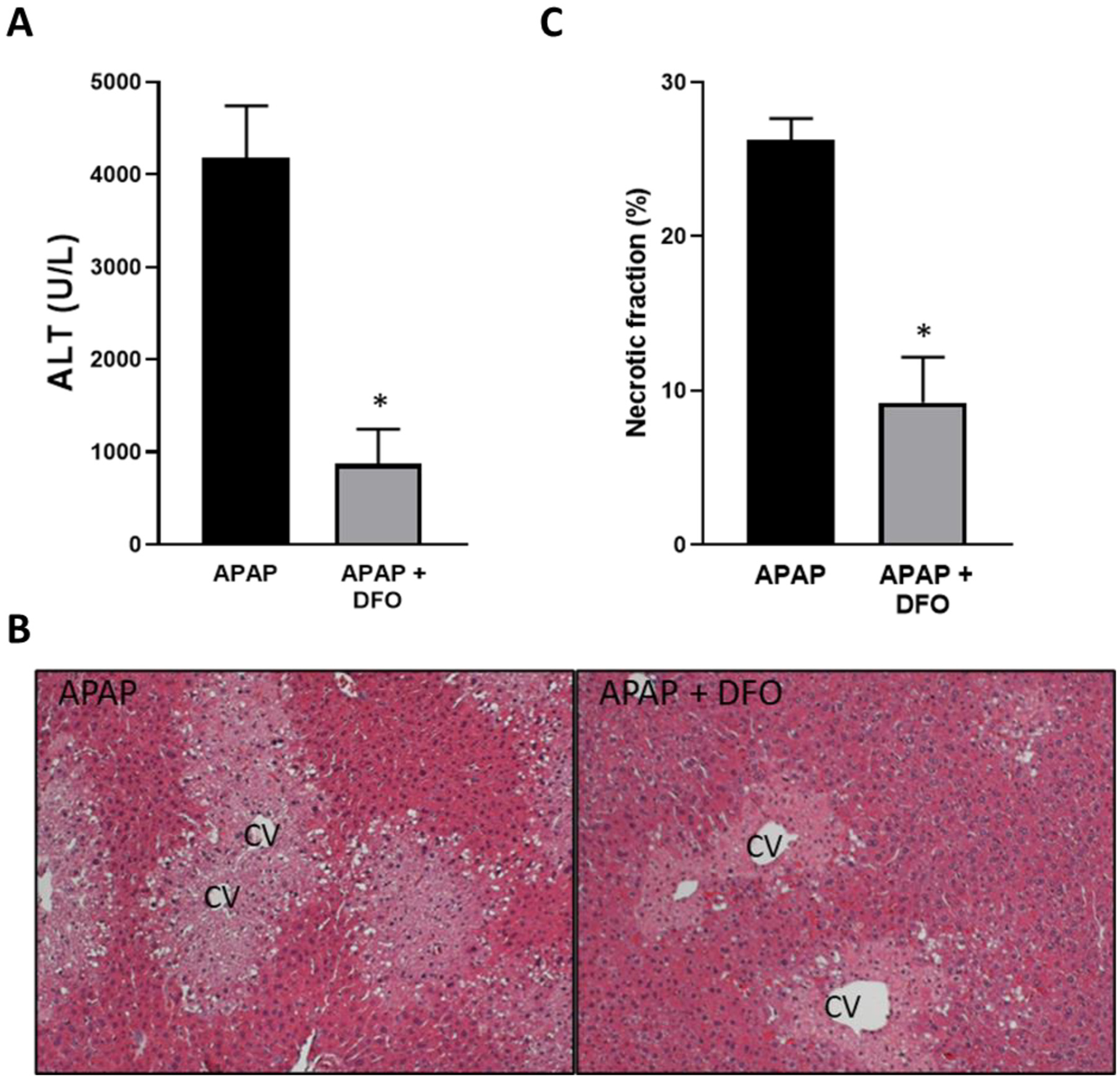
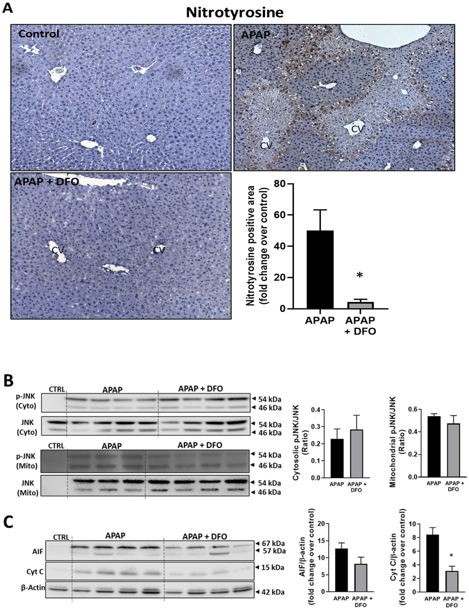
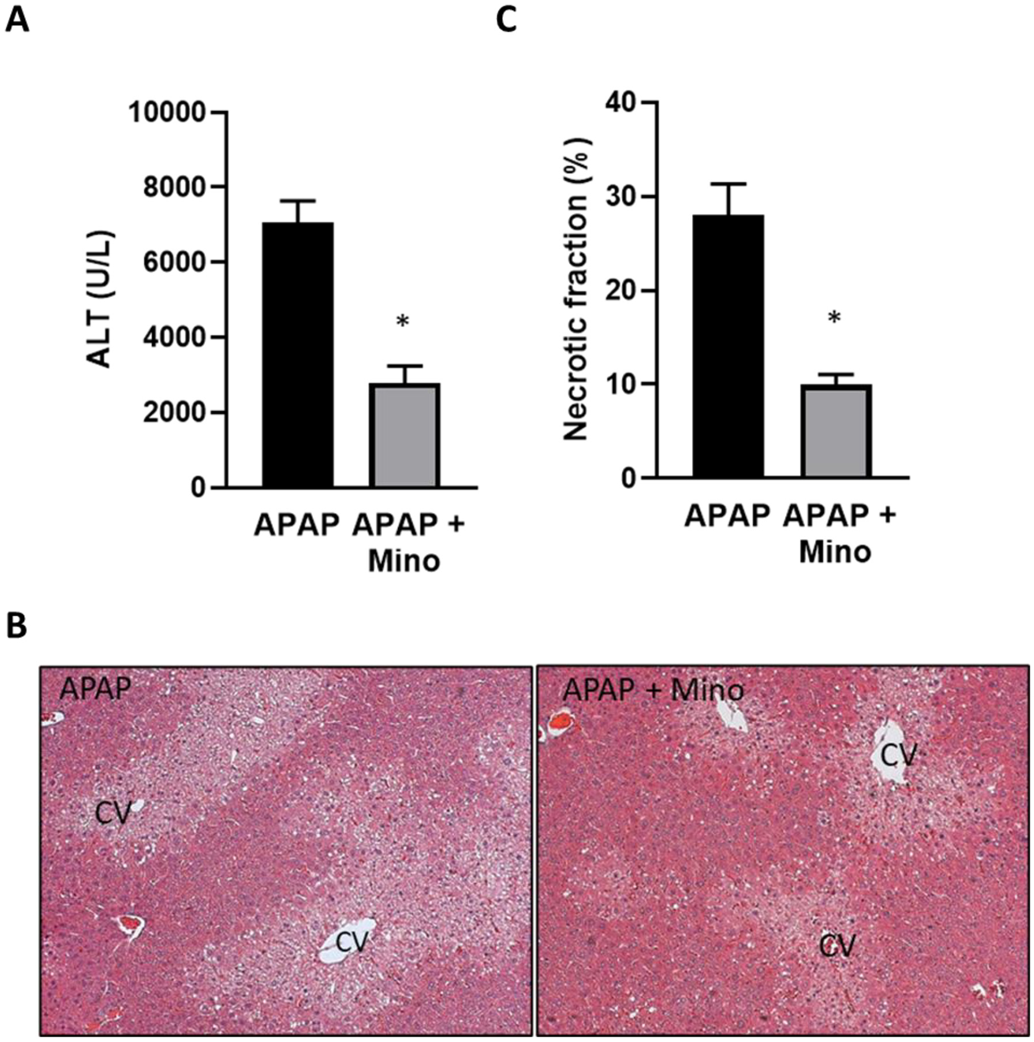
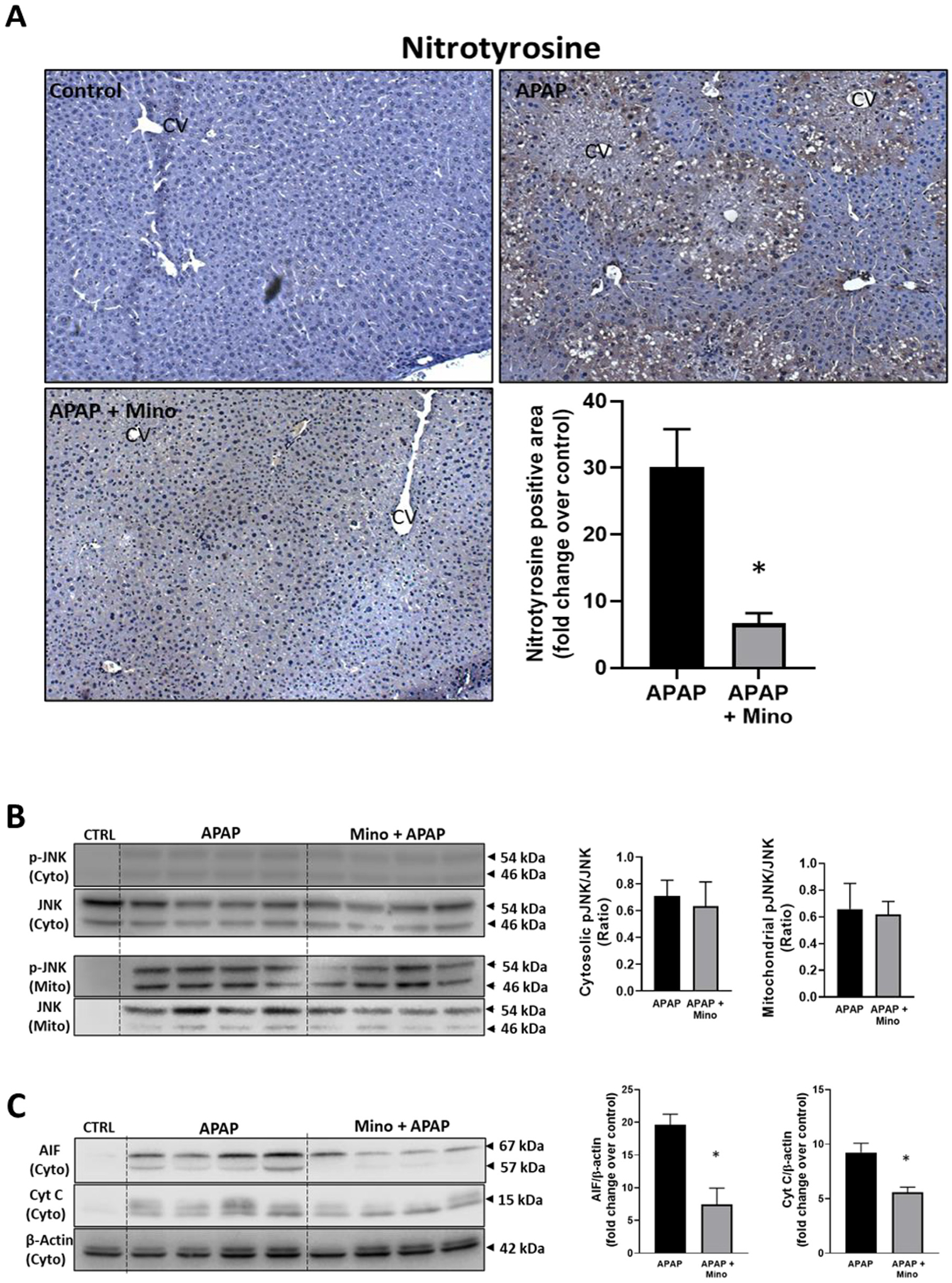
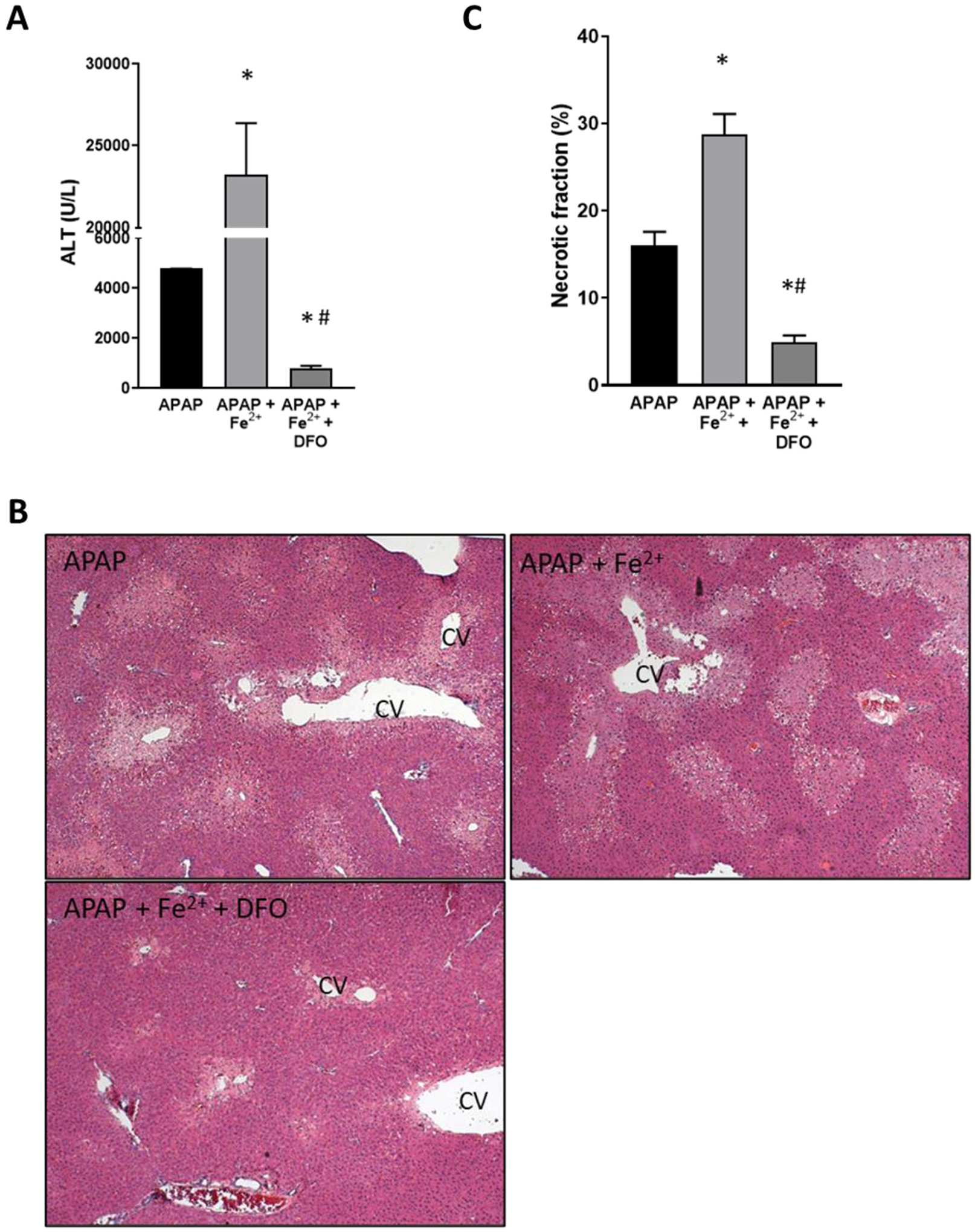
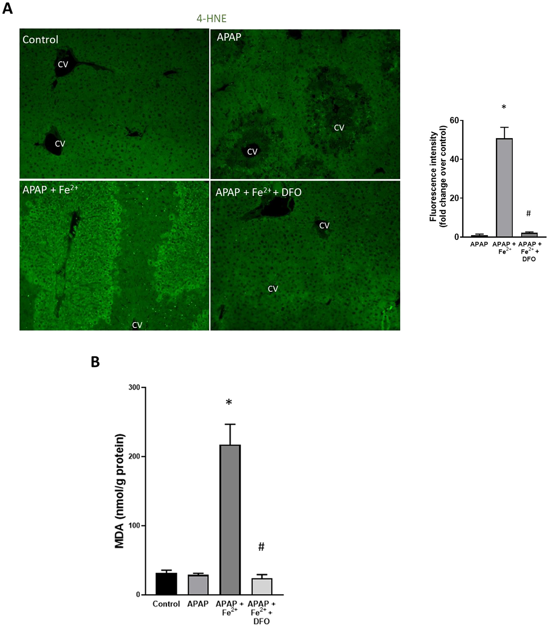
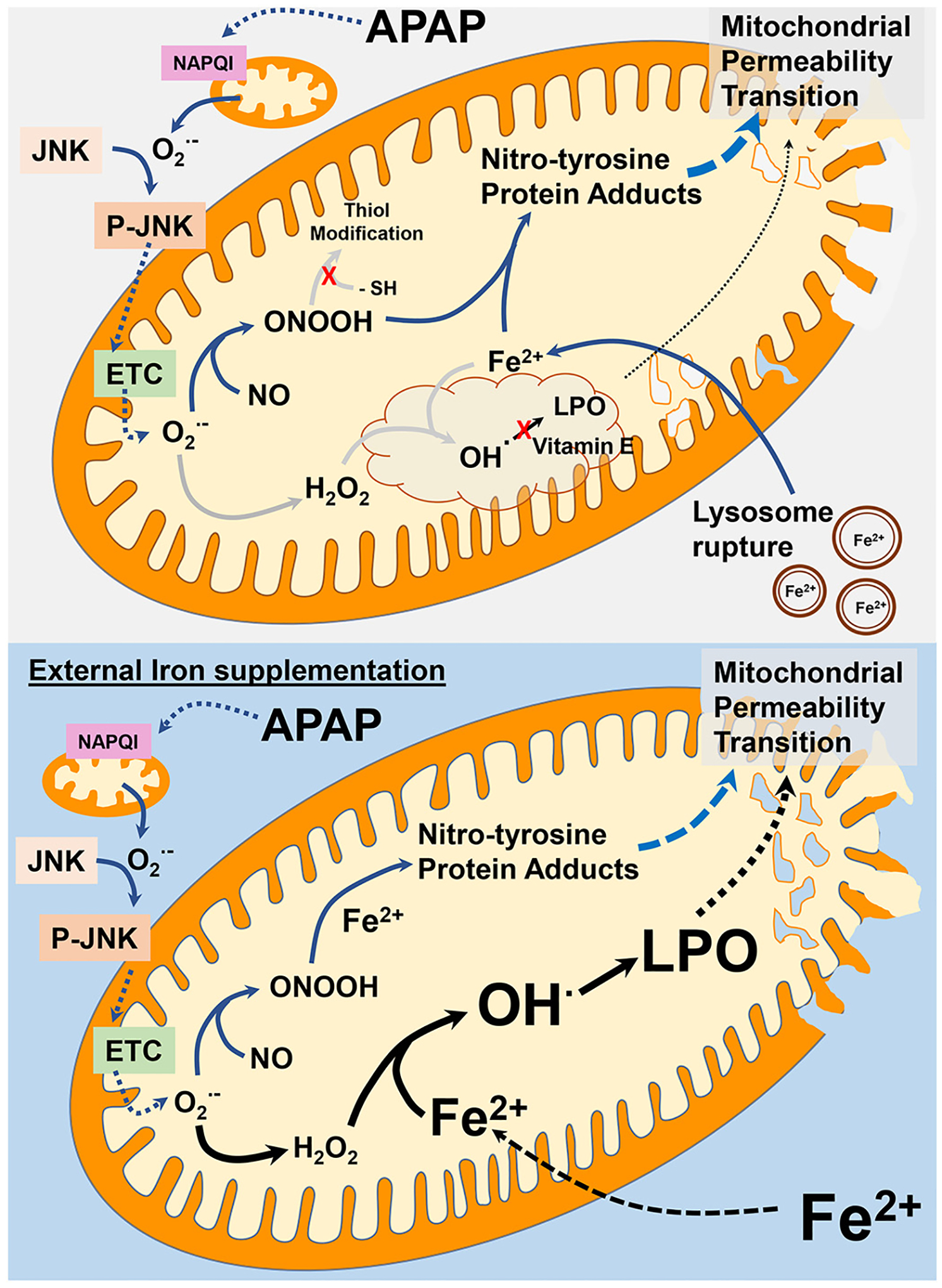
Acetaminophen (APAP) hepatotoxicity, a leading cause of acute liver failure in western countries, is characterized by mitochondrial superoxide and peroxynitrite formation. However, the role of iron, especially as facilitator of lipid peroxidation (LPO), has been controversial. Our aim was to determine the mechanism by which iron promotes cell death in this context. Fasted male C57BL/6J mice were treated with the iron chelator deferoxamine, minocycline (inhibitor of the mitochondrial calcium uniporter) or vehicle 1 h before 300 mg/kg APAP. Deferoxamine and minocycline significantly attenuated APAP-induced elevations in serum alanine amino transferase levels and hepatic necrosis at 6 h. This protection correlated with reduced 3-nitro-tyrosine protein adducts; LPO (malondialdehyde, 4-hydroxynonenal) was not detected. Activation of c-jun N-terminal kinase (JNK) was not affected but mitochondrial release of intermembrane proteins was reduced suggesting that the effect of iron was at the level of mitochondria. Co-treatment of APAP with FeSO4 exacerbated liver injury and protein nitration and triggered significant LPO; all effects were reversed by deferoxamine. Thus, after APAP overdose, iron imported into mitochondria facilitates protein nitration by peroxynitrite triggering mitochondrial dysfunction and cell death. Under these conditions, endogenous defense mechanisms largely prevent LPO. However, after iron overload, protein nitration and LPO contribute to APAP hepatotoxicity.

Keywords: Acetaminophen Hepatotoxicity; Deferoxamine; Ferrous iron; Lipid Peroxidation; Minocycline; Peroxynitrite.


Figures

Fig. 1.
Fig. 1.
Effect of pre-treatment with deferoxamine (DFO) on APAP-induced liver injury. Mice were injected with 200 mg/kg DFO 1 h before 300 mg/kg APAP (i.p.). Blood and liver tissue were collected at 6 h after APAP. (A) Plasma alanine amino transferase (ALT) activities. (B) H&E-stained liver sections. (C) Quantification of areas of necrosis. Bars represent mean ± SEM for n = 5 mice. *p < 0.05 compared to APAP.
Fig. 2.
Fig. 2.
Effect of pre-treatment with DFO on protein nitration, JNK activation and MPTP opening. Mice were injected with 200 mg/kg DFO 1 h before 300 mg/kg APAP (i.p.). Blood and liver tissue were collected at 6 h after APAP. (A) Immunostaining for nitro-tyrosine protein adducts and quantification of positive stained areas. (B) Western blots and densitometric quantification of mitochondrial and cytosolic JNK and p-JNK. (C) Western blots and densitometric quantification of cytosolic AIF and cytochrome c. Bars represent mean ± SEM for n = 3–4 mice. *p < 0.05 compared to APAP.
Fig. 3.
Fig. 3.
Effect of pre-treatment with minocycline on APAP-induced liver injury. Mice were injected with 10 mg/kg minocycline 1 h before 300 mg/kg APAP (i.p.). Blood and liver tissue were collected at 6 h after APAP. (A) Plasma alanine amino transferase (ALT) activities. (B) H&E-stained liver sections. (C) Quantification of areas of necrosis. Bars represent mean ± SEM for n = 5 mice. *p < 0.05 compared to APAP.
Fig. 4.
Fig. 4.
Effect of pre-treatment with minocycline on protein nitration, JNK activation and MPTP opening. Mice were injected with 10 mg/kg minocycline 1 h before 300 mg/kg APAP (i.p.). Blood and liver tissue were collected at 6 h after APAP. (A) Immunostaining for nitro-tyrosine protein adducts and quantification of positive stained areas. (B) Western blots and densitometric quantification of mitochondrial and cytosolic JNK and p-JNK. (C) Western blots and densitometric quantification of cytosolic AIF and cytochrome c. Bars represent mean ± SEM for n = 3–4 mice. *p < 0.05 compared to APAP.
Fig. 5.
Fig. 5.
Effect of co-treatment of FeSO4 with APAP on liver injury. Mice were injected with DFO 200 mg/kg or saline 1 h before receiving i.p. injections of 300 mg/kg APAP with or without 0.15 mmol/kg FeSO4. Blood and liver tissue were collected at 6 h after APAP. (A) Plasma alanine amino transferase (ALT) activities. (B) H&E-stained liver sections. (C) Quantification of areas of necrosis. Bars represent mean ± SEM for n = 5 mice. *p < 0.05 compared to APAP, #p < 0.05 compared to APAP + FeSO4.
Fig. 6.
Fig. 6.
Effect of co-treatment of FeSO4 with APAP on protein nitration, JNK activation and MPTP opening. Mice were injected with DFO 200 mg/kg or saline 1 h before receiving i.p. injections of 300 mg/kg APAP with or without 0.15 mmol/kg FeSO4. Blood and liver tissue were collected at 6 h after APAP. (A) Immunostaining for nitro-tyrosine protein adducts and quantification of positive stained areas. (B) Western blots and densitometric quantification of mitochondrial and cytosolic JNK and p-JNK. (C) Western blots and densitometric quantification of cytosolic AIF and cytochrome c. Bars represent mean ± SEM for n = 3–4 mice. *p < 0.05 compared to APAP, #p < 0.05 compared to APAP + FeSO4.
Fig. 7.
Fig. 7.
Effect of co-treatment of FeSO4 with APAP on lipid peroxidation. Mice were injected with DFO 200 mg/kg or saline 1 h before receiving i.p. injections of 300 mg/kg APAP with or without 0.15 mmol/kg FeSO4. Blood and liver tissue were collected at 6 h after APAP. (A) Immunofluorescence staining for 4-hydroxynonenal (4-HNE) and quantification of fluorescence intensity. (B) Hepatic malondialdehyde (MDA) content. Bars represent mean ± SEM for n = 5 mice. *p < 0.05 compared to APAP, # < 0.05 compared to APAP + FeSO4.
Fig. 8.
Fig. 8.
Role of iron in APAP-induced mitochondrial damage. Under typical APAP overdose conditions, iron mediated lipid peroxidation (LPO) does not play a significant role in APAP hepatotoxicity. Formation of mitochondrial NAPQI protein adducts after APAP metabolism induces superoxide-mediated JNK activation and its mitochondrial translocation. This amplifies mitochondrial superoxide generation through electron transport chain (ETC) dysfunction, which reacts with nitric oxide to produce peroxynitrite. Though peroxynitrite can be scavenged by thiol groups of GSH, this does not occur because of the significant GSH depletion due to NAPQI detoxification. The influx of iron into mitochondria from lysosomal instability instead facilitates peroxynitrite reaction with iron to produce nitro-tyrosine protein adducts, ultimately inducing the mitochondrial permeability transition. Since vitamin E levels are typically maintained after APAP, any hydroxyl and alkoxyl radicals generated by reaction of superoxide derived hydrogen peroxide or fatty acid hydroperoxide with iron are rapidly scavenged, preventing relevant lipid peroxidation. In a situation of significant iron loading, however, these reactions could be overwhelmed by the excess iron which causes extensive lipid peroxidation along with nitro-tyrosine formation to induce the mitochondrial permeability transition.

References

    1. Agarwal R, Hennings L, Rafferty TM, Letzig LG, McCullough S, James LP, MacMillan-Crow LA, Hinson JA, 2012. Acetaminophen-induced hepatotoxicity and protein nitration in neuronal nitric-oxide synthase knockout mice. J. Pharmacol. Exp. Ther 340, 134–142. - PMC - PubMed
    1. Audimoolam VK, Wendon J, Bernal W, Heaton N, O’Grady J, Auzinger G, 2011. Iron and acetaminophen a fatal combination? Transpl. Int 24, e85–e88. - PubMed
    1. Bajt ML, Farhood A, Lemasters JJ, Jaeschke H, 2008. Mitochondrial bax translocation accelerates DNA fragmentation and cell necrosis in a murine model of acetaminophen hepatotoxicity. J. Pharmacol. Exp. Ther 324, 8–14. - PubMed
    1. Banerjee S, Melnyk SB, Krager KJ, Aykin-Burns N, Letzig LG, James LP, Hinson JA, 2015. The neuronal nitric oxide synthase inhibitor NANT blocks acetaminophen toxicity and protein nitration in freshly isolated hepatocytes. Free Radic. Biol. Med 89, 750–757. - PMC - PubMed
    1. Bernal W, Wendon J, 2013. Acute liver failure. N. Engl. J. Med 369, 2525–2534. - PubMed

Publication types