. 2022 Jun;298(6):102008.
doi: 10.1016/j.jbc.2022.102008. Epub 2022 May 2.

Loss of mitochondrial ATPase ATAD3A contributes to nonalcoholic fatty liver disease through accumulation of lipids and damaged mitochondria

Affiliations

Loss of mitochondrial ATPase ATAD3A contributes to nonalcoholic fatty liver disease through accumulation of lipids and damaged mitochondria

Liting Chen et al. J Biol Chem. 2022 Jun.

Abstract

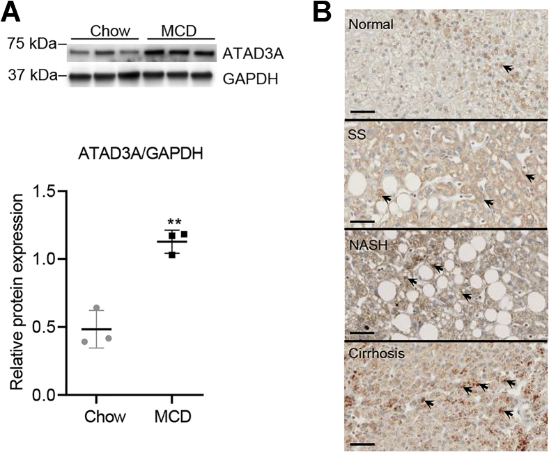
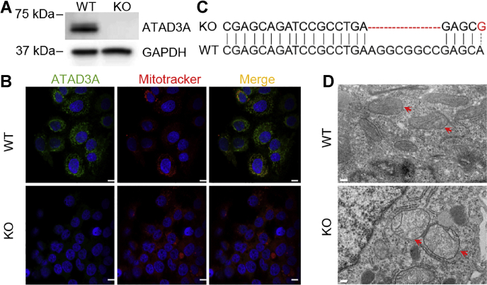
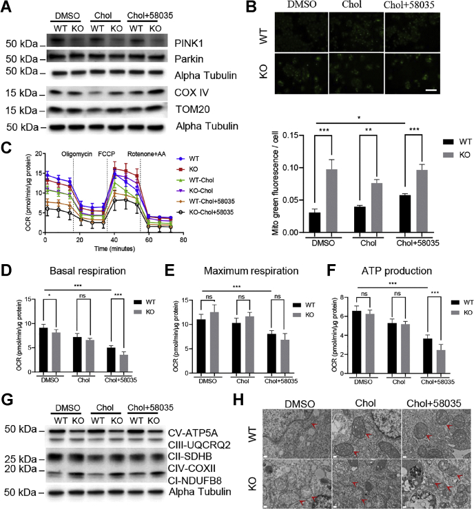
Mitochondrial ATPase ATAD3A is essential for cholesterol transport, mitochondrial structure, and cell survival. However, the relationship between ATAD3A and nonalcoholic fatty liver disease (NAFLD) is largely unknown. In this study, we found that ATAD3A was upregulated in the progression of NAFLD in livers from rats with diet-induced nonalcoholic steatohepatitis and in human livers from patients diagnosed with NAFLD. We used CRISPR-Cas9 to delete ATAD3A in Huh7 human hepatocellular carcinoma cells and used RNAi to silence ATAD3A expression in human hepatocytes isolated from humanized liver-chimeric mice to assess the influence of ATAD3A deletion on liver cells with free cholesterol (FC) overload induced by treatment with cholesterol plus 58035, an inhibitor of acetyl-CoA acetyltransferase. Our results showed that ATAD3A KO exacerbated FC accumulation under FC overload in Huh7 cells and also that triglyceride levels were significantly increased in ATAD3A KO Huh7 cells following inhibition of lipolysis mediated by upregulation of lipid droplet-binding protein perilipin-2. Moreover, loss of ATAD3A upregulated autophagosome-associated light chain 3-II protein and p62 in Huh7 cells and fresh human hepatocytes through blockage of autophagosome degradation. Finally, we show the mitophagy mediator, PTEN-induced kinase 1, was downregulated in ATAD3A KO Huh7 cells, suggesting that ATAD3A KO inhibits mitophagy. These results also showed that loss of ATAD3A impaired mitochondrial basal respiration and ATP production in Huh7 cells under FC overload, accompanied by downregulation of mitochondrial ATP synthase. Taken together, we conclude that loss of ATAD3A promotes the progression of NAFLD through the accumulation of FC, triglyceride, and damaged mitochondria in hepatocytes.

Keywords: ATAD3A; NAFLD; autophagy; cholesterol; fatty acid oxidation; free fatty acid; mitochondrial respiration; mitophagy; triglyceride.


Conflict of interest statement

Conflict of interest The authors declare that they have no conflicts of interest with the contents of this article.

Figures

Figure 1
Figure 1
ATAD3A was upregulated in rats with NASH and human patients with NAFLD.A, immunoblots for ATAD3A in rats fed with chow or MCD diet for 8 weeks (top left); quantification using ImageJ (bottom left). Data are presented as mean ± SD. ∗∗p < 0.01 by unpaired two-tailed student's t test. B, immunohistochemistry for ATAD3A in normal human tissue or patients with SS, NASH, or cirrhosis. A detailed description of the human liver samples used is summarized in Table S1. Black arrows, ATAD3A antibody-stained area. The scale bar represents 50 μm. MCD, methionine-choline deficient; NASH, nonalcoholic steatohepatitis; SS, simple steatosis.
Figure 2
Figure 2
Characterization of ATAD3A KO in Huh7.A, immunoblots for ATAD3A in Huh7 WT and ATAD3A KO cells. B confocal images for ATAD3A (green) and MitoTracker (red) in Huh7 WT and ATAD3A KO cells. The scale bar represents 20 μm. C, next-generation sequencing for ATAD3A identified the deletion of eight base pairs and one base pair mutation at exon 7 of ATAD3A. D, mitochondrial morphology in Huh7 WT and ATAD3A KO cells under TEM. Red arrows, mitochondria. The scale bar represents 0.2 μm. TEM, transmission electron microscopy.
Figure 3
Figure 3
ATAD3A KO exacerbated FC accumulation under FC overload in Huh7 cells.A, cholesterol quantification of ATAD3A KO and WT cells treated with DMSO, cholesterol (Chol), or Chol plus 58035, an acetyl-CoA acetyltransferase inhibitor, for 24 h (n = 3). B, WT or ATAD3A KO Huh7 cells were treated as indicated for 24 h after overnight incubation with DMEM supplemented with 10% of lipoprotein-deficient serum. Cells were fixed and stained with filipin (blue) and Lamin B1 (green) after incubation. White arrows, large puncta stained with filipin. The scale bar represents 130 μm. C, quantification for B. Percentage of area stained with filipin/nucleus for four images of WT or ATAD3A KO Huh7 cells treated as indicated analyzed with ImageJ. D and E, qPCR for HMGR and LXRA mRNA expression normalized to S18 from ATAD3A KO and Huh7 WT cells (n = 3). F, immunoblot analyses for HMGR and LXRA in WT or ATAD3A KO Huh7 cells treated as indicated. Data are presented as mean ± SD. ∗∗∗p < 0.001, ∗p < 0.05 by two-way ANOVA for cholesterol quantification and one-way ANOVA for Filipin quantification and qPCR. FC, free cholesterol; HMGR, 3-hydroxy-3-methylglutaryl-CoA reductase; LXRA, liver X receptor alpha; qPCR, quantitative PCR; TC, total cholesterol.
Figure 4
Figure 4
ATAD3A KO induces TG accumulation via inhibited lipolysis. Treatments are the same as in Figure 3A and B, qPCR analysis of SREBP1c, FAS mRNA expression normalized to S18 using ATAD3A KO and Huh7 WT cells treated as indicated for 24 h after overnight incubation with DMEM supplemented with 10% of lipoprotein-deficient serum (n = 3). C and D, immunoblots for SREBP1c and FAS in WT or ATAD3A KO Huh7 cells treated as indicated. Immunoblots in panel (C) and Figure 3F share the same alpha-Tubulin control because the antibodies in these figures were incubated with proteins separated on the same membrane. E, TG quantification for ATAD3A KO or WT Huh7 cells treated as indicated (n = 3). F, qPCR analysis of PLIN2 mRNA expression normalized to S18 using ATAD3A KO and Huh7 WT cells treated as indicated (n = 3). G, immunoblot analysis for PLIN2 protein expression in human hepatocytes subjected to ATAD3A siRNA or universal control siRNA under the treatment as indicated for 24 h. H and I, qPCR analysis of PPARA and CPT1A mRNA expression normalized to S18 using ATAD3A KO and Huh7 WT cells treated as indicated (n = 3). J, immunoblots for PPARA and CPT1A in WT or ATAD3A KO Huh7 cells treated as indicated. Immunoblots in panels (D) and (J) share the same alpha-Tubulin because the antibodies in (D) and (J) were incubated with proteins separated on the same membrane. Data are presented as mean ± SD. ∗∗∗p < 0.001, ∗∗p < 0.01, ∗p < 0.05 by one-way ANOVA. Chol, cholesterol; CPT1A, carnitine palmitoyltransferase 1A; FAS, fatty acid synthase; KD, knockdown; PLIN2, Perilipin 2; PPARA, peroxisome proliferator activated receptor a; qPCR, quantitative PCR; SREBP1c, plasminogen activator inhibitor 1 RNA-binding protein 1c; TG, triglyceride.
Figure 5
Figure 5
ATAD3A KO blocks autophagy and induces cell death under FC overload in Huh7 cells and human hepatocytes. Immunoblots for LC3-I, LC3-II, and p62 in WT or ATAD3A KO Huh7 (A), and ATAD3A, LC3-I, LC3-II, and p62 in KD human hepatocytes isolated from HLCM (C) treated as indicated for 24 h. Immunoblots in panel (C) and Figure 4G share the same GAPDH control because the antibodies in these figures were incubated with proteins separated on the same membrane. B, qPCR analysis of p62 mRNA expression normalized to S18 using ATAD3A KO and Huh7 WT cells treated as indicated (n = 3). D, TEM for WT or ATAD3A KO Huh7 cells treated as indicated for 24 h. Red arrows, autophagolysosome. The scale bar represents 0.5 μm. E and F, immunoblots for LC3-I, LC3-II, and p62 for WT or ATAD3A KO Huh7 cells pretreated with 3-MA or bafilomycin A1 for 1 h before cholesterol (Chol) or Chol plus 58035 treatment for 24 h. G, MTT (n = 6) for WT or ATAD3A KO Huh7 cells treated as indicated. H, cell count under the microscope for human hepatocytes with ATAD3A siRNA or universal control siRNA treated as indicated for 24 h (n = 3). Data are presented as mean ± SD. ∗∗∗p < 0.001, ∗∗p < 0.01, ∗p < 0.05 by one-way ANOVA. HLCM: humanized liver chimeric mice; FC, free cholesterol; KD, knockdown; MTT, 3-(4,5-dimethylthiazol-2-yl)-2,5-diphenyltetrazolium bromide; qPCR, quantitative PCR; RFU, relative fluorescent units; TEM, transmission electron microscopy.
Figure 6
Figure 6
ATAD3A KO inhibits PINK1-Parkin–dependent mitophagy and mitochondrial respiration in Huh7 cells.A, immunoblots for PINK1, Parkin, COX IV, and TOM20 in WT or ATAD3A KO Huh7 treated with DMSO, Chol, or Chol plus 58035 for 24 h. B, Mitotracker green staining (upper) and quantification (lower) for WT or ATAD3A KO Huh7 treated as indicated. The scale bar represents 130 μm. C, OCR Seahorse assay for Huh7 WT and ATAD3A KO cells treated as indicated. DF, basal respiration, maximum respiration, and ATP production of Huh7 WT and ATAD3A KO cells treated as indicated. G, immunoblots for oxidative phosphorylation enzymes in WT or ATAD3A KO Huh7 cells treated as indicated. H, representative mitochondrial structure TEM images of ATAD3A KO Huh7 cells or WT cells. Red arrows, mitochondria. The scale bar represents 0.2 μm. Data are presented as mean ± SD. ∗∗∗p < 0.001, ∗∗p < 0.01, ∗p < 0.05 by one-way ANOVA. Chol, cholesterol; OCR, oxygen consumption rate; PINK1, PTEN-induced kinase 1; ROS, reactive oxygen species; ns, not significant; TEM, transmission electron microscopy.
Figure 7
Figure 7
Schematic representation of the influence of FC and loss of ATAD3A on autophagy, mitophagy, and lipids. FC overload induces autophagy but blocks mitophagy and reduces PLIN2 expression and TG amount in cells. The loss of ATAD3A suppresses both autophagy and mitophagy, upregulates PLIN2, and accumulates TG in cells under normal conditions or when FC is overloaded. Collectively, ATAD3A deletion contributes to NAFLD through TG accumulation and compromised mitochondrial quality control in hepatocytes. FC, free cholesterol; PLIN2: perilipin 2; TG, triglyceride.

References

    1. Younossi Z., Anstee Q.M., Marietti M., Hardy T., Henry L., Eslam M., et al. Global burden of NAFLD and NASH: trends, predictions, risk factors and prevention. Nat. Rev. Gastroenterol. Hepatol. 2018;15:11–20. - PubMed
    1. Huang T.D., Behary J., Zekry A. Non-alcoholic fatty liver disease: a review of epidemiology, risk factors, diagnosis and management. Intern. Med. J. 2020;50:1038–1047. - PubMed
    1. Arguello G., Balboa E., Arrese M., Zanlungo S. Recent insights on the role of cholesterol in non-alcoholic fatty liver disease. Biochim. Biophys. Acta. 2015;1852:1765–1778. - PubMed
    1. Cao S.S., Kaufman R.J. Endoplasmic reticulum stress and oxidative stress in cell fate decision and human disease. Antioxid. Redox Signal. 2014;21:396–413. - PMC - PubMed
    1. Pang L., Liu K., Liu D., Lv F., Zang Y., Xie F., et al. Differential effects of reticulophagy and mitophagy on nonalcoholic fatty liver disease. Cell Death Dis. 2018;9:90. - PMC - PubMed

Publication types

MeSH terms

Substances