. 2022 May 5;13(1):2460.
doi: 10.1038/s41467-022-29978-y.

Recycling of memory B cells between germinal center and lymph node subcapsular sinus supports affinity maturation to antigenic drift

Affiliations

Recycling of memory B cells between germinal center and lymph node subcapsular sinus supports affinity maturation to antigenic drift

Yang Zhang et al. Nat Commun. .

Abstract

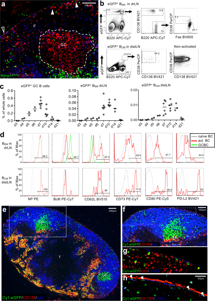
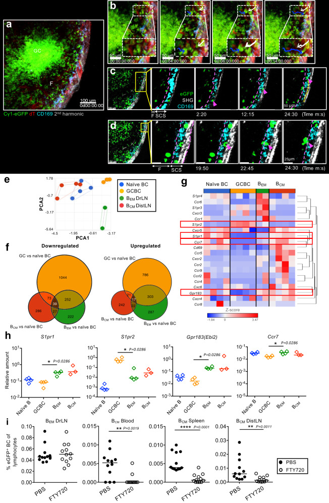
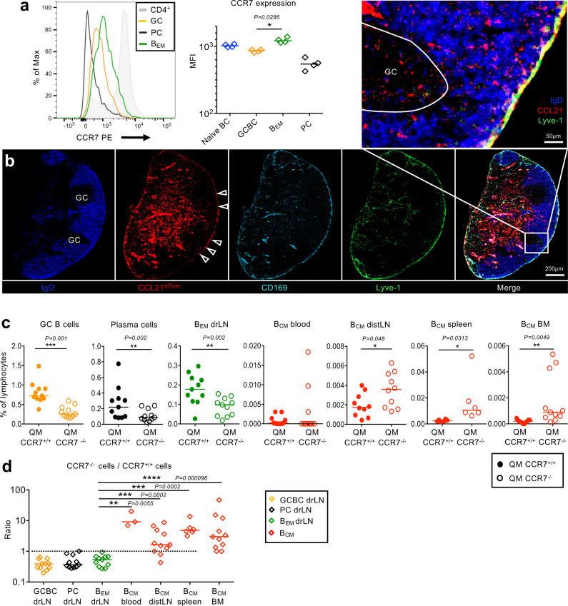
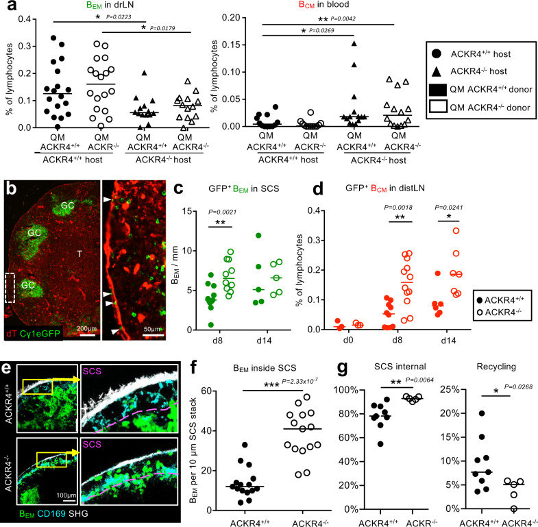
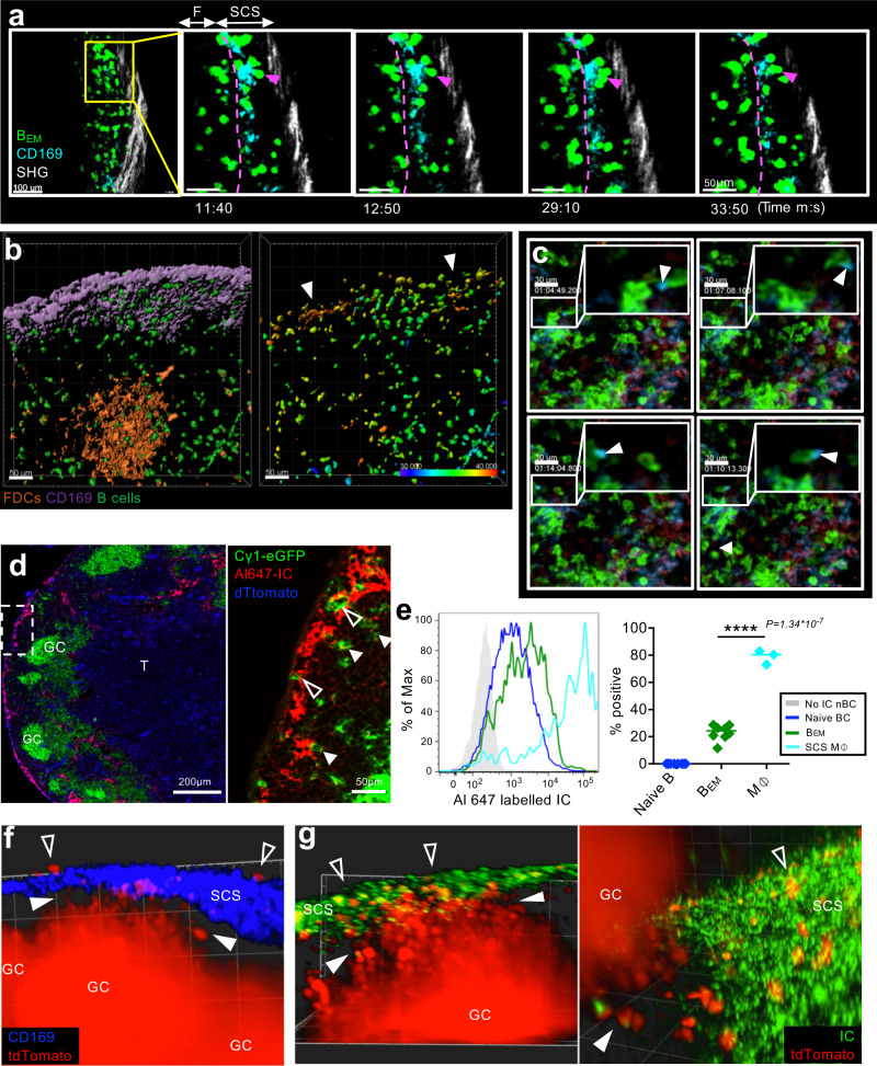
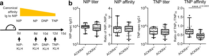
Infection or vaccination leads to the development of germinal centers (GC) where B cells evolve high affinity antigen receptors, eventually producing antibody-forming plasma cells or memory B cells. Here we follow the migratory pathways of B cells emerging from germinal centers (BEM) and find that many BEM cells migrate into the lymph node subcapsular sinus (SCS) guided by sphingosine-1-phosphate (S1P). From the SCS, BEM cells may exit the lymph node to enter distant tissues, while some BEM cells interact with and take up antigen from SCS macrophages, followed by CCL21-guided return towards the GC. Disruption of local CCL21 gradients inhibits the recycling of BEM cells and results in less efficient adaption to antigenic variation. Our findings thus suggest that the recycling of antigen variant-specific BEM cells and transport of antigen back to GC may support affinity maturation to antigenic drift.


Conflict of interest statement

The authors declare no competing interests.

Figures

Fig. 1
Fig. 1. Appearance of antigen-activated memory-like B cells in drLN and distLN.
a Location of B1-8i/k−/−/Blimp1GFP/Cdt1mKO2 B cells in drLN 6 d after immunization. Antigen-specific B cells in the G0/G1 phase of cell cycle (red) inside the GC (dashed line) and in the follicle (F) close to the SCS (arrow heads). Interfollicular region (open triangle). Blimp-1+ PCs are eGFP (green). Hoechst33342-labeled naïve T cells (blue). Scale bar: 100 μm. Representative image of 3 lymph nodes. b Gating of eGFP+ BEM in drLN and BCM and distLN 8 d after immunization of recipients of NP-specific Cγ1Cre QM mTmG B cells. c Kinetic of eGFP+ B cell appearance in drLN and distLN. Data merged from two independent experiments (n = 2–3). Each datapoint represents one animal. d Memory B cell typical markers on BEM and BCM in drLN and distLN. e drLN of recipient of QM Cγ1Cre mTmG cells 8 d after immunization. T zone (T). f Enlargement of box in e showing the eGFP+ BEM close to the subcapsular sinus, g Ki-67 expression in BEM, h same area showing BEM location below the LYVE1+ ER-TR7-ve SCS floor endothelium and inside the SCS (arrowheads). Image is a representative of three lymph nodes.
Fig. 2
Fig. 2. BEM movement in the drLN.
a Intravital observation of popliteal lymph node from Cγ1Cre mTmG mice 8 d after NP-CGG foot immunization (see suppl. movie 1). b Still images showing eGFP+ BEM entering the SCS (blue arrow) and reentering the lymph node follicle from the SCS (white arrow). c Images showing an eGFP+ B cells migrating along the SCS. d Images showing a BEM reentering the lymph node follicle. Representative of 7 Cγ1Cre mTmG mice (n = 7). RNA-Seq data from B cell populations sorted from drLN 8 d after immunization of recipients of Cγ1-Cre QM mTmG B cells. e Principal component analysis of global gene expression in sorted populations, f Numbers of differentially expressed genes. g Heat map of selected chemotactic receptors among mRNA. Genes were hierarchically clustered by Euclidean distance measure. Data are from two independent experiments with four samples (each sample from pooled popliteal lymph nodes from three mice). h S1pr1, S1pr2, Ebi2, and Ccr7 mRNA expression analyzed by qRT-PCR. Each diamond represents pooled lymph nodes from four mice. All values are relative to B2m mRNA. Two-tailed Mann-Whitney test, *: p = 0.0286. Data representative of three independent experiments (total n = 12). i BEM in drLN and BCM in blood, spleen, and distLN 8 d after immunization. Mice received FTY720 over 2 d before collection. Each dot represents one mouse, data merged from three independent experiments (total n = 12). Two-tailed Mann-Whitney test, **: p < 0.01, ****: p < 0.0001.
Fig. 3
Fig. 3. CCR7-dependent migration of memory B cells.
a CCR7 expression on different lymphocyte subsets measured by flow cytometry. Each symbol represents one animal. Two-tailed Mann-Whitney test, *p = 0.0286. b drLN from a heterozygous Ccl21tdTom/wt reporter mouse 8 d after foot immunization. The SCS ceiling endothelium is missing. Images show dTomato expression in red, and IgD (blue), CD169 (turquoise) and Lyve-1 (green)-specific antibody staining. Arrow heads: Ccl21 expression in SCS. Enlarged box in shows Ccl21a expression under and in the SCS floor endothelium. Image is a representative from 4 lymph nodes. c GC B cells, plasma cells (PC), BEM from drLN 8 d after foot immunization of recipients of a mix of QM mT CCR7+/+ and QM eYFP CCR7−/− B cells. BCM in blood, distLN, spleen, and bone marrow (BM). Two- tailed Wilcoxon matched-pairs test. d Ratio of CCR7−/− to CCR7+/+ QM B cells in different tissues. Two-tailed unpaired t test. Each symbol represents one animal. Data are from two independent experiments with 5–6 mice each.
Fig. 4
Fig. 4. Local ACKR4-generated chemokine gradients at the SCS direct migration of memory B cells.
a Frequency of QM ACKR4+/+ and QM ACKR4−/− BEM in drLN or BCM in blood of ACKR4+/+ and ACKR4−/− hosts. Data merged from three independent experiments with 4–5 mice each. Two-tailed Mann-Whitney test. b drLN from an Cγ1-Cre mTmG mouse 8 d after immunization. Arrows: eGFP+ BEM inside the SCS. Representative image of nine lymph nodes. c eGFP+ BEM in the SCS of drLN in Cγ1Cre mTmG ACKR4+/+ or ACKR4−/− mice at d8 and d14 after foot immunization. d BCM in distLN in ACKR4+/+ or ACKR4−/− mice after foot immunization. Data in c and d are merged from two independent experiments with 3–5 mice each. Two-tailed Mann-Whitney test. e Intravital observation of BEM entry into SCS in Cγ1Cre mTmG ACKR4+/+ or ACRK4−/− drLN. f Quantitation of intravital eGFP+ BEM in SCS of ACKR4+/+ or ACKR4−/− drLN. Each dot represents cell counts per field of view of a 10 μm Z stack. Pooled data from imaging of three ACKR4+/+ and two ACKR4−/− popliteal LNs. Two-tailed unpaired t test, ***P = 2.33 × 10−7. g Automated B cell tracking showing fraction of BEM tracks that are inside the SCS or recycling from the SCS into the lymph node parenchyma. Images from five ACKR4+/+ and 2 ACKR4−/− popliteal LNs were analyzed. Each symbol corresponds to the average fraction of tracks from one video, total n of tracks = 2478. One-tailed unpaired t test, *p = 0.0268; **p = 0.0064.
Fig. 5
Fig. 5. BEM interaction with antigen on SCS macrophages.
a Intravital observation of interactions of eGFP+ BEM (green) and SCS macrophages (CD169 turquois). b Intravital Ca2+ levels in BEM. Left: Surface rendering of CD169-stained macrophages (purple) and GC (orange, CD21/35Atto590), B1-8hi TN-XXL+ B cells (green). Right: FRET intensity in BEM of same frame with color-coded mean FRET intensity. Arrowheads: clusters of FRET-positive BEM in contact with SCS macrophages. Animation in movie 6. Scale bar 50 µm. c Intravital observation (clockwise from top left) of BEM (green) in SCS containing CD169+ material (blue, arrowhead). d drLN from an Cγ1Cre mTmG mouse 10 min after foot injection with Alexa647 labeled immune complex (IC). Open Arrow heads: eGFP+ BEM in SCS. Closed arrow heads: Alexa647-IC colocalizing with eGFP+ BEM in follicle. Representative image of three lymph nodes. e Left: Alexa647-IC on CD11b+ CD169+ SCS macrophage (green), eGFP+ BEM (red), naïve B cells (blue), or naïve B cells without Alexa647 injection (gray). Right: Percent Alexa647-IC positive BEM or macrophages compared to naïve B cells. Overton subtraction from two independent experiments with three mice each. SCS M⏀: CD169+ subcapsular sinus macrophages. Two-tailed unpaired Student’s t test, ****p = 1.34 × 10−7. f Light-sheet microscopy of live explanted drLN, showing S1PR2tdTom BEM (red) in located between GC and SCS (closed arrowheads). SCS macrophages (blue). Some BEM are seen in SCS above the SCS layer (open arrowheads). See also movie 8. g Same as (f) showing Alexa488 labeled IC (green) in the SCS and S1pr2 expression dependent tdTomato positive BEM (red) between GC and SCS (closed arrowheads) or in contact with IC in SCS (open arrowheads). Representative image of four lymph nodes. See also Suppl. movies 9, 10.
Fig. 6
Fig. 6. BEM interaction with antigen in SCS affects response to antigenic variants.
a Design of antigenic drift experiment using variants of the NIP hapten. b NIP-specific and TNP-specific IgG1 antibody titer and affinity in ACKR4+/− and ACKR4−/− mice. Merged data from 4 independent experiments with five mice each (total n = 20). Boxes show medians and 50% range, whiskers: 5–95%. Symbols represent individual mice. Two-tailed Mann-Whitney test, ****p < 0.0001.

References

    1. Zhang Y, Garcia-Ibanez L, Toellner K-M. Regulation of germinal centre B cell differentiation. Immunol. Rev. 2016;270:8–19. doi: 10.1111/imr.12396. - DOI - PMC - PubMed
    1. Yoshida T, et al. Memory B and memory plasma cells. Immunol. Rev. 2010;237:117–139. doi: 10.1111/j.1600-065X.2010.00938.x. - DOI - PubMed
    1. Weisel FJ, Zuccarino-Catania GV, Chikina M, Shlomchik MJ. A temporal switch in the germinal center determines differential output of memory B and plasma cells. Immunity. 2016;44:116–130. doi: 10.1016/j.immuni.2015.12.004. - DOI - PMC - PubMed
    1. Zhang Y, et al. Plasma cell output from germinal centers is regulated by signals from Tfh and stromal cells. J. Exp. Med. 2018;215:1227–1243. doi: 10.1084/jem.20160832. - DOI - PMC - PubMed
    1. Laidlaw BJ, et al. The Eph-related tyrosine kinase ligand Ephrin-B1 marks germinal center and memory precursor B cells. J. Exp. Med. 2017;214:639–649. doi: 10.1084/jem.20161461. - DOI - PMC - PubMed

Publication types