. 2022 May 5;12(1):7326.
doi: 10.1038/s41598-022-11033-x.

FGF21-FGFR4 signaling in cardiac myocytes promotes concentric cardiac hypertrophy in mouse models of diabetes

Affiliations

FGF21-FGFR4 signaling in cardiac myocytes promotes concentric cardiac hypertrophy in mouse models of diabetes

Christopher Yanucil et al. Sci Rep. .

Abstract

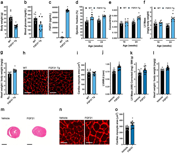
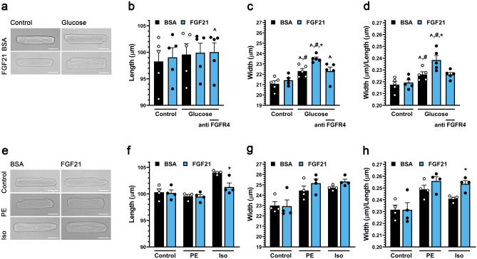
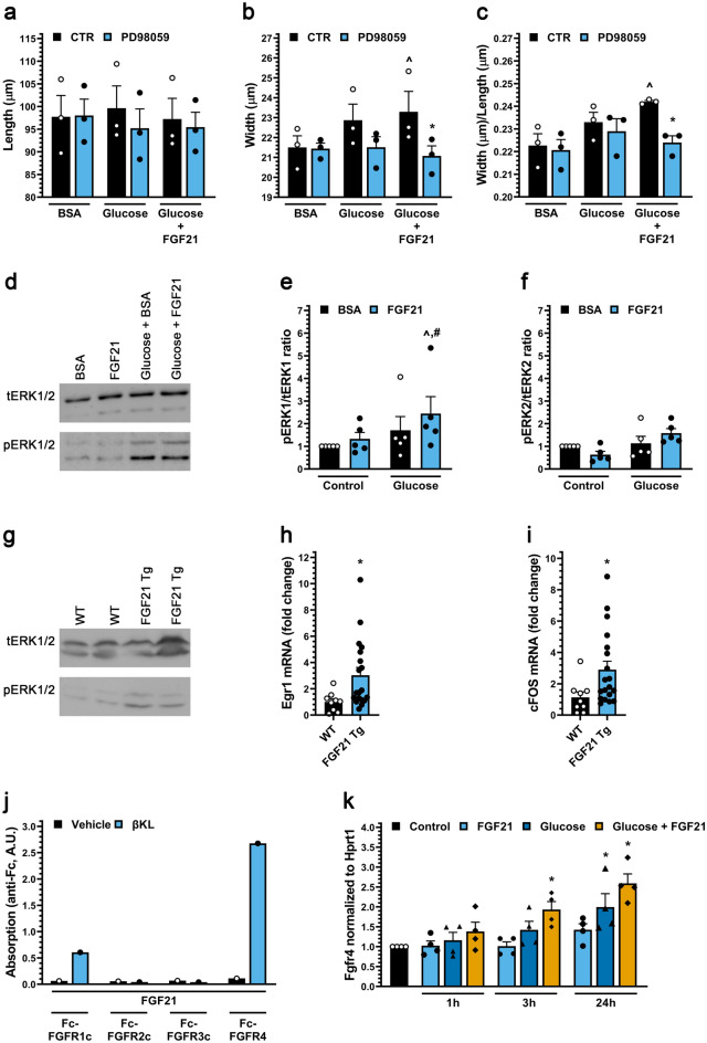
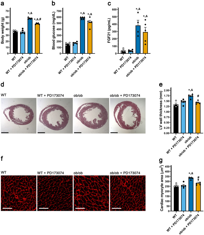
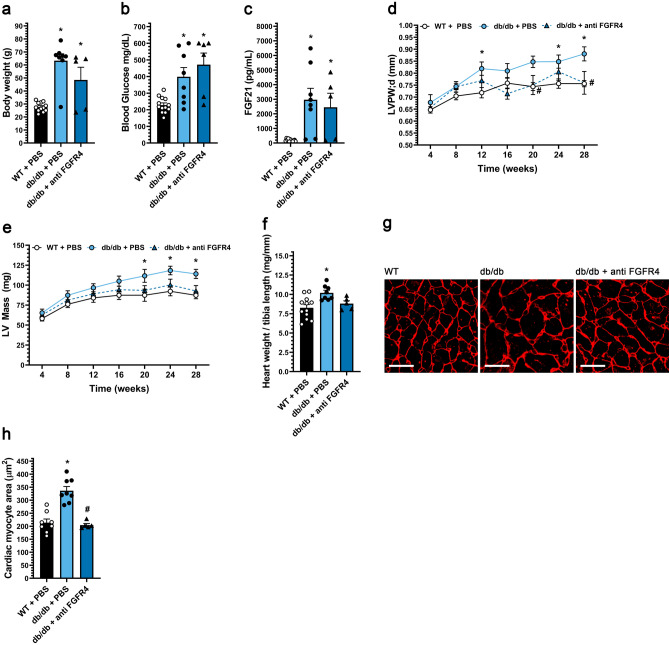
Fibroblast growth factor (FGF) 21, a hormone that increases insulin sensitivity, has shown promise as a therapeutic agent to improve metabolic dysregulation. Here we report that FGF21 directly targets cardiac myocytes by binding β-klotho and FGF receptor (FGFR) 4. In combination with high glucose, FGF21 induces cardiac myocyte growth in width mediated by extracellular signal-regulated kinase 1/2 (ERK1/2) signaling. While short-term FGF21 elevation can be cardio-protective, we find that in type 2 diabetes (T2D) in mice, where serum FGF21 levels are elevated, FGFR4 activation induces concentric cardiac hypertrophy. As T2D patients are at risk for heart failure with preserved ejection fraction (HFpEF), we propose that induction of concentric hypertrophy by elevated FGF21-FGFR4 signaling may constitute a novel mechanism promoting T2D-associated HFpEF such that FGFR4 blockade might serve as a cardio-protective therapy in T2D. In addition, potential adverse cardiac effects of FGF21 mimetics currently in clinical trials should be investigated.


Conflict of interest statement

C.F. and D.K. have served as consultants for Bayer, and C.F. also for Calico Labs. C.Y. and C.F. are inventors on two pending patents (PCT/US2019/049211; PCT/US19/49161) and they are co-founders of a startup biotech company (Alpha Young LLC). C.F. is also the CSO of Alpha Young LLC. C.F. has a patent on FGFR inhibition (European Patent No. 2723391). C.F. and A.G. received honoraria for publishing a book (“FGF23”, Elsevier, ISBN9780128180365). A.F. is vice-president of L&F Health LLC and the scientist co-founder and a shareholder of ZyVersa Therapeutics Inc and River 3 Renal Corp. X.L., K.S., E.C.M., J.L., I.C., B.C., K.H., D.W., A.R.W., A.S., J.M.R. and M.S.K. have no competing interest.

Figures

Figure 1
Figure 1
Systemic FGF21 elevation in mice induces concentric cardiac hypertrophy. (ai) Analysis of FGF21 transgenic (Tg) mice and wild-type littermates at 24 weeks of age unless otherwise indicated. (a) Body weight, (b) blood glucose levels, and (c) serum FGF21 levels. (d) Ejection fraction, (e) concentricity [relative wall thickness = (LVAW;d + LVPW;d)/LVID;d], and (f) left ventricular (LV) mass, as determined by echocardiography at 16 and 24 weeks of age. (g) Heart weight/body weight ratio, (h, i) wheat germ agglutinin staining of LV tissue (scale bar = 25 µm) and cardiac myocyte cross-section area. (jo) Wildtype mice were injected i.v. with FGF21 or vehicle for five consecutive days before analysis on the 6th day. (j) LV anterior wall thickness (diastole), and (k) LV mass/body weight ratio, as determined by echocardiography. (l) Gravimetric heart weight/body weight ratio. (m) Hematoxylin and eosin-stained transverse heart sections (scale bar = 2 mm). (n, o) Wheat germ agglutinin staining of LV tissue (scale bar = 25 µm) and cardiac myocyte cross-section area. Statistical significance was determined by 2-way ANOVA with post-hoc testing with Sidak's multiple comparisons test (df), or by two-tailed t-test. All values are expressed as mean ± SEM. (a, b) N = 9; (c) N = 8–9; (df) N = 8, *p ≤ 0.05 vs. WT of same age; (g, i) N = 8, *p ≤ 0.05 vs. WT; (jl, o), N = 10, *p ≤ 0.05 vs. vehicle. For the complete set of echocardiography parameters see Supplementary Tables 1 and 3.
Figure 2
Figure 2
FGF21 induces hypertrophic growth of cultured cardiac myocytes in the presence of high glucose via FGFR4. (a) Transmitted light images of primary adult rat ventricular myocytes (ARVMs) treated with BSA (control), and mouse recombinant FGF21 (25 ng/ml) with or without 10 mM increased glucose (final 5.6 or 15.6 mM) for 48 h (scale bar = 25 µm), and (bd) myocyte length, width, and width to length ratio. FGFR4-specific blocking antibody (anti-FGFR4; 10 mg/ml) was included as indicated. (e) Images of ARVMs treated with BSA (control), and FGF21 (25 ng/ml) with or without phenylephrine (PE; 20 µM) or isoproterenol (Iso; 10 µM) for 48 h (scale bar = 25 µm), and (fh) myocyte length, width, and width to length ratio. Comparison between groups was performed in form of a one-way (bd, fh) ANOVA followed by post-hoc Tukey test. All values are expressed as mean ± SEM. (bd) N = 5, ^p ≤ 0.05 vs. BSA Control, #p ≤ 0.05 vs. FGF21 Control, *p ≤ 0.05 vs. Glucose Control. (fh) N = 4, *p ≤ 0.05 vs. respective Control.
Figure 3
Figure 3
FGF21 activates ERK1/2 in cultured cardiac myocytes in the presence of high glucose and in heart tissue of mice. (ac) Length, width and width to length ratio for ARVMs treated with BSA (control), FGF21 (25 ng/ml), increased glucose and/or the MEK inhibitor PD98059 (20 µM) for 48 h. Bars and colored symbols indicate average mean and means of independent experiments using different myocyte preparations, respectively. (df) Western blot analysis of ARVMs treated with BSA (control) or mouse recombinant FGF21 (25 ng/ml) with or without 10 mM glucose for 6 h. ERK1 is p44 and ERK2 is p42. (g) Analysis of cardiac tissue from FGF21 Tg mice and wild-type littermates at 8–12 weeks of age by Western blotting. (h, i) qRT-PCR for Egr-1 and c-Fos mRNA using total RNA from heart tissue of FGF21 Tg mice and wild-type littermates at 8–12 weeks of age. (j) Binding of 1 µg of soluble β-klotho (βKL) or PBS, 500 ng of Fc-tagged FGFR 1c, 2c, 3c, or 4 to 96-well plates coated with 200 ng of FGF21. (k) qRT-PCR for FGFR4 mRNA using total RNA isolated from ARVMs treated with BSA (control), FGF21 (25 ng/ml), and/or increased glucose (15.6 mM total). Comparison between groups was performed in form of a one-way (ac, k) or two-way (ef) ANOVA followed by post-hoc Tukey test or a two tailed t-test (h, i). All values are expressed as mean ± SEM. (ac) N = 3, ^p ≤ 0.05 vs. BSA CTR, *p ≤ 0.05 vs. Glucose + FGF21 CTR; (e, f) N = 5, ^p ≤ 0.05 vs. BSA CTR, #p ≤ 0.05 vs. FGF21 CTR; (h, i) N = 9−19, *p ≤ 0.05 vs. WT; (k) N = 4, *p ≤ 0.05 vs. CTR. All Western blots are cropped, and original blots are presented in Supplementary Fig. 3.
Figure 4
Figure 4
Systemic pan-FGFR blockade protects diabetic mice from cardiac hypertrophy. Three-month old ob/ob and wildtype mice were injected daily with 1 mg/kg PD173074 or vehicle solution for six weeks followed by endpoint analysis. (a) Body weight, (b) blood glucose levels, and (c) serum FGF21 levels. (d) Representative images of H&E-stained transverse heart sections (scale bar = 2 mm) and (e) quantification of the thickness of the LV wall in these images. (f) Wheat germ agglutinin-stained LV tissue (scale bar = 25 µm), and (g) quantification of cardiac myocyte area. Comparison between groups was performed in form of a one-way ANOVA followed by a post-hoc Tukey test (ac, e, g). All values are expressed as mean ± SEM. (ac, e, g) N = 4–5, *p ≤ 0.05 vs. WT, &p ≤ 0.05 vs. WT + PD173074 of same age, #p ≤ 0.05 vs. ob/ob of same age.
Figure 5
Figure 5
FGFR4 blockade protects diabetic mice from pathologic cardiac remodeling. db/db mice and wild-type (WT) littermates were treated for 24 weeks with either FGFR4 blocking antibody (25 mg/kg) or vehicle (PBS) on a bi-weekly basis, starting at 4 weeks of age. (a) Body weight, (b) blood glucose levels, and (c) serum FGF21 levels. (d) left ventricular (LV) posterior wall thickness in diastole, and (e) LV mass from 4 weeks until 28 weeks of age, as determined by serial echocardiography. (f) Gravimetric heart weight/tibia length ratio, (g) wheat germ agglutinin-stained LV tissue section (scale bar = 25 µm), and (h) cardiac myocyte cross-section area. Comparison between groups was performed in form of a one-way ANOVA (ac, f, h) or a 2-way ANOVA (d, e) followed by a post-hoc Tukey test. All values are expressed as mean ± SEM. (a, c, f) N = 5–12; (b) N = 6–13; (h) N = 5–9, *p ≤ 0.05 vs. WT, #p ≤ 0.05 vs. db/db. (d, e) N = 6–13, *p ≤ 0.05 vs. WT of same age, #p ≤ 0.05 vs. db/db of same age. For the complete set of echocardiography parameters see Supplementary Table 3.

References

    1. Jia G, Hill MA, Sowers JR. Diabetic cardiomyopathy: An update of mechanisms contributing to this clinical entity. Circ. Res. 2018;122:624–638. doi: 10.1161/CIRCRESAHA.117.311586. - DOI - PMC - PubMed
    1. Devereux RB, et al. Impact of diabetes on cardiac structure and function: the strong heart study. Circulation. 2000;101:2271–2276. doi: 10.1161/01.CIR.101.19.2271. - DOI - PubMed
    1. Aneja A, Tang WH, Bansilal S, Garcia MJ, Farkouh ME. Diabetic cardiomyopathy: Insights into pathogenesis, diagnostic challenges, and therapeutic options. Am. J. Med. 2008;121:748–757. doi: 10.1016/j.amjmed.2008.03.046. - DOI - PubMed
    1. Virani SS, et al. Heart disease and stroke statistics-2021 update: A report from the American Heart Association. Circulation. 2021;143:e254–e743. doi: 10.1161/CIR.0000000000000950. - DOI - PubMed
    1. Mishra S, Kass DA. Cellular and molecular pathobiology of heart failure with preserved ejection fraction. Nat. Rev. Cardiol. 2021 doi: 10.1038/s41569-020-00480-6. - DOI - PMC - PubMed

MeSH terms

Substances