. 2022 May 5;12(1):7331.
doi: 10.1038/s41598-022-10994-3.

Therapeutic and tectonic keratoplasty with simple cryopreserved remnants of donor corneas: an 11 year retrospective case series

Affiliations

Therapeutic and tectonic keratoplasty with simple cryopreserved remnants of donor corneas: an 11 year retrospective case series

Jae-Gon Kim et al. Sci Rep. .

Abstract

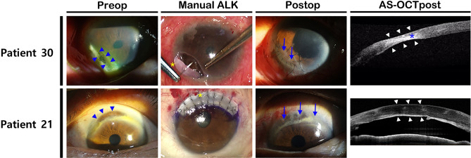
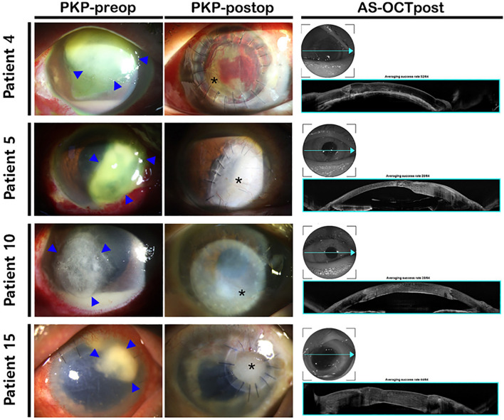
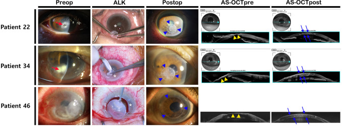
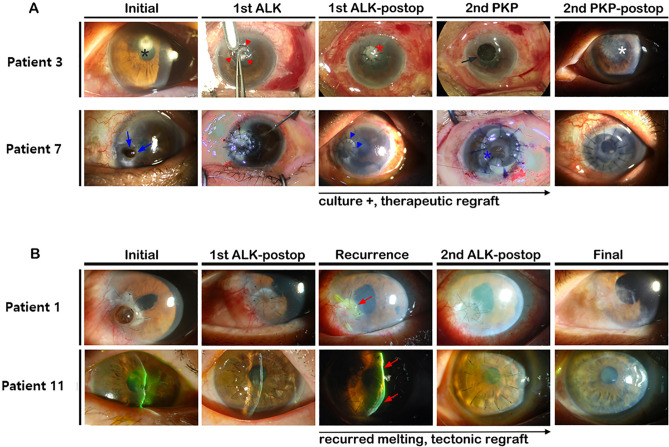
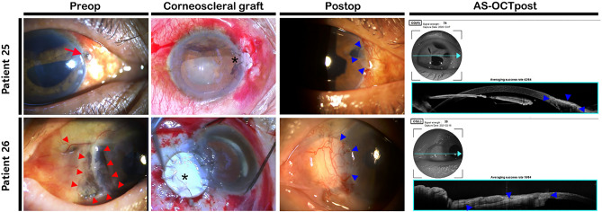
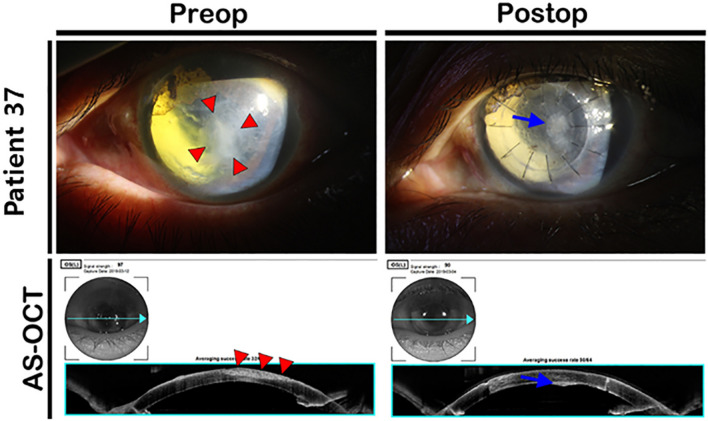
This study sought to describe the use of deep-frozen donor corneal remnants preserved after keratoplasty procedures for therapeutic or tectonic keratoplasty without subsequent optical keratoplasty. This single-center retrospective consecutive case series analyzed the electronic medical records of patients who had undergone therapeutic or tectonic keratoplasty using deep-frozen donor remains preserved in Optisol-GS, for the past 11 years at Keimyung University Dongsan Medical Center. Fifty-five surgical cases in 46 patients were included. Twenty-three surgical cases in 18 patients underwent therapeutic keratoplasty for refractory infectious corneal ulcer. Complete eradication of primary infection was achieved in 14 patients (77.8%). Tectonic keratoplasty were performed 32 cases in 28 patients. Twenty-seven of 28 patients were ultimately able to maintain anatomical integrity (96.4%). Mean uncorrected visual acuity improved from 1.77 ± 0.94 preoperatively to 1.31 ± 0.95 at the last follow-up postoperatively in the tectonic graft group by logarithm of the minimal angle of resolution (P = 0.002). There were no cases of graft rejection. Keratoplasty using cryopreserved donor tissue is a suitable surgical alternative for infectious or non-infectious corneal ulcers in elderly patients or patients with poor general condition. It could be a viable alternative to overcome the shortage of corneal donors.


Conflict of interest statement

The authors declare no competing interests.

Figures

Figure 1
Figure 1
Cases of impending perforation at the peripheral cornea. (Upper) A case of marginal corneal melting with exposed Descemet membrane (blue arrowheads) due to chronic dacryocystitis. A manually trimmed graft was applied (blue arrows). Although the grafted cornea was slightly absorbed after tectonic keratoplasty due to sustained inflammation, further thinning did not occur after therapeutic dacrycystorhinostomy (white arrowheads). (Lower) A case of tectonic deep anterior lamellar keratoplasty due to Terrien marginal degeneration (blue arrowheads). Because extensive transplantation of the peripheral cornea was required, lamellar keratoplasty by fresh donors was contraindicated due to the risk of transplant rejection. The recipient stroma was removed up to the Descemet membrane level with manual dissection, and the corneal rim remaining after PKP was cut and transplanted (blue arrows). AS-OCT was performed 1 year postoperatively, revealing that corneal thickness was well maintained (white arrowheads). Preop—preoperative; ALK—anterior lamellar keratoplasty; Postop—postoperative; AS-OCTpost—postoperative anterior segment optical coherence tomography; PKP—penetrating keratoplasty; AS-OCT—anterior segment optical coherence tomography.
Figure 2
Figure 2
Representative images of refractory infectious keratitis and therapeutic PKP using cryopreserved cornea grafting. Although a large area of opaque cornea remained after PKP, the infection focuses were completely removed, and the anatomical integrity of the eye was successfully maintained. Optical keratoplasty was not performed because the patients did not desire further surgery. Postoperative AS-OCT revealed good restoration of corneal integrity. PKP—penetrating keratoplasty; Preop—preoperative; Postop—postoperative; AS-OCTpost—postoperative anterior segment optical coherence tomography; AS-OCT—anterior segment optical coherence tomography.
Figure 3
Figure 3
Representative cases of tectonic ALK using cryopreserved cornea. In the slit-lamp image, wide and deep ulceration was present (red arrows). During tectonic graft, careful dissection was performed as deeply as possible to remove the melted cornea (white asterisks). After surgery, the occurrence of corneal opacity was relatively low, yielding excellent cosmetic results (blue arrowheads). In preoperative AS-OCT, extremely thinned cornea was identified (yellow arrowheads). After keratoplasty, AS-OCT revealed good approximation of donor graft and recovery of corneal anatomy with curvature (blue arrows). Preop—preoperative; ALK—anterior lamellar keratoplasty; Postop—postoperative; AS-OCTpre—preoperative anterior segment optical coherence tomography; AS-OCTpost—postoperative anterior segment optical coherence tomography; AS-OCT—anterior segment optical coherence tomography.
Figure 4
Figure 4
Representative images of patients who underwent repeated therapeutic or tectonic keratoplasty. (A) Patient 3 was a 77-year-old female with hepatocellular carcinoma and chronic obstructive pulmonary disease. Patient 7 was an 83-year-old female with uncontrolled diabetes and asthma. Both elderly patients were using steroid inhalers and had weakened immunity, so aggressive treatments were used throughout management of the case. A prompt therapeutic ALK was performed, but candidiasis recurred at the graft–host junction. To eradicate fungi, additional wide and full thickness therapeutic PKP was performed. (B) Patient 1 and 11. Primary tectonic keratoplasty was performed due to impending corneal perforation. Due to recurrence of corneal melting related to underlying systemic disease, a second lamellar graft was performed. ALK—anterior lamellar keratoplasty; Postop—postoperative; PKP—penetrating keratoplasty.
Figure 5
Figure 5
Kaplan–Meier curve for enucleated patients. When surgical success was defined as maintaining ocular integrity for more than 12 months, all patients that underwent enucleation were enucleated within 2 months after surgery, and all enucleations were performed within 1 month in the therapuetic keratoplasty group. In the group treated with therapeutic keratoplasty, the period of graft survival was significantly shorter, which eventually led to enucleation or eviceration. P value calculated using the Logrank test.
Figure 6
Figure 6
Representative cases of corneoscleral graft. Patient 25 presented with a relatively large diameter corneoscleral penetration site at the 3 o’clock position (red arrow). A corneoscleral graft using a remnant rim of corneal donor remnant tissue was well fitted to the penetration site (asterisk, top row). Postoperative slit-lamp examination and AS-OCT revealed good cosmesis with well-formed corneoscleral integrity (blue arrowheads). Patient 26 presented with widespread corneoscleral melting related to necrotizing sclerokeratitis (red arrowheads). Partial thickness corneoscleral graft and conjunctival rotation autograft were performed (asterisk). The grafts aligned to the corneal and scleral borders of the limbus, and had good cosmetic and anatomical outcomes (blue arrowheads). Preop—preoperative; Postop—postoperative; AS-OCTpost—postoperative anterior segment optical coherence tomography; AS-OCT—anterior segment optical coherence tomography.
Figure 7
Figure 7
Cosmetic DALK for thick central corneal opacity. Preoperatively, a distinctive thick central corneal opacity was present (red arrowheads). After DALK using cryopreserved cornea, corneal opacity was significantly decreased. A thin opacity was observed near the Descemet membrane, but corneal clarity was significantly improved. Preop—preoperative; Postop—postoperative; AS-OCT—anterior segment optical coherence tomography; DALK—deep anterior lamellar keratoplasty.

Similar articles

Cited by

References

    1. Boruchoff SA, Donshik PC. Medical and surgical management of corneal thinnings and perforations. Int. Ophthalmol. Clin. 1975;15:111–123. doi: 10.1097/00004397-197501540-00010. - DOI - PubMed
    1. Portnoy SL, Insler MS, Kaufman HE. Surgical management of corneal ulceration and perforation. Surv. Ophthalmol. 1989;34:47–58. doi: 10.1016/0039-6257(89)90129-X. - DOI - PubMed
    1. Stamate AC, Tătaru CP, Zemba M. Update on surgical management of corneal ulceration and perforation. Rom. J. Ophthalmol. 2019;63:166–173. doi: 10.22336/rjo.2019.25. - DOI - PMC - PubMed
    1. Singhal D, et al. Surgical alternatives to keratoplasty in microbial keratitis. Surv. Ophthalmol. 2021;66:290–307. doi: 10.1016/j.survophthal.2020.08.004. - DOI - PubMed
    1. Gain P, et al. Global survey of corneal transplantation and eye banking. JAMA Ophthalmol. 2016;134:167–173. doi: 10.1001/jamaophthalmol.2015.4776. - DOI - PubMed