. 2022 May 5;12(1):7375.
doi: 10.1038/s41598-022-10801-z.

Reversal of diabetic-induced myopathy by swimming exercise in pregnant rats: a translational intervention study

Collaborators, Affiliations

Reversal of diabetic-induced myopathy by swimming exercise in pregnant rats: a translational intervention study

Bruna B Catinelli et al. Sci Rep. .

Abstract

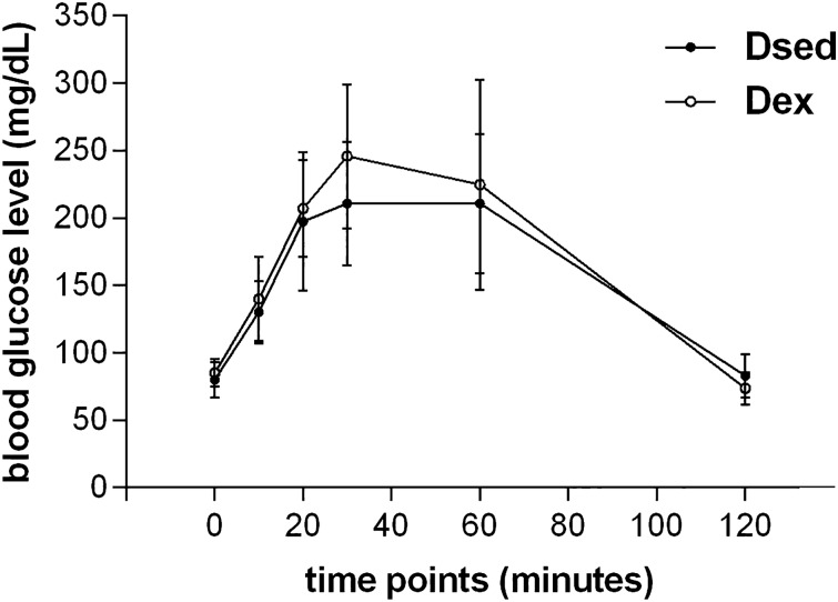
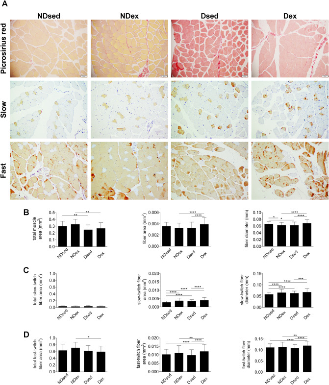
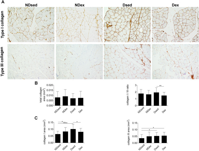
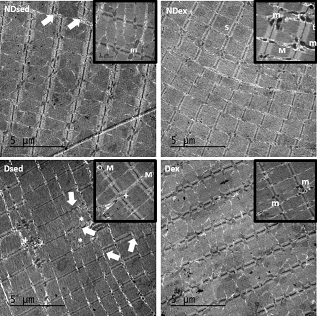
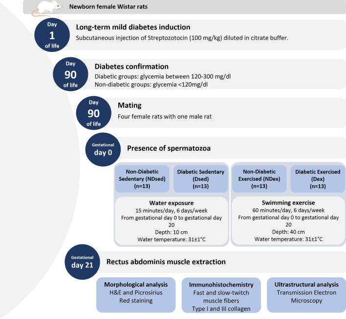
Gestational diabetes mellitus (GDM) plus rectus abdominis muscle (RAM) myopathy predicts long-term urinary incontinence (UI). Atrophic and stiff RAM are characteristics of diabetes-induced myopathy (DiM) in pregnant rats. This study aimed to determine whether swimming exercise (SE) has a therapeutic effect in mild hyperglycemic pregnant rats model. We hypothesized that SE training might help to reverse RAM DiM. Mild hyperglycemic pregnant rats model was obtained by a unique subcutaneous injection of 100 mg/kg streptozotocin (diabetic group) or citrate buffer (non-diabetic group) on the first day of life in Wistar female newborns. At 90 days of life, the rats are mated and randomly allocated to remain sedentary or subjected to a SE protocol. The SE protocol started at gestational day 0 and consisted of 60 min/day for 6 days/week in a period of 20 days in a swim tunnel. On day 21, rats were sacrificed, and RAM was collected and studied by picrosirius red, immunohistochemistry, and transmission electron microscopy. The SE protocol increased the fiber area and diameter, and the slow-twitch and fast-twitch fiber area and diameter in the diabetic exercised group, a finding was also seen in control sedentary animals. There was a decreased type I collagen but not type III collagen area and showed a similar type I/type III ratio compared with the control sedentary group. In conclusion, SE during pregnancy reversed the RAM DiM in pregnant rats. These findings may be a potential protocol to consider in patients with RAM damage caused by GDM.


Conflict of interest statement

The authors declare no competing interests.

Figures

Figure 1
Figure 1
Oral glucose tolerance test. Blood glucose level before the test and 10, 20, 30, 60 and 120 min after the administration of intragastric glucose solution in Dsed and Dex groups.
Figure 2
Figure 2
Histological sections of rat rectus abdominis muscle (RAM), obtained from CellSens Dimension (Olympus Corporation®) Version 1.16 image analysis software—(https://www.olympus-lifescience.com/en/software/cellsens/). (A) RAM samples were taken from non-diabetic sedentary (NDsed), non-diabetic exercised (NDex), diabetic sedentary (Dsed) and diabetic exercised (Dex) rats as described in “Materials and methods”. RAM samples were stained with picrosirius red and immunohistochemistry against myosin heavy chain of slow and fast RAM fibers. (B) Picrosirius red staining for total muscle area, fiber area, and fiber diameter in RAM samples as in (A). (C) Immunohistochemistry for total slow-twitch fiber area,slow-twitch fiber area, and slow-twitch fiber diameter as in (A). (D) Immunohistochemistry for total fast-twitch fiber area,fast-twitch fiber area, and fast-twitch fiber diameter as in (A). Values are means ± S.D. (n = 5 animals/group. *p < 0.05, **p < 0.01, ***p < 0.001 and ****p < 0.0001. Scale bar: 50 µm. Magnification: ×20.
Figure 3
Figure 3
Immunohistochemical sections of rat rectus abdominis muscle (RAM), obtained from CellSens Dimension (Olympus Corporation®) Version 1.16 image analysis software—(https://www.olympus-lifescience.com/en/software/cellsens/). (A) RAM samples were taken from non-diabetic sedentary (NDsed), non-diabetic exercised (NDex), diabetic sedentary (Dsed) and diabetic exercised (Dex) rats as described in Materials and methods. RAM samples were stained with Picrosirius red and immunohistochemistry using antibodies against type I and III collagen on RAM fibers. (B) Picrosirius red staining for total collagen area and immunohistochemistry for type I/III ratio. (C) Immunohistochemistry for type I and III collagen area as in (A). Values are means ± S.D. (n = 5 animals/group. *p < 0.05, **p < 0.01, ***p < 0.001 and ****p < 0.0001. Scale bar: 50 µm. Magnification: ×20.
Figure 4
Figure 4
Electron micrographs of rectus abdominis muscle (RAM). Samples of RAM were obtained from non-diabetic sedentary (NDsed), non-diabetic exercised (NDex), diabetic sedentary (Dsed), and diabetic exercised (Dex) rats. Magnifications (20.000 x) show a detailled area of the micrograph. The micrographs show disorganized Z lines (white arrows), sarcomeres disruption areas (asterisk), intermyofibrillar mitochondria (m), myelin figures (M), organized triads (t) and an increase in collagen deposition ( +). Scale bar: 5 µm.
Figure 5
Figure 5
Experimental sequence of groups.

References

    1. Standards of Medical Care in Diabetes American Diabetes Association. 2. Classification and diagnosis of diabetes. Diabetes Care. 2019;42:13–28. - PubMed
    1. Hernández-ochoa EO, Llanos P, Lanner JT. The underlying mechanisms of diabetic myopathy. J. Diabetes Res. 2017;2017:3–6. - PMC - PubMed
    1. Perry CGR, Hawke TJ. Diabetic myopathy : Current molecular understanding of this novel neuromuscular disorder. Curr. Opin. Neurol. 2017 doi: 10.1097/WCO.0000000000000479. - DOI - PubMed
    1. Rudge MVC, et al. Study protocol to investigate biomolecular muscle profile as predictors of long-term urinary incontinence in women with gestational diabetes mellitus. BMC Pregnancy Childb. 2020;20:1–14. doi: 10.1186/s12884-020-2749-x. - DOI - PMC - PubMed
    1. Barbosa AMP, et al. Urinary incontinence and vaginal squeeze pressure two years post-cesarean delivery in primiparous women with previous gestational diabetes mellitus. Clinics. 2011;66:1341–1346. - PMC - PubMed

Publication types