Review
. 2022 Jul;44(4):475-484.
doi: 10.1007/s00281-022-00942-8. Epub 2022 May 5.

Harnessing the liver to induce antigen-specific immune tolerance

Affiliations
Review

Harnessing the liver to induce antigen-specific immune tolerance

Cornelia Gottwick et al. Semin Immunopathol. 2022 Jul.

Abstract

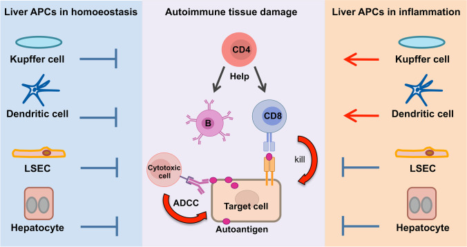
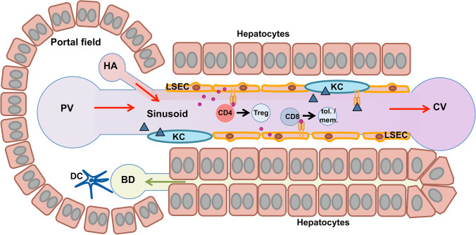
Autoimmune diseases develop when the adaptive immune system attacks the body's own antigens leading to tissue damage. At least 80 different conditions are believed to have an autoimmune aetiology, including rheumatoid arthritis, type I diabetes, multiple sclerosis or systemic lupus erythematosus. Collectively, autoimmune diseases are a leading cause of severe health impairment along with substantial socioeconomic costs. Current treatments are mostly symptomatic and non-specific, and it is typically not possible to cure these diseases. Thus, the development of more causative treatments that suppress only the pathogenic immune responses, but spare general immunity is of great biomedical interest. The liver offers considerable potential for development of such antigen-specific immunotherapies, as it has a distinct physiological capacity to induce immune tolerance. Indeed, the liver has been shown to specifically suppress autoimmune responses to organ allografts co-transplanted with the liver or to autoantigens that were transferred to the liver. Liver tolerance is established by a unique microenvironment that facilitates interactions between liver-resident antigen-presenting cells and lymphocytes passing by in the low blood flow within the hepatic sinusoids. Here, we summarise current concepts and mechanisms of liver immune tolerance, and review present approaches to harness liver tolerance for antigen-specific immunotherapy.

Keywords: Antigen presentation; Autoimmune disease; Immune tolerance; Immunotherapy; Nanomedicine; Scavenger cells.


Conflict of interest statement

This work was supported by the Deutsche Forschungsgemeinschaft—DFG (SFB841), the German Federal Ministry of Education and Research (16GW0051, 13XP5079C) and the European Regional Development Fund (OpToPas). CG has no relevant financial or non-financial interests to disclose. AC and JH are inventors of a patent related to this work (EP 2780036 (B1)). JH has consulted Topas Therapeutics GmbH without compensation.

Figures

Fig. 1
Fig. 1
Liver antigen-presenting cells as regulators of autoimmunity. Autoimmune diseases are caused by an adaptive immune response to autoantigens producing damage of target cells or organs. Autoantibodies produced by autoreactive B cells can cause tissue damage indirectly by binding to self-antigens and subsequently activating cytotoxic effector cells by antibody-dependent cell-mediated cytotoxicity (ADCC). Alternatively, cell damage is directly caused by autoreactive CD8 T cells releasing cytotoxic activities upon recognition of self-peptides on MHC I molecules of the target cell. In some cases, CD4 T cells can also become cytotoxic, but they are more relevant for providing help to autoreactive B cells and CD8 T cells upon recognition of self-peptides on MHC II molecules. Typically, CD4 T cell help is required for the development of autoimmune diseases and to maintain damaging autoimmune responses. In homoeostatic conditions, liver antigen-presenting cells, including Kupffer cells, dendritic cells and liver sinusoidal endothelial cells (LSECs), which take up and present autoantigen peptides on MHC molecules to autoreactive T cells, but also hepatocytes can induce T cell tolerance and suppress inflammatory activities, offering opportunities for therapy. However, under inflammatory conditions and liver injury, Kupffer cells and dendritic cells become critical drivers of pathogenic lymphocyte activation and inflammation. In contrast, tolerance-induction by LSECs and hepatocytes is considerably more robust, and it was shown that LSECs remain tolerogenic unless virally infected
Fig. 2
Fig. 2
Hepatic antigen-presenting cells in anatomical context. Blood flow (red arrows) enters the liver sinusoids through the portal vein (PV) and the hepatic artery (HA) and leaves through the central vein (CV). The hepatic sinusoids are lined by the liver sinusoidal endothelial cells (LSECs), which are scavenger cells clearing the blood from small particles and macromolecules by receptor-mediated endocytosis. LSECs present collected antigens to lymphocytes, producing a state of immune tolerance. CD4 effector T cells (CD4) can be transformed into regulatory T cells (Treg) through TGF-beta signals. CD8 T cells (CD8) can become tolerant or memory T cells (tol./mem.). Kupffer cells (KCs) reside in the lumen of the hepatic sinusoids and facilitate the removal of larger blood-borne particles by phagocytosis. They also present collected antigens to lymphocytes, producing tolerance. Dendritic cells (DCs) predominantly locate in the portal fields, and often close to bile ducts (BD), where they function as sentinels guarding the integrity of the biliary epithelium. DCs are antigen-presenting cells, producing tolerance in homoeostatic conditions, but readily promote inflammation upon sensing of cell damage or infection. As LSECs and KCs are the predominant cells in sinusoidal blood, it is easier to target those than liver DCs with vectors or carriers for antigen-specific immunotherapy

References

    1. Davidson A, Diamond B. Autoimmune diseases. N Engl J Med. 2001;345:340–350. doi: 10.1056/NEJM200108023450506. - DOI - PubMed
    1. NIH Autoimmune Diseases Coordinating Committee (2005) Progress in autoimmune diseases research. https://www.niaid.nih.gov/sites/default/files/adccfinal.pdf. Accessed 13 January 2022
    1. Dinse GE, Parks CG, Weinberg CR, Co CA, Wilkerson J, Zeldin DC, Chan EKL, Miller FW. Increasing prevalence of antinuclear antibodies in the United States. Arthritis Rheumatol. 2020;72:1026–1035. doi: 10.1002/art.41214. - DOI - PMC - PubMed
    1. Goebels N, Hofstetter H, Schmidt S, Brunner C, Wekerle H, Hohlfeld R. Repertoire dynamics of autoreactive T cells in multiple sclerosis patients and healthy subjects: epitope spreading versus clonal persistence. Brain. 2000;123:508–518. doi: 10.1093/brain/123.3.508. - DOI - PubMed
    1. Culina S, Lalanne AI, Afonso G, Cerosaletti K, Pinto S, Sebastiani G, Kuranda K, Nigi L, Eugster A, Østerbye T, Maugein A, McLaren JE, Ladell K, Larger E, Beressi JP, Lissina A, Appay V, Davidson HW, Buus S, Price DA, Kuhn M, Bonifacio E, Battaglia M, Caillat-Zucman S, Dotta F, Scharfmann R, Kyewski B, Mallone R, ImMaDiab Study Group Islet-reactive CD8+ T cell frequencies in the pancreas, but not in blood, distinguish type 1 diabetic patients from healthy donors. Sci Immunol. 2018;3:eaao4013. doi: 10.1126/sciimmunol.aao4013. - DOI - PMC - PubMed

Publication types