

Ultrastructural and biochemical classification of pathogenic tau, α-synuclein and TDP-43
- PMID: 35513543
- PMCID: PMC9107452
- DOI: 10.1007/s00401-022-02426-3
Ultrastructural and biochemical classification of pathogenic tau, α-synuclein and TDP-43
Erratum in
-
Correction to: Ultrastructural and biochemical classification of pathogenic tau, α-synuclein and TDP-43.Acta Neuropathol. 2022 Jul;144(1):165. doi: 10.1007/s00401-022-02439-y. Acta Neuropathol. 2022. PMID: 35593889 Free PMC article. No abstract available.
Abstract
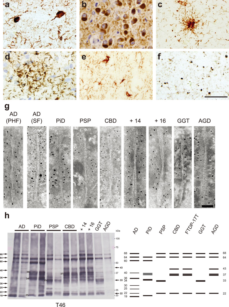
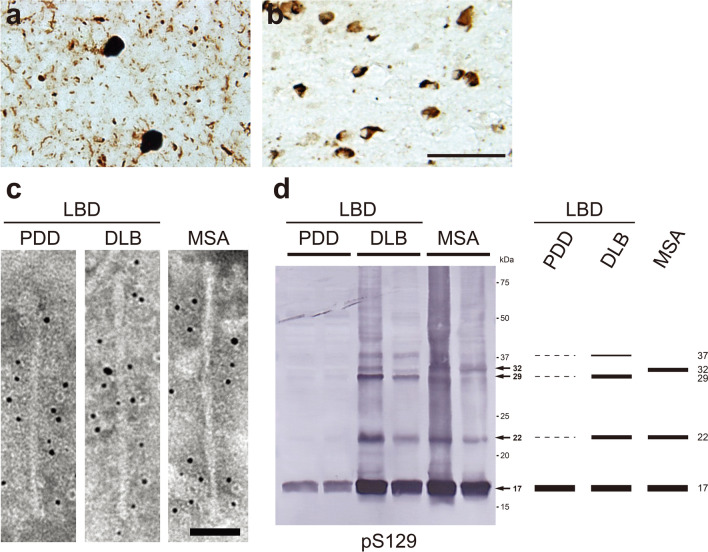
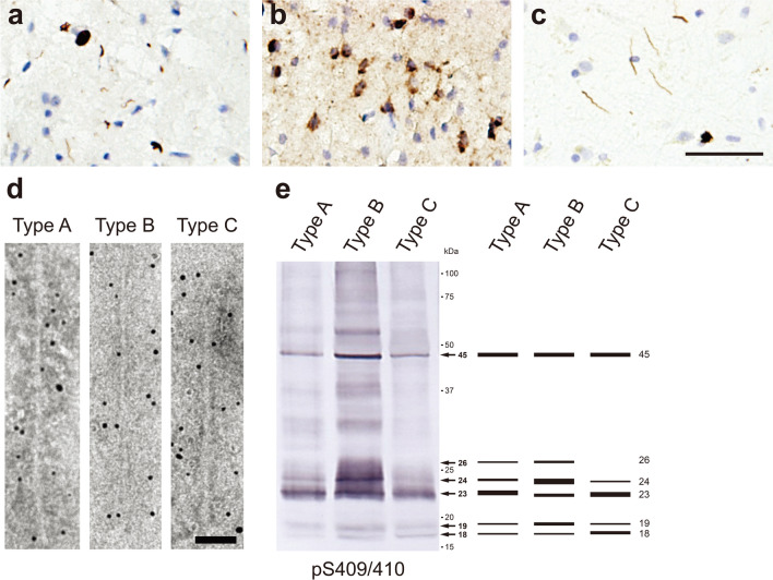
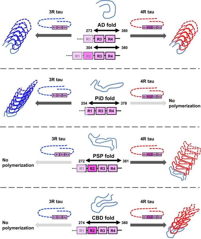
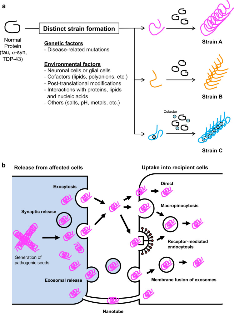
Intracellular accumulation of abnormal proteins with conformational changes is the defining neuropathological feature of neurodegenerative diseases. The pathogenic proteins that accumulate in patients' brains adopt an amyloid-like fibrous structure and exhibit various ultrastructural features. The biochemical analysis of pathogenic proteins in sarkosyl-insoluble fractions extracted from patients' brains also shows disease-specific features. Intriguingly, these ultrastructural and biochemical features are common within the same disease group. These differences among the pathogenic proteins extracted from patients' brains have important implications for definitive diagnosis of the disease, and also suggest the existence of pathogenic protein strains that contribute to the heterogeneity of pathogenesis in neurodegenerative diseases. Recent experimental evidence has shown that prion-like propagation of these pathogenic proteins from host cells to recipient cells underlies the onset and progression of neurodegenerative diseases. The reproduction of the pathological features that characterize each disease in cellular and animal models of prion-like propagation also implies that the structural differences in the pathogenic proteins are inherited in a prion-like manner. In this review, we summarize the ultrastructural and biochemical features of pathogenic proteins extracted from the brains of patients with neurodegenerative diseases that accumulate abnormal forms of tau, α-synuclein, and TDP-43, and we discuss how these disease-specific properties are maintained in the brain, based on recent experimental insights.
Keywords: Prion-like propagation; Strains; Synucleinopathy; TDP-43; TDP-43 proteinopathy; Tau; Tauopathy; α-Synuclein.
© 2022. The Author(s).
Figures
Similar articles
-
Experimental models of prion-like protein propagation.Neuropathology. 2020 Oct;40(5):460-466. doi: 10.1111/neup.12656. Epub 2020 Jun 1. Neuropathology. 2020. PMID: 32478452 Review.
-
Prion-like properties of assembled TDP-43.Curr Opin Neurobiol. 2020 Apr;61:23-28. doi: 10.1016/j.conb.2019.11.018. Epub 2019 Dec 18. Curr Opin Neurobiol. 2020. PMID: 31862626 Review.
-
Prion-like mechanisms in neurodegenerative diseases.Nat Rev Neurosci. 2010 Mar;11(3):155-9. doi: 10.1038/nrn2786. Epub 2009 Dec 23. Nat Rev Neurosci. 2010. PMID: 20029438 Free PMC article. Review.
-
Prion-like propagation of α-synuclein in neurodegenerative diseases.Prog Mol Biol Transl Sci. 2019;168:323-348. doi: 10.1016/bs.pmbts.2019.07.005. Epub 2019 Jul 31. Prog Mol Biol Transl Sci. 2019. PMID: 31699325 Review.
-
[The Prion-like Mechanism in Neurodegenerative Diseases-Current Studies and Future Prospects].Brain Nerve. 2016 Oct;68(10):1197-1204. doi: 10.11477/mf.1416200573. Brain Nerve. 2016. PMID: 27703107 Review. Japanese.
Cited by
-
Modulation of Tau Pathology in Alzheimer's Disease by Dietary Bioactive Compounds.Int J Mol Sci. 2024 Jan 9;25(2):831. doi: 10.3390/ijms25020831. Int J Mol Sci. 2024. PMID: 38255905 Free PMC article. Review.
-
Effect of Electric Field on α-Synuclein Fibrils: Revealed by Molecular Dynamics Simulations.Int J Mol Sci. 2023 Mar 28;24(7):6312. doi: 10.3390/ijms24076312. Int J Mol Sci. 2023. PMID: 37047286 Free PMC article.
-
A case of argyrophilic grain disease with an initial clinical diagnosis of Parkinson's disease.J Neurol. 2024 Dec;271(12):7628-7632. doi: 10.1007/s00415-024-12688-4. Epub 2024 Oct 15. J Neurol. 2024. PMID: 39402237 No abstract available.
-
Hunting for the cause: Evidence for prion-like mechanisms in Huntington's disease.Front Neurosci. 2022 Aug 24;16:946822. doi: 10.3389/fnins.2022.946822. eCollection 2022. Front Neurosci. 2022. PMID: 36090278 Free PMC article. Review.
-
Exercise suppresses neuroinflammation for alleviating Alzheimer's disease.J Neuroinflammation. 2023 Mar 19;20(1):76. doi: 10.1186/s12974-023-02753-6. J Neuroinflammation. 2023. PMID: 36935511 Free PMC article. Review.
References
-
- Ahmed Z, Doherty KM, Silveira-Moriyama L, Bandopadhyay R, Lashley T, Mamais A, Hondhamuni G, Wray S, Newcombe J, O'Sullivan SS, et al. Globular glial tauopathies (GGT) presenting with motor neuron disease or frontotemporal dementia: an emerging group of 4-repeat tauopathies. Acta Neuropathol. 2011;122:415–428. doi: 10.1007/s00401-011-0857-4. - DOI - PubMed
-
- Al-Hilaly YK, Foster BE, Biasetti L, Lutter L, Pollack SJ, Rickard JE, Storey JMD, Harrington CR, Xue WF, Wischik CM, et al. Tau (297–391) forms filaments that structurally mimic the core of paired helical filaments in Alzheimer's disease brain. FEBS Lett. 2020;594:944–950. doi: 10.1002/1873-3468.13675. - DOI - PMC - PubMed
Publication types
MeSH terms
Substances
Grants and funding
LinkOut - more resources
Full Text Sources
Medical
