. 2022 Sep;27(6):541-551.
doi: 10.1007/s00775-022-01941-8. Epub 2022 May 5.

Rational inhibitor design for Pseudomonas aeruginosa salicylate adenylation enzyme PchD

Affiliations

Rational inhibitor design for Pseudomonas aeruginosa salicylate adenylation enzyme PchD

Catherine L Shelton et al. J Biol Inorg Chem. 2022 Sep.

Abstract

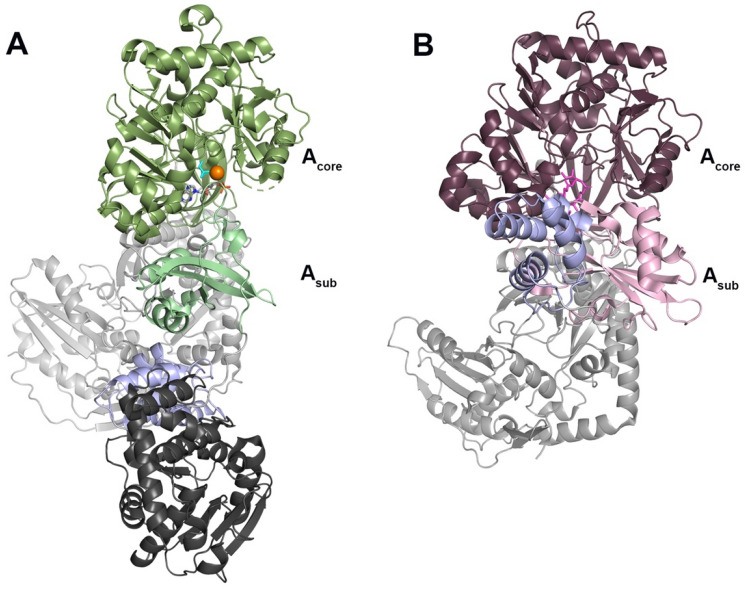
Pseudomonas aeruginosa is an increasingly antibiotic-resistant pathogen that causes severe lung infections, burn wound infections, and diabetic foot infections. P. aeruginosa produces the siderophore pyochelin through the use of a non-ribosomal peptide synthetase (NRPS) biosynthetic pathway. Targeting members of siderophore NRPS proteins is one avenue currently under investigation for the development of new antibiotics against antibiotic-resistant organisms. Here, the crystal structure of the pyochelin adenylation domain PchD is reported. The structure was solved to 2.11 Å when co-crystallized with the adenylation inhibitor 5'-O-(N-salicylsulfamoyl)adenosine (salicyl-AMS) and to 1.69 Å with a modified version of salicyl-AMS designed to target an active site cysteine (4-cyano-salicyl-AMS). In the structures, PchD adopts the adenylation conformation, similar to that reported for AB3403 from Acinetobacter baumannii.

Keywords: Adenylation domain; Antibiotic resistance; Inhibitor design; Pseudomonas aeruginosa; Pyochelin.


Conflict of interest statement

The authors declare no conflict of interest.

Figures

Fig. 1
Fig. 1
A PchD catalyzes the adenylation of salicylate as the first step in pyochelin biosynthesis. B Salicyl-AMS, originally described by Ferreras et al. as an inhibitor of siderophore producing NRPS adenylation domains in M. tuberculosis and Y. pestis has been modified by the addition of a cyano group at C4 of the salicylate ring to produce 4-cyano-salicyl-AMS
Fig. 2
Fig. 2
A PchD bound to salicyl-AMS (salmon) shown in midnight blue. B PchD bound to 4-cyanosalicylAMS (orange) shown in skyblue
Fig. 3
Fig. 3
A The adenylation conformation represented by the full NRPS module from AB3403 (4ZXI) with the adenylation domain shown in green, the condensation domain shown in light grey, the PCP domain shown in light purple, and the thioesterase domain shown in dark grey. Ligands shown in the adenylation domain include AMP (grey), Mg2+ (orange) and glycine (cyan) (B). The thioester-forming conformation represented by the EntF NRPS module (5T3D) with the adenylation domain shown in plum/pink, the condensation domain shown in light grey, and the PCP domain shown in light purple. The mechanism-based inhibitor Ser-AVS is shown in fuchsia
Fig. 4
Fig. 4
A PchD (blue) closely aligns with the adenylating conformation demonstrated by the AB3403 adenylation domain (green). 4-cyano-salicyl-AMS ligand of PchD shown in teal. B The EntF adenylation domain (Acore:plum, Asub: pink) demonstrates the thioester-forming conformation and was co-crystalized with the ligand serine adenosine vinylsulfonamide (magenta). The Asub of MbtA (wheat; Acore: orange) does not align with either the C adenylation or the D thioester conformation
Fig. 5
Fig. 5
A Relative position of Cys250 to the salicyl-AMS inhibitor. B Relative position of the 4-cyano-salicyl-AMS inhibitor. Inhibitor densities are shown with a polder map drawn at 3σ
Fig. 6
Fig. 6
DhbE adenylation domain bound to DHB-adenylate (1MDB) shown in yellow overlayed with both salicyl-AMS PchD (salmon inhibitor, midnight blue structure) and 4-cyano-salicyl-AMS bound PchD (orange inhibitor, skyblue structure). Residues conferring specificity for salicylate are Cys250 and Ile347. For the DhbE structure these positions are a conserved serine and a valine which are hypothesized to more readily accommodate the second hydroxyl of DHB
Fig. 7
Fig. 7
Contacts between active site residues and the inhibitor

References

    1. Skaar EP. The battle for iron between bacterial pathogens and their vertebrate hosts. PLoS Pathog. 2010;6(8):e1000949. doi: 10.1371/journal.ppat.1000949. - DOI - PMC - PubMed
    1. Braud A, Hannauer M, Mislin GL, Schalk IJ. The Pseudomonas aeruginosa pyochelin-iron uptake pathway and its metal specificity. J Bacteriol. 2009;191(11):3517–3525. doi: 10.1128/JB.00010-09. - DOI - PMC - PubMed
    1. Schalk IJ, Hannauer M, Braud A. New roles for bacterial siderophores in metal transport and tolerance. Environ Microbiol. 2011;13(11):2844–2854. doi: 10.1111/j.1462-2920.2011.02556.x. - DOI - PubMed
    1. Cox CD. Effect of pyochelin on the virulence of Pseudomonas aeruginosa. Infect Immun. 1982;36(1):17–23. doi: 10.1128/iai.36.1.17-23.1982. - DOI - PMC - PubMed
    1. Sokol PA. Surface expression of ferripyochelin-binding protein is required for virulence of Pseudomonas aeruginosa. Infect Immun. 1987;55(9):2021–2025. doi: 10.1128/iai.55.9.2021-2025.1987. - DOI - PMC - PubMed

Publication types

LinkOut - more resources