. 2022 Sep;26(9):5663-5672.
doi: 10.1007/s00784-022-04521-x. Epub 2022 May 5.

Effect of voxel size in cone-beam computed tomography on surface area measurements of dehiscences and fenestrations in the lower anterior buccal region

Affiliations

Effect of voxel size in cone-beam computed tomography on surface area measurements of dehiscences and fenestrations in the lower anterior buccal region

B J van Leeuwen et al. Clin Oral Investig. 2022 Sep.

Abstract

Objectives: This study aims to assess whether different voxel sizes in cone-beam computed tomography (CBCT) affected surface area measurements of dehiscences and fenestrations in the mandibular anterior buccal region.

Materials and methods: Nineteen dry human mandibles were scanned with a surface scanner (SS). Wax was attached to the mandibles as a soft tissue equivalent. Three-dimensional digital models were generated with a CBCT unit, with voxel sizes of 0.200 mm (VS200), 0.400 mm (VS400), and 0.600 mm (VS600). The buccal surface areas of the six anterior teeth were measured (in mm2) to evaluate areas of dehiscences and fenestrations. Differences between the CBCT and SS measurements were determined in a linear mixed model analysis.

Results: The mean surface area per tooth was 88.3 ± 24.0 mm2, with the SS, and 94.6 ± 26.5 (VS200), 95.1 ± 27.3 (VS400), and 96.0 ± 26.5 (VS600), with CBCT scans. Larger surface areas resulted in larger differences between CBCT and SS measurements (- 0.1 β, SE = 0.02, p < 0.001). Deviations from SS measurements were larger with VS600, compared to VS200 (1.3 β, SE = 0.05, P = 0.009). Fenestrations were undetectable with CBCT.

Conclusions: CBCT imaging magnified the surface area of dehiscences in the anterior buccal region of the mandible by 7 to 9%. The larger the voxel size, the larger the deviation from SS measurements. Fenestrations were not detectable with CBCT.

Clinical relevance: CBCT is an acceptable tool for measuring dehiscences but not fenestrations. However, CBCT overestimates the size of dehiscences, and the degree of overestimation depends on the actual dehiscence size and CBCT voxel size employed.

Keywords: Accuracy; Cone-beam computed tomography; Dehiscence; Fenestration; Reliability.


Conflict of interest statement

The authors declare no competing interests.

Figures

Fig. 1
Fig. 1
Preparing the mandible for CBCT scanning. A Dental modeling wax was attached to the buccal side of the mandible to serve as a soft tissue equivalent. B, C The mandible was positioned in the center of the scanning table of the CBCT machine, in the same orientation as a live patient would be oriented, aligned with vertical and horizontal laser guides
Fig. 2
Fig. 2
Preparing the datasets for analysis. A Anterior view of the three CBCT scans (VS600, VS400, VS200) superimposed on the SS; after alignment, the superimposed images were saved in one dataset. B Posterior view of the superimposed dataset (SS, VS200, VS400, VS600), shown after digitally removing the lingual surfaces of the six anterior teeth and the lingual side of the mandibular body. C Anterior view shows the buccal surfaces of each of the six anterior teeth; colors show the entire surfaces included in the tooth surface area calculations. D An example of a mandible with fenestrations (colored spots) on the buccal side of the mandible. The surface areas of the fenestrations were determined
Fig. 3
Fig. 3
Inability of CBCT to detect fenestrations. (Top left) SS image (Pri) of a mandible shows six fenestrations detected by the observers (see Fig. 2D for colored rendition); (top right and bottom right and left) CBCT images of the same mandible show that the fenestrations were not detected on CBCT scans with resolutions of VS200 (voxel size 0.200 mm), VS400 (voxel size 0.400 mm), or VS600 (voxel size 0.600 mm)
Fig. 4
Fig. 4
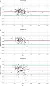
Bland–Altman plots show the degrees of agreement on the dehiscence surface area measurements (mm2) between scanner measurements (SS) and CBCT measurements. Dots represent the difference between SS and CBCT measurements. The red line indicates the mean difference between the two imaging results, and the green lines show the 95% limits of agreement. A VS200 (voxel size 0.200 mm), B VS400 (voxel size 0.400 mm), C VS600 (voxel size 0.600 mm)
Fig. 5
Fig. 5
Plot shows that, as the SS surface area measurement increases, the difference between the SS and the CBCT (VS600, VS400, and VS200) surface area measurements increases. The slope is negative, based on the regression coefficients found in the linear mixed model analysis. X-axis, SS measurements; Y-axis, SS measurement — CBCT measurement

References

    1. Wehrbein H, Bauer W, Diedrich P. Mandibular incisors alveolar bone and symphysis after orthodontic treatment A retrospective study. Am J Orthod Dentofacial Orthop. 1996;110(3):239–246. doi: 10.1016/S0889-5406(96)80006-0. - DOI - PubMed
    1. Sheng Y, Guo HM, Bai YX, Li S. Dehiscence and fenestration in anterior teeth : comparison before and after orthodontic treatment. J Orofac Orthop. 2020;81(1):1–9. doi: 10.1007/s00056-019-00196-4. - DOI - PubMed
    1. Artun J, Krogstad O. Periodontal status of mandibular incisors following excessive proclination A study in adults with surgically treated mandibular prognathism. Am J Orthod Dentofacial Orthop. 1987;91(3):225–232. doi: 10.1016/0889-5406(87)90450-1. - DOI - PubMed
    1. Yared KF, Zenobio EG, Pacheco W. Periodontal status of mandibular central incisors after orthodontic proclination in adults. Am J Orthod Dentofacial Orthop. 2006;130(1):6.e1–8. doi: 10.1016/j.ajodo.2006.01.015. - DOI - PubMed
    1. Wennström JL, Lindhe J, Sinclair F, Thilander B. Some periodontal tissue reactions to orthodontic tooth movement in monkeys. J Clin Periodontol. 1987;14(3):121–129. doi: 10.1111/j.1600-051x.1987.tb00954.x. - DOI - PubMed