

Fully-Automatic 3D Intuitive Visualization of Age-Related Macular Degeneration Fluid Accumulations in OCT Cubes
- PMID: 35513586
- PMCID: PMC9582110
- DOI: 10.1007/s10278-022-00643-6
Fully-Automatic 3D Intuitive Visualization of Age-Related Macular Degeneration Fluid Accumulations in OCT Cubes
Abstract
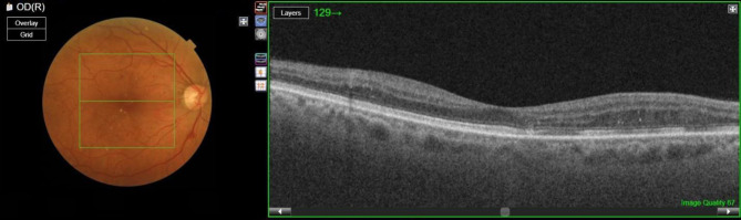
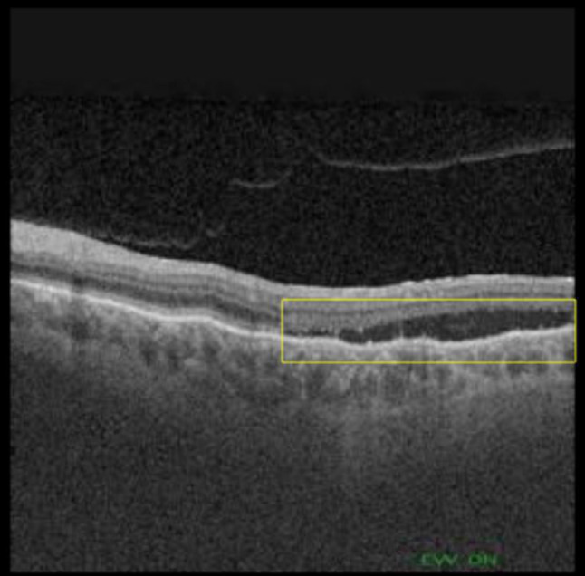
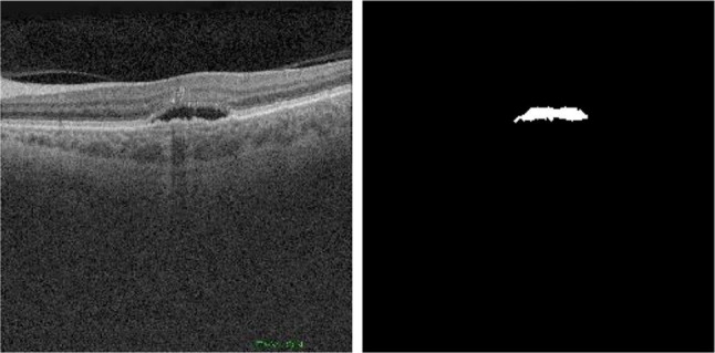
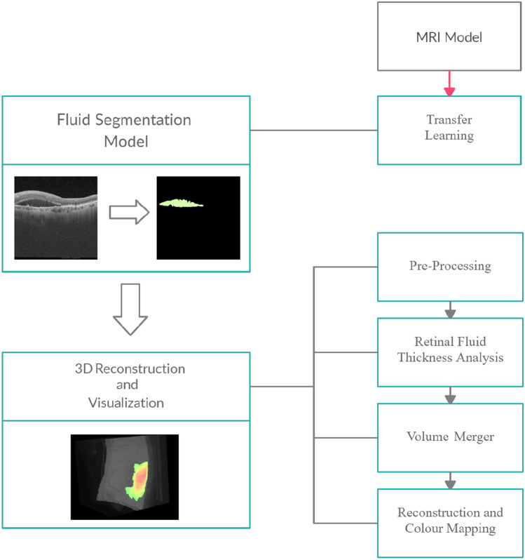
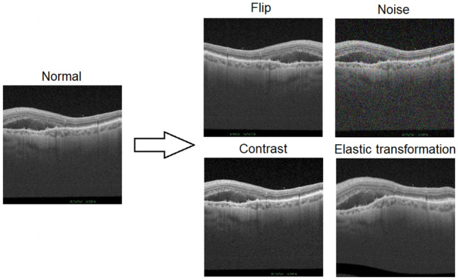
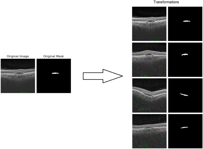
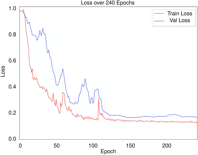
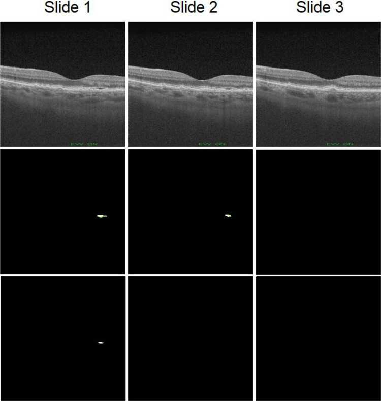
Age-related macular degeneration is the leading cause of vision loss in developed countries, and wet-type AMD requires urgent treatment and rapid diagnosis because it causes rapid irreversible vision loss. Currently, AMD diagnosis is mainly carried out using images obtained by optical coherence tomography. This diagnostic process is performed by human clinicians, so human error may occur in some cases. Therefore, fully automatic methodologies are highly desirable adding a layer of robustness to the diagnosis. In this work, a novel computer-aided diagnosis and visualization methodology is proposed for the rapid identification and visualization of wet AMD. We adapted a convolutional neural network for segmentation of a similar domain of medical images to the problem of wet AMD segmentation, taking advantage of transfer learning, which allows us to work with and exploit a reduced number of samples. We generate a 3D intuitive visualization where the existence, position and severity of the fluid were represented in a clear and intuitive way to facilitate the analysis of the clinicians. The 3D visualization is robust and accurate, obtaining satisfactory 0.949 and 0.960 Dice coefficients in the different evaluated OCT cube configurations, allowing to quickly assess the presence and extension of the fluid associated to wet AMD.
Keywords: 3D visualization; Age-related macular degeneration; Computer-aided diagnosis; Optical Coherence Tomography.
© 2022. The Author(s).
Conflict of interest statement
The authors declare no conflicts of interest.
Figures
Similar articles
-
Weakly supervised lesion localization for age-related macular degeneration detection using optical coherence tomography images.PLoS One. 2019 Apr 5;14(4):e0215076. doi: 10.1371/journal.pone.0215076. eCollection 2019. PLoS One. 2019. PMID: 30951557 Free PMC article.
-
Automatic detection of retinal regions using fully convolutional networks for diagnosis of abnormal maculae in optical coherence tomography images.J Biomed Opt. 2019 May;24(5):1-9. doi: 10.1117/1.JBO.24.5.056003. J Biomed Opt. 2019. PMID: 31111697 Free PMC article.
-
U-Net-Based Segmentation of Current Imaging Biomarkers in OCT-Scans of Patients with Age Related Macular Degeneration.Stud Health Technol Inform. 2023 May 18;302:947-951. doi: 10.3233/SHTI230315. Stud Health Technol Inform. 2023. PMID: 37203542
-
A view of the current and future role of optical coherence tomography in the management of age-related macular degeneration.Eye (Lond). 2017 Jan;31(1):26-44. doi: 10.1038/eye.2016.227. Epub 2016 Nov 25. Eye (Lond). 2017. PMID: 27886184 Free PMC article. Review.
-
Optical coherence tomography angiography in the management of age-related macular degeneration.Curr Opin Ophthalmol. 2018 May;29(3):217-225. doi: 10.1097/ICU.0000000000000469. Curr Opin Ophthalmol. 2018. PMID: 29538181 Review.
Cited by
-
Artificial intelligence in age-related macular degeneration: state of the art and recent updates.BMC Ophthalmol. 2024 Mar 15;24(1):121. doi: 10.1186/s12886-024-03381-1. BMC Ophthalmol. 2024. PMID: 38491380 Free PMC article. Review.
-
Artificial intelligence for diagnosing exudative age-related macular degeneration.Cochrane Database Syst Rev. 2024 Oct 17;10(10):CD015522. doi: 10.1002/14651858.CD015522.pub2. Cochrane Database Syst Rev. 2024. PMID: 39417312
References
-
- Bourne R, Jonas J, Flaxman S, Keeffe J, Leasher J, Naidoo K, Parodi M, Pesudovs K, Price H, White R, Wong T, Resnikoff S, Taylor H. Prevalence and causes of vision loss in high-income countries and in Eastern and Central Europe: 1990–2010. British Journal of Ophthalmology. 2014;98(5):629–638. doi: 10.1136/bjophthalmol-2013-304033. - DOI - PubMed
-
- “Vision impairment and blindness”, Who.int, 2020. [Online]. Available: https://www.who.int/news-room/fact-sheets/detail/blindness-and-visual-im.... [Accessed: 22- May- 2021]
-
- R. Gallego-Pinazo, R. Dolz-Marco and M. Díaz-Llopis, “Hacia la nueva clasificación de la degeneración macular asociada a la edad basada en la tomografía de coherencia óptica de dominio espectral”, Archivos de la Sociedad Española de Oftalmología, vol. 87, no. 8, pp. 247–252, 2012. - PubMed
Publication types
MeSH terms
Grants and funding
- PI17/00940/Instituto de Salud Carlos III
- DTS18/00136/Instituto de Salud Carlos III
- RTI2018-095894-B-I00/Ministerio de Ciencia e Innovación y Universidades (ES)
- PID2019-108435RB-I00/Ministerio de Ciencia e Innovación, Gobierno de España
- ED431C 2020/24/Consellería de Cultura, Educación e Universidade, Xunta de Galicia (ES)
LinkOut - more resources
Full Text Sources
Medical
