Review
. 2022 Oct;38(10):1229-1247.
doi: 10.1007/s12264-022-00859-0. Epub 2022 May 5.

Neuronal Death Mechanisms and Therapeutic Strategy in Ischemic Stroke

Affiliations
Review

Neuronal Death Mechanisms and Therapeutic Strategy in Ischemic Stroke

Rui Mao et al. Neurosci Bull. 2022 Oct.

Abstract

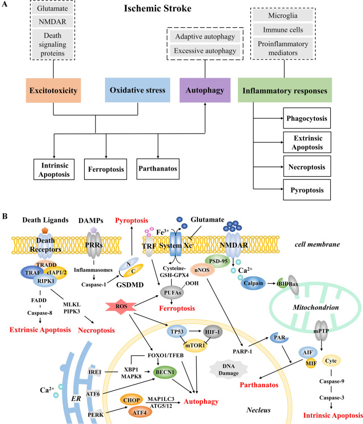
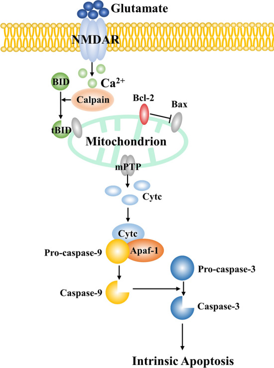
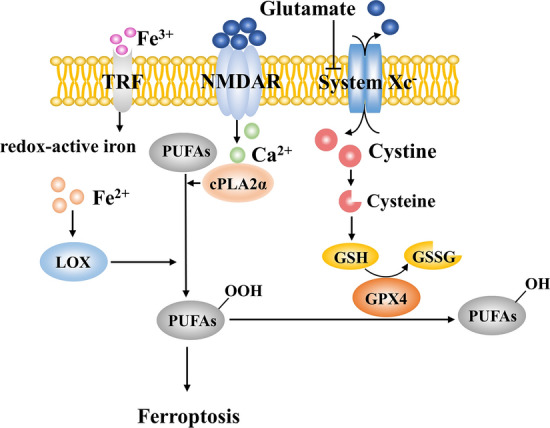
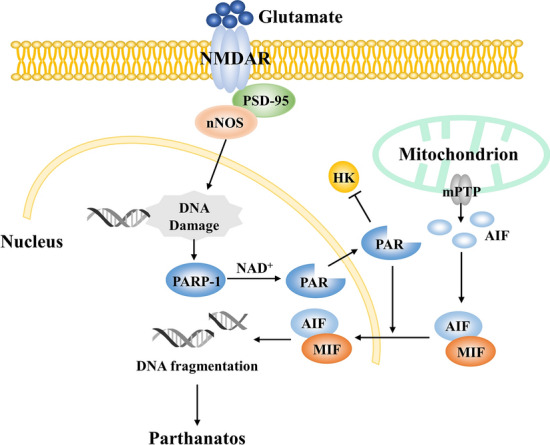
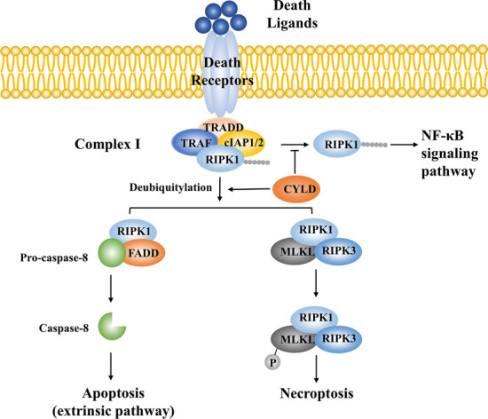
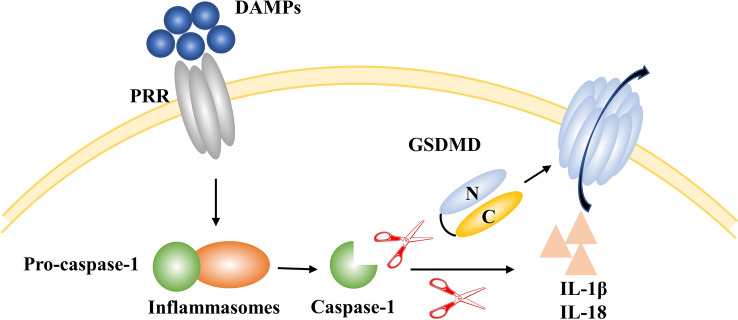
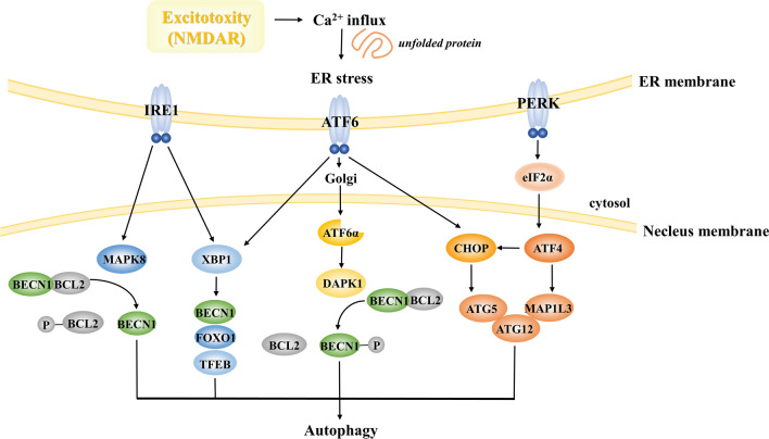
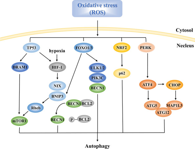
Ischemic stroke caused by intracranial vascular occlusion has become increasingly prevalent with considerable mortality and disability, which gravely burdens the global economy. Current relatively effective clinical treatments are limited to intravenous alteplase and thrombectomy. Even so, patients still benefit little due to the short therapeutic window and the risk of ischemia/reperfusion injury. It is therefore urgent to figure out the neuronal death mechanisms following ischemic stroke in order to develop new neuroprotective strategies. Regarding the pathogenesis, multiple pathological events trigger the activation of cell death pathways. Particular attention should be devoted to excitotoxicity, oxidative stress, and inflammatory responses. Thus, in this article, we first review the principal mechanisms underlying neuronal death mediated by these significant events, such as intrinsic and extrinsic apoptosis, ferroptosis, parthanatos, pyroptosis, necroptosis, and autophagic cell death. Then, we further discuss the possibility of interventions targeting these pathological events and summarize the present pharmacological achievements.

Keywords: Ischemic stroke; Mechanisms; Neuronal death; Therapeutic strategy.


Conflict of interest statement

All authors claim that there are no conflicts of interest.

Figures

Fig. 1
Fig. 1
A The main structure of this review. The content in the dashed frame represents the relevant therapeutic targets. B A simple schematic of several death mechanisms in ischemic stroke.
Fig. 2
Fig. 2
Apoptosis by the intrinsic/mitochondrial pathway during cerebral ischemia. Increased extracellular glutamate activates NMDARs, causing excessive Ca2+ influx. Activated calpain cleaves BID to tBID, which interacts with Bax and helps to form the mPTP. Cytc is released from these pores and combines with Apaf-1 and pro-caspase-9. The apoptosome stepwise activates the executioner caspase-3, and consequently induces apoptosis. Apaf‐1, apoptotic protein‐activating factor‐1; Bcl‐2, B‐cell leukemia/lymphoma 2; BID, Bcl-2 interacting domain; Cytc, cytochrome C; mPTP, mitochondrial permeability transition pore; NMDA, N‐methyl‐d‐aspartate; tBID, truncated Bid.
Fig. 3
Fig. 3
Ferroptosis during cerebral ischemia. Normally, GPX4 uses GSH to catalyze lipid hydroperoxides into alcohols. Increased extracellular glutamate inhibits the activity of System Xc as well as the cysteine-GSH-GPX4 axis. Meanwhile, Ca2+ overload activates cPLA2α to provide substrates for lipid peroxidation. Fe2+-activated lipoxygenase (LOX) also participates in this process. Increased redox-active iron and overwhelming lipid peroxidation contribute to ferroptosis. cPLA2α, cytosolic phospholipase A2α; GPX4, γ‐L‐glutamyl‐L‐cysteinylglycine peroxidase 4; GSH, glutathione; GSSG, oxidized glutathione; LOX, lipoxygenase; PUFA, polyunsaturated fatty acid; TRF, transferrin.
Fig. 4
Fig. 4
Parthanatos during cerebral ischemia. PSD-95 links NMDARs to nNOS. Neuronal DNA injury that is caused by oxidative or nitrosative stress activates PARP-1, which uses NAD+ to synthesize PAR. PAR inhibits HK and promotes the nuclear translocation of AIF. The mPTP allows the release of AIF into the cytoplasm where it binds to MIF. The AIF–MIF complex enters the nucleus and mediates DNA fragmentation. HK, hexokinase; MIF, migration inhibitory factor; NAD+, nicotinamide adenine dinucleotide; nNOS, nitric oxide synthase; PAR, poly (ADP-ribose); PARP-1, poly (ADP-ribose) polymerase-1; PSD-95, postsynaptic density protein‐95.
Fig. 5
Fig. 5
Death ligands induce apoptosis and necroptosis. Death ligands (TNF-α, FASL, and TRAIL) bind to death receptors. CYLD de-ubiquitylates RIPK1 and the rest of complex I recruits FADD. FADD homodimerizes caspase-8, resulting in apoptosis. RIPK1 activation contributes to the formation of the RIPK1–RIPK3–MLKL complex. RIPK3-mediated phosphorylation of MLKL induces necroptosis. Death receptors also triggers NF-κB signaling via RIPK1 ubiquitylation by cIAP1/2. CYLD inhibits this process. FADD, Fas-associated death domain; cIAP1/2, cellular inhibitor of apoptosis 1/2; MLKL, mixed lineage kinase domain-like; RIPK, receptor interacting serine/threonine kinases; TRADD, tumor necrosis factor receptor type 1-associated death domain; TRAF, TNFR-associated factor.
Fig. 6
Fig. 6
Pyroptosis during ischemic stroke. DAMPs bind to PRRs, resulting in the formation of inflammasomes and the production of caspase-1. On the one hand, caspase-1 cleaves GSDMD to form pores on the cell membrane, resulting in content release and pyroptosis. On the other hand, caspase-1 cleaves pro-IL-1β and pro-IL-18 to form active IL-1β and IL-18, causing inflammatory responses. GSDMD, gasdermin D; DAMPs, damage-associated molecular patterns; PRR, pattern recognition receptor.
Fig. 7
Fig. 7
Autophagy induced by excitotoxicity. NMDAR leads to Ca2+ influx, causing unfolded protein and ER stress. The three transmembrane sensors IRE1, ATF6, and PERK activate a complex cascade with autophagic induction. IRE1 upregulates BECN1 via MAPK8 and XBP1. XBP1 also transactivates FOXO1 and TFEB, bypassing BECN1, and induces autophagy. PERK upregulates ATF4 and CHOP via the eIF2α axis, then enhances the transcription of MAP1LC3, ATG5, and ATG12. ATF6 upregulates the DAPK1-mediated phosphorylation of BECN1 and is believed to activate both XBP1 and CHOP. IRE1, inositol-requiring enzyme 1; ATF, activating transcription factor; PERK, PKR-like endoplasmic reticulum kinase; MAPK8, mitogen-activated protein kinase 8; XBP1, box-binding protein 1; FOXO1, forkhead box O1; TFEB, transcription factor EB; CHOP, CCAAT/enhancer binding protein homologous protein; eIF2α, eukaryotic initiation factor 2α; MAP1LC3, microtubule associated protein 1 light chain 3 beta; DAPK1, death-associated protein kinase 1.
Fig. 8
Fig. 8
Autophagy induced by oxidative stress. Increased ROS upregulates nuclear p53, transactivates DRAM1, inhibits mTOR1, and activates HIF-1. Besides, HIF-1 due to oxygen deprivation triggers the transcription of BNIP3 and NIX. BNIP3 also inhibits mTOR1 via inhibiting Rheb. The FOXO family, especially FOXO1 and FOXO3, upregulates ATGs including ULK1/2, BECN1, and PIK3C, as well as BNIP3. ROS also upregulates NRF2 to enhance the levels of p62 and upregulates PERK as shown in Fig. 6. DRAM1, damage-regulated autophagy modulator1; mTOR1, mechanistic target of rapamycin complex 1; HIF-1, hypoxia inducible factor 1; BNIP3, BCL2/adenovirus E1B 19 kDa interacting protein 3; NIX, BNIP3-like; Rheb, Ras homolog protein enriched in brain; FOXO, forkhead box O; ULK1/2, Unc-51 like autophagy activating kinase; NRF2, NF-E2-related factor 2.

Similar articles

Cited by

References

    1. Gorelick PB. The global burden of stroke: Persistent and disabling. Lancet Neurol. 2019;18:417–418. doi: 10.1016/S1474-4422(19)30030-4. - DOI - PubMed
    1. Fricker M, Tolkovsky AM, Borutaite V, Coleman M, Brown GC. Neuronal cell death. Physiol Rev. 2018;98:813–880. doi: 10.1152/physrev.00011.2017. - DOI - PMC - PubMed
    1. Ge Y, Chen WL, Axerio-Cilies P, Wang YT. NMDARs in cell survival and death: Implications in stroke pathogenesis and treatment. Trends Mol Med. 2020;26:533–551. doi: 10.1016/j.molmed.2020.03.001. - DOI - PubMed
    1. Lau A, Tymianski M. Glutamate receptors, neurotoxicity and neurodegeneration. Pflugers Arch. 2010;460:525–542. doi: 10.1007/s00424-010-0809-1. - DOI - PubMed
    1. Wu QJ, Tymianski M. Targeting NMDA receptors in stroke: New hope in neuroprotection. Mol Brain. 2018;11:15. doi: 10.1186/s13041-018-0357-8. - DOI - PMC - PubMed

Substances