

Longitudinal MRI-visible perivascular space (PVS) changes with long-duration spaceflight
- PMID: 35513698
- PMCID: PMC9072425
- DOI: 10.1038/s41598-022-11593-y
Longitudinal MRI-visible perivascular space (PVS) changes with long-duration spaceflight
Abstract
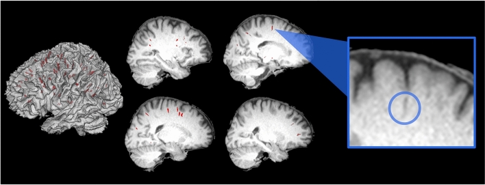
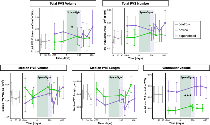
Humans are exposed to extreme environmental stressors during spaceflight and return with alterations in brain structure and shifts in intracranial fluids. To date, no studies have evaluated the effects of spaceflight on perivascular spaces (PVSs) within the brain, which are believed to facilitate fluid drainage and brain homeostasis. Here, we examined how the number and morphology of magnetic resonance imaging (MRI)-visible PVSs are affected by spaceflight, including prior spaceflight experience. Fifteen astronauts underwent six T1-weighted 3 T MRI scans, twice prior to launch and four times following their return to Earth after ~ 6-month missions to the International Space Station. White matter MRI-visible PVS number and morphology were calculated using an established, automated segmentation algorithm. We validated our automated segmentation algorithm by comparing algorithm PVS counts with those identified by two trained raters in 50 randomly selected slices from this cohort; the automated algorithm performed similarly to visual ratings (r(48) = 0.77, p < 0.001). In addition, we found high reliability for four of five PVS metrics across the two pre-flight time points and across the four control time points (ICC(3,k) > 0.50). Among the astronaut cohort, we found that novice astronauts showed an increase in total PVS volume from pre- to post-flight, whereas experienced crewmembers did not (p = 0.020), suggesting that experienced astronauts may exhibit holdover effects from prior spaceflight(s). Greater pre-flight PVS load was associated with more prior flight experience (r = 0.60-0.71), though these relationships did not reach statistical significance (p > 0.05). Pre- to post-flight changes in ventricular volume were not significantly associated with changes in PVS characteristics, and the presence of spaceflight associated neuro-ocular syndrome (SANS) was not associated with PVS number or morphology. Together, these findings demonstrate that PVSs can be consistently identified on T1-weighted MRI scans, and that spaceflight is associated with PVS changes. Specifically, prior spaceflight experience may be an important factor in determining PVS characteristics.
© 2022. The Author(s).
Conflict of interest statement
NB, IK, YD, and AM were employed by the company KBR. The remaining authors (KE, SR, HM, DS, ML, RR, SW, JB, LS, JI, RS, and JP) declare that the research was conducted in the absence of any commercial or financial relationships that could be construed as a potential conflict of interest.
Figures

References
-
- Hupfeld KE, McGregor HR, Lee JK, Beltran NE, Kofman IS, De Dios YE, Reuter-Lorenz PA, Riascos RF, Pasternak O, Wood SJ, Bloomberg JJ, Mulavara AP, Seidler RD. The impact of 6 and 12 months in space on human brain structure and intracranial fluid shifts. Cereb. Cortex Commun. 2020;1:tgaa023. doi: 10.1093/texcom/tgaa023. - DOI - PMC - PubMed
Publication types
MeSH terms
Grants and funding
LinkOut - more resources
Full Text Sources
Research Materials
