Review
. 2022 Sep;23(9):583-602.
doi: 10.1038/s41580-022-00480-z. Epub 2022 May 5.

Mechanics and functional consequences of nuclear deformations

Affiliations
Review

Mechanics and functional consequences of nuclear deformations

Yohalie Kalukula et al. Nat Rev Mol Cell Biol. 2022 Sep.

Abstract

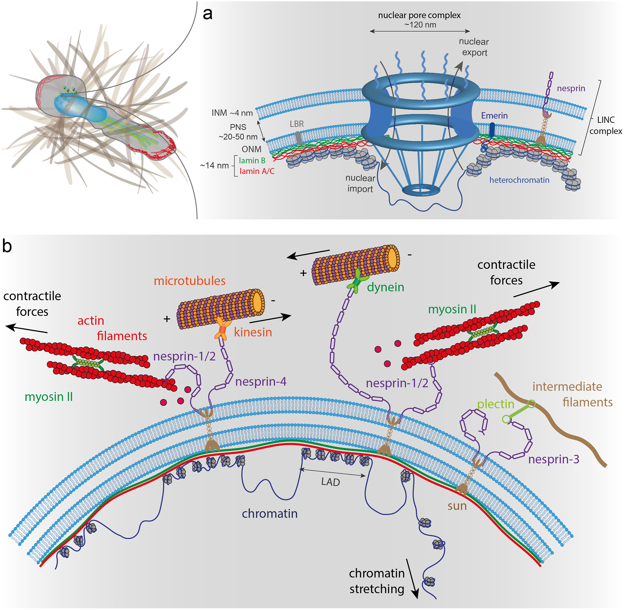
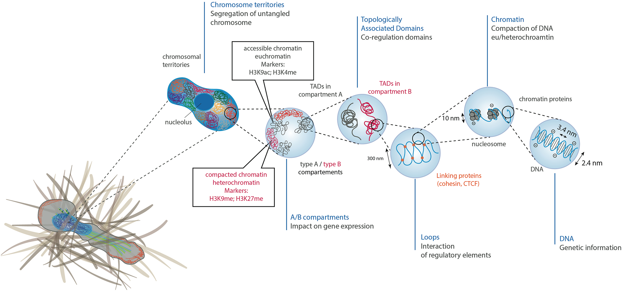
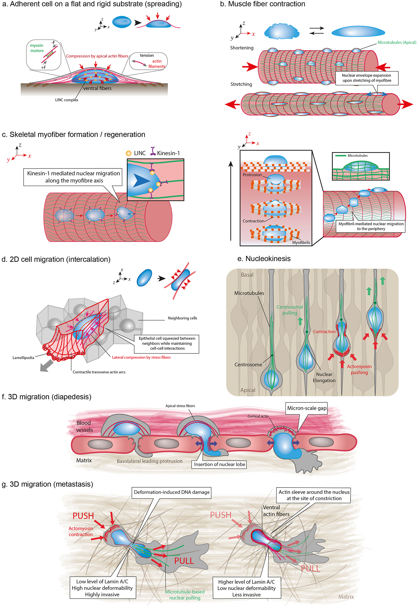
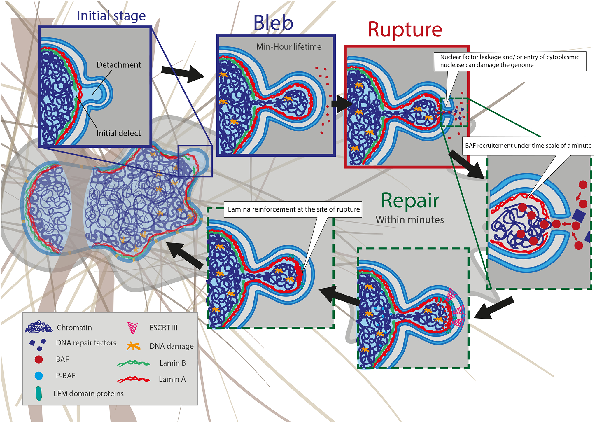
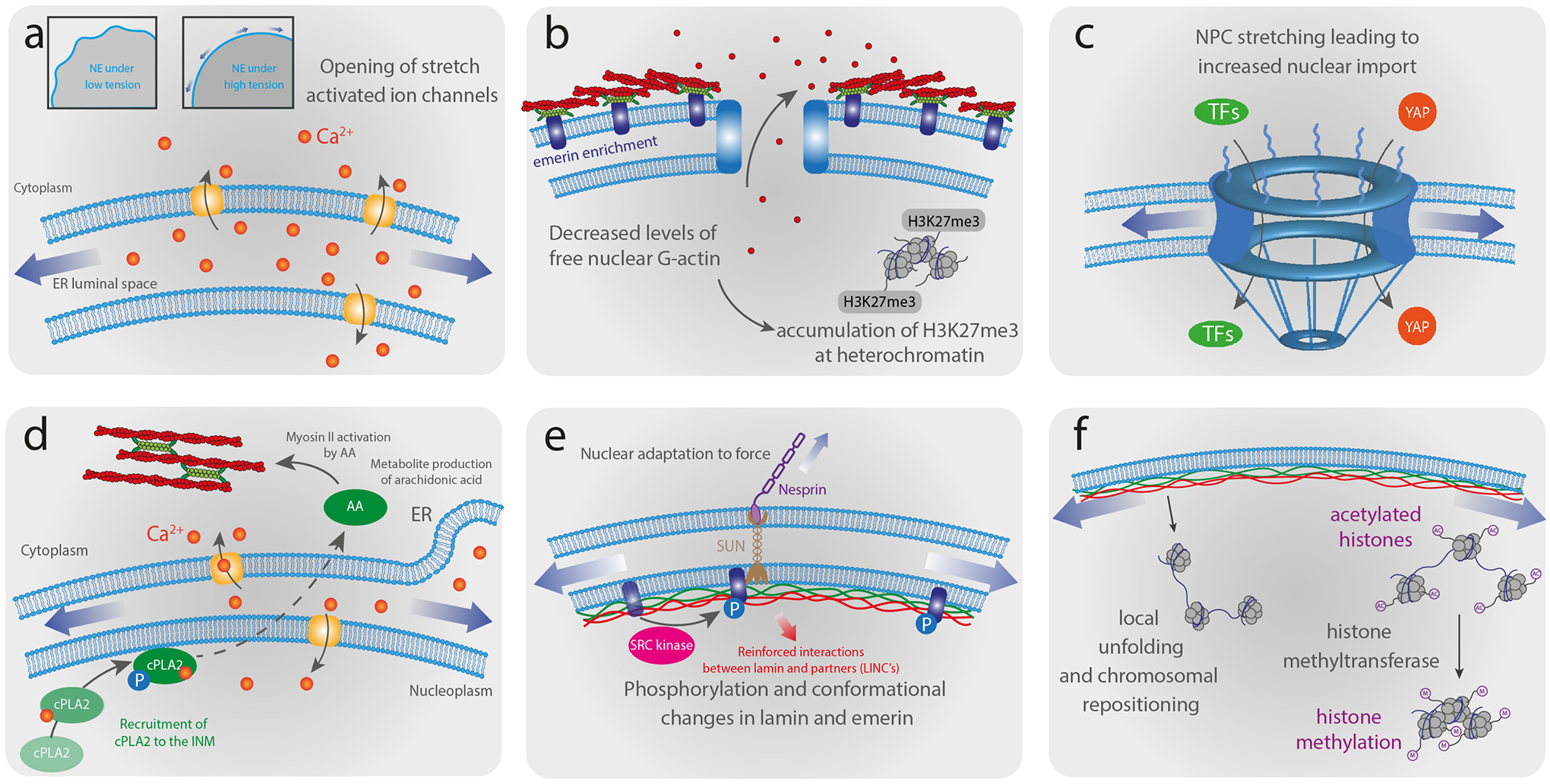
As the home of cellular genetic information, the nucleus has a critical role in determining cell fate and function in response to various signals and stimuli. In addition to biochemical inputs, the nucleus is constantly exposed to intrinsic and extrinsic mechanical forces that trigger dynamic changes in nuclear structure and morphology. Emerging data suggest that the physical deformation of the nucleus modulates many cellular and nuclear functions. These functions have long been considered to be downstream of cytoplasmic signalling pathways and dictated by gene expression. In this Review, we discuss an emerging perspective on the mechanoregulation of the nucleus that considers the physical connections from chromatin to nuclear lamina and cytoskeletal filaments as a single mechanical unit. We describe key mechanisms of nuclear deformations in time and space and provide a critical review of the structural and functional adaptive responses of the nucleus to deformations. We then consider the contribution of nuclear deformations to the regulation of important cellular functions, including muscle contraction, cell migration and human disease pathogenesis. Collectively, these emerging insights shed new light on the dynamics of nuclear deformations and their roles in cellular mechanobiology.


Figures

Figure 1 –
Figure 1 –. The nuclear envelope and nucleo-skeletal interactions.
(a) The nuclear envelope (NE) is composed of the outer (ONM) and inner (INM) nuclear membranes, which form a double lipid bilayer. The nuclear lamina is attached to the INM and in close contact with condensed chromatin, while nuclear pore complexes (NPCs) are surrounded by less condensed chromatin. The genomic regions connected to the lamina are lamina-associated chromatin domains (LADs), which have low transcriptional activity. The nuclear interior is connected to cytoskeletal filaments by nesprins and SUN domain proteins. Nesprin-1 and nesprin-2 bind to actin filaments, whereas nesprin-3 interacts with intermediate filaments. Nesprins-1, −2, and −4 can interact with microtubules via kinesin and dynein molecular motors. (b) NPCs allow controlled nuclear import and export of large molecules. The nuclear lamina meshwork composed of A-type and B-type lamins binds to the INM. Lamins, along with other INM proteins, such as LBR and emerin, anchor chromatin to the NE. Nesprins, ONM, SUN domain proteins and INM form together the LINC complex.
Figure 2 –
Figure 2 –. Chromatin organization and consequences of nuclear deformations in DNA organization.
Chromosomal DNA is packaged inside the cell nucleus with the help of histones. At the simplest level, chromatin is a double-stranded helical structure of DNA. The negatively charged DNA double helix is complexed with histones, which are positively charged proteins, to form nucleosomes. Inside the interphase nucleus, chromosomes occupy distinct territories (highlighted by different colors). Within each chromosome territory, the chromatin is folded into multiple loops and segregated into two distinct compartments: compartment A clustered around nucleolus and nuclear bodies (permissive region, in grey), and compartment B (repressive region, in red) located at the nuclear periphery. Within compartments, chromatin is further partitioned into topologically associating domains (TADs), which have preferential intradomain interactions compared to interdomain interactions with the neighboring cis chromatin domains. Histone methylation, particularly at residues H3K9 and H3K27, are often associated with heterochromatin, whereas histone acetylation, particularly at residue H3K9 or histone methylation at residue H3K4 are typically associated with euchromatin.
Figure 3 –
Figure 3 –. Physiological sources of nuclear deformations.
(a) Actomyosin contraction (in red) produces tension in actin fibers spanning the nucleus (in blue), which are connected to the NE via LINC complexes (in orange). Tension in apical actomyosin fibers generates vertical compressive forces that result in nuclear flattening. (b) Contraction and stretching of myofibers induce nuclear deformations, including NE wrinkling and expansion. Apical microtubules (in green) form cage-like structure around nuclei and exert compressive forces during myofibers elongation. (c) Formation and regeneration of skeletal myofibers require migration of nuclei along the myofiber axis through the interplay between LINC complex and microtubule associated motors such as kinesin-1. Myofibril contraction drives nuclear movement from the center to the periphery of the myofiber during muscle fiber maturation. This process requires myofibrils to exert contractile forces on the nucleus, resulting in large nuclear deformations. (d) Epithelial cell intercalation within dense tissues requires cellular elongation and nuclear deformation. Lateral compressive forces are exerted on both nuclear sides by ventral fibers, which are thick actomyosin bundles connected from their both ends to focal adhesions at the bottom of the cell. (e) Nucleokinesis events are observed during the development of the neuroepithelium of the central nervous system and is accompanied by considerable nuclear deformations. This mechanism occurs in densely packed tissues and involves pulling forces on the nucleus exerted by a microtubule cage towards the centrosome and pushing forces at the cell rear generated by actomyosin contraction, depending on the system. In mammals, microtubules exert pulling forces on the nuclear lamina through LINC complexes that move the nucleus towards the centrosome. (f) Immune cells and tumor cells can breach the endothelial barrier of blood vessels by inserting protrusion between or inside endothelial cells. Transendothelial migration through the small gaps (can be associated with a nuclear softening. Migration through the small openings (a few micrometers in diameter) is associated with large nuclear deformations. (g) Migrating cells translocate and deform their nucleus through narrow ECM pores or in between cells by using a combination of “push” and “pull” mechanisms. Nuclear deformations result from the balance between the amount and direction of the applied cytoskeletal force and the mechanical properties of the nucleus. Nuclear translocation requires both rear and front actomyosin contraction leading to pushing/pulling forces, respectively. At the front, microtubule motors are recruited to generate pulling forces. Together, the balance of forces results in the forward movement of the nucleus through the narrow constriction. High level of lamin A/C results in stiffer nuclei and highly invasive cells, whereas actin sleeve can be recruited at the site of the constriction to locally deform stiffer nuclei with higher level of lamin AC/ during the translocation of less invasive cells
Figure 4 –
Figure 4 –. Nuclear envelope rupture and repair.
Migration through confined environments or external compression of cells can result in nuclear membrane bleb formation and NE rupture. The nuclear membrane rupture process is typically initiated by the formation of a nuclear membrane extrusion, or bleb. Blebs from at sites with high nuclear membrane curvature and where an initial defect in the nuclear lamina exists. Blebs are driven by increased hydrostatic pressure within the nucleus. Initially, only the nuclear membrane detaches from the lamina. Later, lamin A/C and chromatin can enter the bleb. The lifetime of blebs can be minutes to hours, but the rupture itself is usually quite short, on the orders of minutes. Blebs can have varying size and can contain chromatin or are just fluid filled. Nuclear blebs typically lack lamin B (green) and NPCs. Continued nuclear compression by confinement from the extracellular matrix, apical actin stress fibers, cell contractions, or external compression results in bleb expansion until the nuclear membranes in the bleb exceeds a critical threshold and ruptures, leading to the leakage of soluble proteins from the nucleoplasm into the cytoplasm and uncontrolled influx of cytoplasmic proteins into the nucleus. Following NE rupture, barrier-to-autointegration factor (BAF) is rapidly (< min) recruited to initiate NE repair. The recruitment of ESCRT III complexes further contributes to resealing the nuclear membranes. The process of repair/rescue is typically completed within 10–15 min and often associated with recruitment of lamin A/C to the site of rupture. Although the NE rupture is resealed, the bleb/protrusion often persists and is not fully resorbed.
Figure 5 –
Figure 5 –. Schematic illustration of nuclear mechanoresponses and mechanosensing.
(a) High tension exerted on the NE during nuclear deformations induces unfolding of the wrinkled NE and the opening of stretch-activated ion channels. (b) Deformation of the nucleus induces enrichment of non-muscle myosin and emerin at the ONM. Relocalization of emerin to the ONM promotes perinuclear actin polymerization that leads to decreased levels of free nuclear monomeric actin, thereby reducing global transcriptional activity and increasing heterochromatin formation. The decrease of emerin at the INM leads to a loss of H3K9me2 and heterochromatin maintain their silenced state by recruiting H3K27me3. (c) Increased nuclear membrane tension stretch NPCs, leading to increased nuclear import of transcription factors (TFs) and mechanoresponsive transcriptional activators, such as YAP. (d) High NE tension resulting from nuclear deformation induces nuclear membrane unfolding, subsequent calcium release and the recruitment of cytosolic phospholipase A2 (cPLA2) activated by phosphorylation to the nuclear periphery, which promotes production of arachidonic acid (AA). The activation of the cPLA2-AA pathway leads to myosin II via AA-mediated Rhoa activation recruitment to the cell cortex, increasing actomyosin contractility. (e) Nuclear deformation induces phosphorylation of emerin and conformational changes in lamin A/C, which can alter the interaction with binding partners and induce further signaling events or recruit other proteins to the NE. (f) Forces acting on the nucleus may reposition or locally unfold chromatin domains, altering their transcriptional activity, and modulate the methylation level of histones by methyltransferases, regulating the transcriptional activity.

Similar articles

Cited by

References

    1. Lammerding J Mechanics of the Nucleus. Compr Physiol 1, 783–807 (2011). - PMC - PubMed
    1. Szczesny SE & Mauck RL The Nuclear Option: Evidence Implicating the Cell Nucleus in Mechanotransduction. J. Biomechl Eng 139, (2017). - PMC - PubMed
    1. Long JT & Lammerding J Nuclear Deformation Lets Cells Gauge Their Physical Confinement. Dev. Cell 56, 156–158 (2021). - PubMed
    1. Thomas CH, Collier JH, Sfeir CS & Healy KE Engineering gene expression and protein synthesis by modulation of nuclear shape. Proc. Natl Acad. Sci. U.S.A 99, 1972–1977 (2002). - PMC - PubMed
    1. Skinner BM & Johnson EEP Nuclear morphologies: their diversity and functional relevance. Chromosoma 126, 195–212 (2017). - PMC - PubMed

Publication types