. 2022 May 5;41(1):166.
doi: 10.1186/s13046-022-02376-4.

Interleukin-6 mediated inflammasome activation promotes oral squamous cell carcinoma progression via JAK2/STAT3/Sox4/NLRP3 signaling pathway

Affiliations

Interleukin-6 mediated inflammasome activation promotes oral squamous cell carcinoma progression via JAK2/STAT3/Sox4/NLRP3 signaling pathway

Li Xiao et al. J Exp Clin Cancer Res. .

Abstract

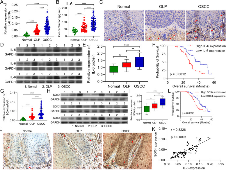
Background: Interleukin-6 (IL-6) has been reported to be critical in oral squamous cell carcinoma (OSCC). However, the set of pathways that IL-6 might activate in OSCC are not fully understood.

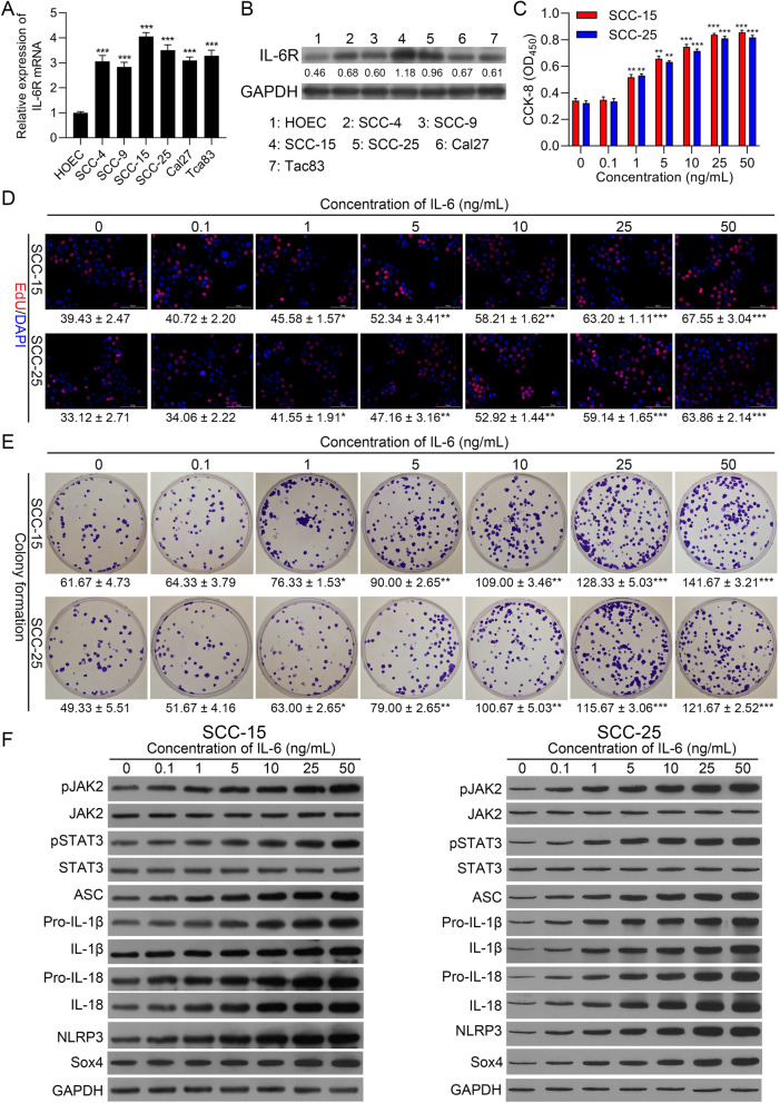
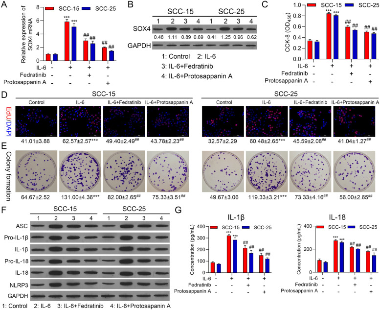
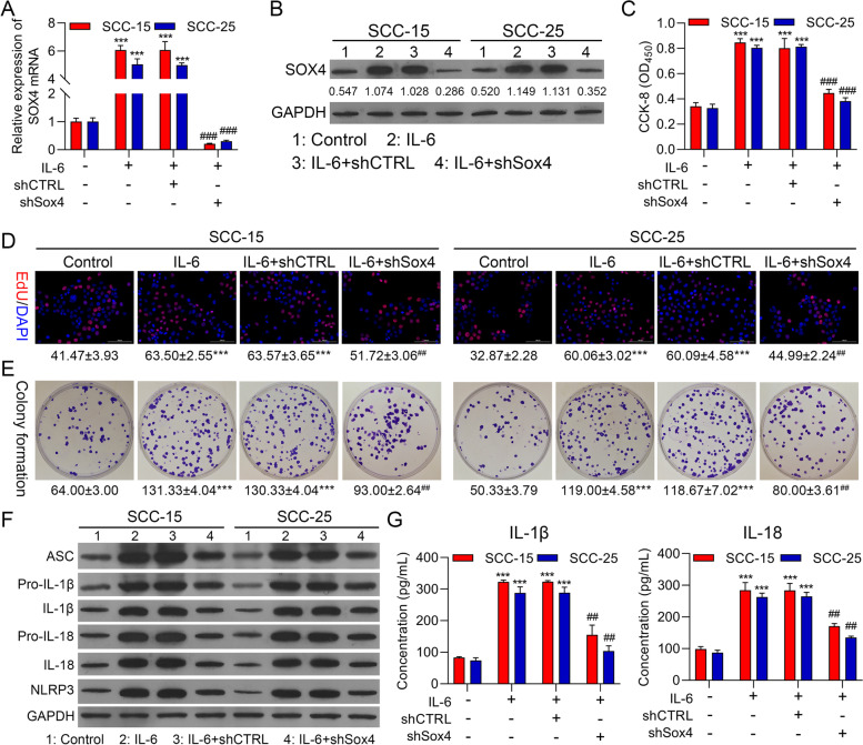
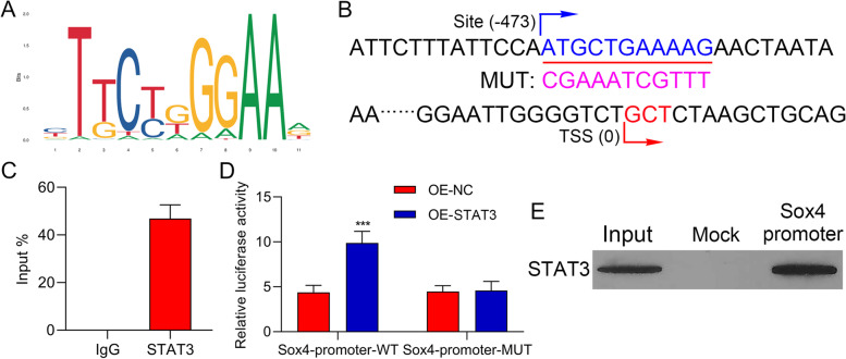
Methods: IL-6 and Sox4 expressions were first determined with RT-qPCR, ELISA, Western blot, or immunohistochemistry in OSCC tissues, and correlations between IL-6 and Sox4 expression and patient pathological characteristics were examined, and Kaplan-Meier approach was employed for evaluating the prognostic utility in OSCC patients. CCK-8, EdU stain and colony formation assays were utilized to test cell proliferation in vitro. Mechanistically, downstream regulatory proteins of IL-6 were verified through chromatin immunoprecipitation, luciferase reporter, pull-down, and the rescued experiments. Western blot was used for detecting protein expression. A nude mouse tumorigenicity assay was used to confirm the role of IL-6 and Sox4 in vivo.

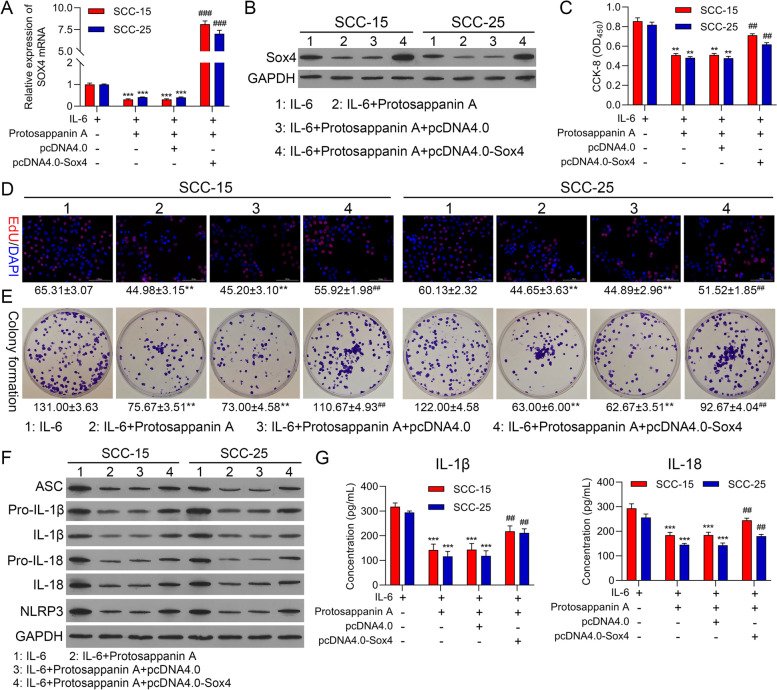
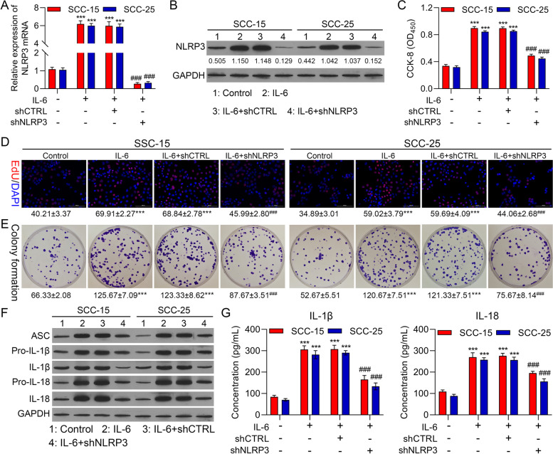
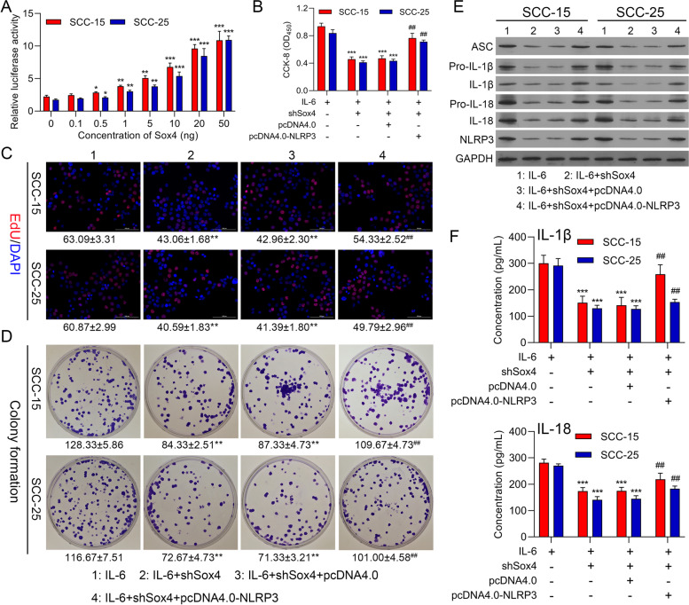
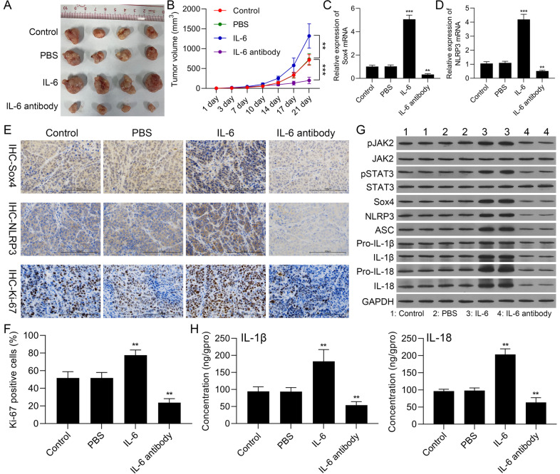
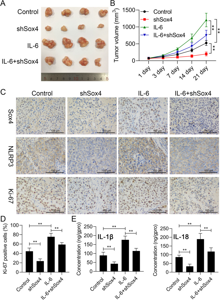
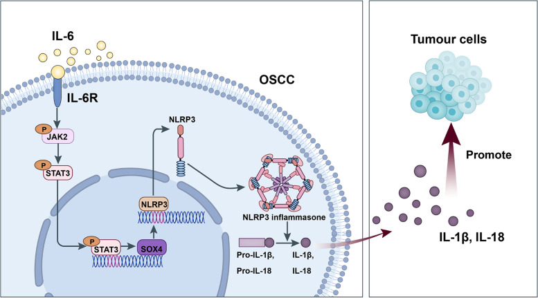
Results: IL-6 was upregulated in OSCC tissues, and Sox4 expression was positively correlated with IL-6 expression. High IL-6 and Sox4 expression was closely related to tumor size, TNM stage, and a poorer overall survival. Besides, IL-6 could accelerate OSCC cell proliferation by activating inflammasome via JAK2/STAT3/Sox4/NLRP3 pathways in vitro and in vivo. Furthermore, STAT3 played as a transcription factor which positively regulated Sox4, and IL-6 promotes Sox4 expression by activating JAK2/STAT3 pathway. Moreover, through the rescue experiments, we further confirmed that IL-6 could promote proliferation and NLRP3 inflammasome activation via JAK2/STAT3/Sox4 pathway in OSCC cells. Finally, knockdown of Sox4 suppressed OSCC growth in vivo, and antagonized the acceleration of IL-6 on tumor growth.

Conclusions: We confirmed that IL-6 plays an oncogenic role in OSCC progression by activating JAK2/STAT3/Sox4/NLRP3 pathway, which might be the therapeutic targets for OSCC remedy.

Keywords: IL-6; JAK2; NLRP3 inflammasome; NLRP3 pathway; Oral squamous cell carcinoma; STAT3; Sox4.


Conflict of interest statement

The authors declare that they have no competing interests.

Figures

Fig. 1
Fig. 1
IL-6 and Sox4 were memorably up-regulated, and positively correlated in OSCC tissues. (A) RT-qPCR analysis of IL-6 in normal, OLP, and OSCC tissues. (B) The content of IL-6 was tested with ELISA kit in the serum of normal, OLP, and OSCC patients. (C) IL-6 expression was determined via IHC in normal, OLP, and OSCC tissues. (D and E) Western blotting analysis of IL-6 in normal, OLP, and OSCC (developed after follow-up) tissues, which were from the same patient (n = 9). After diagnosis of OLP, OLP tissue and adjacent normal tissue were taken, and OSCC tissue was taken after follow-up. (F) The overall survival of IL-6 expression in OSCC patients. (H) The level of Sox4 was confirmed via RT-qPCR. (H and I) Sox4 expression levels were assessed with Western blot in normal, OLP, and OSCC tissues, which also were from the same patient (n = 9). (L) The overall survival of Sox4 expression in OSCC patients. (J) Immunohistochemical staining of Sox4 expression in normal, OLP, and OSCC tissues. (K) Correlation analysis of IL-6 and Sox4 (r = 0.8226). ** P < 0.01, *** P < 0.001, **** P < 0.0001
Fig. 2
Fig. 2
Introduction of IL-6 dramatically induced proliferation and activated JAK2/STAT3/Sox4/NLRP3 pathway in OSCC cells. IL-6R expression was credited through RT-qPCR (A) and Western blot (B) in oral epithelial cells (HOEC) and OSCC cell lines (SCC-4, SCC-9, SCC-15, SCC-25, Cal27 and Tca83). SCC-15 and SCC-25 cells were induced by 0, 0.1, 1, 5, 10, 25, 50 ng/mL IL-6, and cell proliferation was monitored by applying (C) CCK-8, (D) EdU staining and (E) colony formation. (F) Western blot was conducted to identify the impacts of IL-6 on pJAK2, JAK2, pSTAT3, STAT3, ASC, pro-IL-1β, IL-1β, pro-IL-18, IL-18, and NLRP3 in OSCC cells. ** P < 0.01, ***P < 0.001
Fig. 3
Fig. 3
IL-6 observably accelerated proliferation and NLRP3 inflammasome activation by JAK2/STAT3 pathway in OSCC cells. SCC-15 and SCC-25 cells were processed with 5 μmol/L JAK2 inhibitor (Fedratinib) and 25 μmol/L STAT3 inhibitor (Protosappanin A), and induced by 25 ng/ml IL-6 for 24 h. The expression change of Sox4 was monitored using RT-qPCR (A) and Western blot (B). Cell proliferation was also tested using (C) CCK-8, (D) EdU staining and (E) colony formation. (F) The levels of inflammation-related proteins were assessed with Western blot. (G) ELISA kits were adopted to examine the concentration of IL-1β and IL-18 in the processed OSCC cells. *** P < 0.001 vs. control group; ## P < 0.01 vs. IL-6 group
Fig. 4
Fig. 4
Silencing of Sox4 markedly suppressed proliferation and NLRP3 inflammasome activation in IL-6-treated OSCC cells. shCTRL and shSox4 were constructed and transfected into SCC-15 and SCC-25 cells, which were also disposed of 25 ng/ml IL-6. RT-qPCR (A) and Western blot (B) displayed the change in Sox4 expression. Cell proliferation was confirmed with (C) CCK-8, (D) EdU staining and (E) colony formation. (F) Western blot was adopted to verify the expression changes of inflammation-related proteins. (G) The changes of IL-1β and IL-18 levels were also evaluated with ELISA kits. *** P < 0.001 vs. IL-6 group; ## P < 0.01, ### P < 0.001 vs. IL-6 + shCTRL group. CTRL, control
Fig. 5
Fig. 5
Upregulation of Sox4 prominently resisted blocking STAT3 mediated suppression of proliferation and NLRP3 inflammasome activation in IL-6-induced OSCC cells. SCC-15 and SCC-25 cells were dealt with 25 ng/ml IL-6 or 25 μmol/L Protosappanin A, and transfected with Sox4 overexpression plasmid. The expression change of Sox4 was confirmed with RT-qPCR (A) and Western blot (B). (C) CCK-8, (D) EdU staining and (E) colony formation was applied to exhibit the change in cell proliferation. (F) Western blotting analysis of the inflammation-related proteins. (G) ELISA analysis of IL-1β and IL-18 levels. ** P < 0.01, *** P < 0.001 vs. IL-6 group; ## P < 0.01, ### P < 0.001 vs. IL-6 + Protosappanin A group
Fig. 6
Fig. 6
STAT3 could signally regulate Sox4 expression in a targeted manner. (A) Identification sites on STAT3 gene were exhibited. (B) Binding sites of STAT3 on Sox4 promoter and the sequence of mutations. (C) The regulatory relationship between STAT3 and Sox4 was certified with ChIP. (D) The targeted regulation of Sox4 by STAT3 was verified with Dual luciferase reporter gene assay. (E) DNA Pull-down was applied to test the regulatory correlation between STAT3 expression and Sox4 promoter. *** P < 0.001
Fig. 7
Fig. 7
Knockdown of NLRP3 notably weakened proliferation and NLRP3 inflammasome activation in IL-6-mediated OSCC cells. NLRP3 was silenced in SCC-15 and SCC-25 cells, which were addressed with 25 ng/ml IL-6. NLRP3 expression was assessed through RT-qPCR (A) and western blot (B). Cell proliferation was determined via (C) CCK-8, (D) EdU staining and (E) colony formation. (F) The expressions of the inflammation-related proteins were monitored with Western blot. (G) ELISA kits were applied to identify IL-1β and IL-18 levels. *** P < 0.001 vs. IL-6 group; ### P < 0.001 vs. IL-6 + shCTRL group
Fig. 8
Fig. 8
Sox4 silencing dramatically repressed proliferation and NLRP3 inflammasome activation by downregulating NLRP3 in IL-6-induced OSCC cells. (A) The influence of Sox4 on NLRP3 promoter activity was examined using Dual luciferase reporter gene assay, * P < 0.05, ** P < 0.01, *** P < 0.001. (B) CCK-8, (C) EdU staining, and (D) colony formation were conducted to test cell proliferation in IL-6-treated SCC-15 and SCC-25 cells, which were transfected with shSox4 or/and NLRP3 overexpression plasmid, respectively. (E) Western blot was utilized to investigate the impacts of Sox4 silencing and NLRP3 overexpression on the levels of the inflammation-related proteins in IL-6-induced OSCC cells. (F) IL-1β and IL-18 levels were analyzed using ELISA kits in the processed OSCC cells. *** P < 0.001 vs. IL-6 group; ## P < 0.01 vs. IL-6 + shSox4 group
Fig. 9
Fig. 9
IL-6 notably accelerated tumor growth and activated JAK2/STAT3-Sox4-NLRP3 pathway in nude mouse xenografts of OSCC. SCC-15 cells were applied to construct nude mouse xenografts, which were intervened with IL-6 or anti-IL-6. (A) Subcutaneous tumors were dissected and photographed at the end of day 21. (B) Tumor growth curve was presented after xenotransplantation. Sox4 (C) and NLRP3 (D) expressions were certified with RT-qPCR. (E) Immunohistochemical staining of Sox4, NLRP3, and Ki-67 expressions in the tumors. (F) Ki-67 positive cells were counted. (G) Western blotting analysis of JAK2, STAT3, Sox4, and NLRP3 inflammasome activation-related proteins. (H) IL-1β and IL-18 levels were confirmed using ELISA kits in the serum of nude mice. ** P < 0.01, *** P < 0.001
Fig. 10
Fig. 10
Sox4 knockdown obviously weakened the induction of IL-6 on the tumor growth and inflammation in nude mouse xenografts of OSCC. SCC-15 cells processed with shSox4 or/and IL-6 were subcutaneously injected into nude mice. (A) After 3 weeks, groups of tumors were presented. (B) The volume of the tumor was counted in each group. (C) Immunohistochemical staining of Sox4, NLRP3, and Ki-67 expressions in each group. (D) Ki-67 positive cells were calculated. (E) ELISA kits were applied to analyze IL-1β and IL-18 levels in the serum of nude mice
Fig. 11
Fig. 11
Schematic overview of the mechanistic basis for the observed study results. IL-6 plays an oncogenic role in OSCC progression by activating JAK2/STAT3/Sox4/NLRP3 pathway. IL-6 could accelerate OSCC cell proliferation by activating inflammasome via JAK2/STAT3/Sox4/NLRP3 pathways. STAT3 played as a transcription factor which positively regulated Sox4, and IL-6 promotes Sox4 expression by activating JAK2/STAT3 pathway. IL-6 could promote proliferation and NLRP3 inflammasome activation via JAK2/STAT3/Sox4 pathway in OSCC cells

Similar articles

Cited by

References

    1. Wang Y, Hu H, Wang Q, Li Z, Zhu Y, Zhang W, et al. The level and clinical significance of 5-hydroxymethylcytosine in oral squamous cell carcinoma: An immunohistochemical study in 95 patients. Pathol Res Pract. 2017;213(8):969–974. - PubMed
    1. Panarese I, Aquino G, Ronchi A, Longo F, Montella M, Cozzolino I, et al. Oral and Oropharyngeal squamous cell carcinoma: prognostic and predictive parameters in the etiopathogenetic route. Expert Rev Anticancer Ther. 2019;19(2):105–119. - PubMed
    1. Johnson DE, Burtness B, Leemans CR, Lui VWY, Bauman JE, Grandis JR. Head and neck squamous cell carcinoma. Nat Rev Dis Primers. 2020;6(1):92. - PMC - PubMed
    1. Mei Z, Huang J, Qiao B, Lam AK. Immune checkpoint pathways in immunotherapy for head and neck squamous cell carcinoma. Int J Oral Sci. 2020;12(1):16. - PMC - PubMed
    1. Rose-John S. Interleukin-6 Family Cytokines. Cold Spring Harb Perspect Biol. 2018;10(2):a028415. - PMC - PubMed

MeSH terms