

Toxicity and Local Tolerance of a Novel Spike Protein RBD Vaccine Against SARS-CoV-2, Produced Using the C1 Thermothelomyces Heterothallica Protein Expression Platform
- PMID: 35514116
- PMCID: PMC9128004
- DOI: 10.1177/01926233221090518
Toxicity and Local Tolerance of a Novel Spike Protein RBD Vaccine Against SARS-CoV-2, Produced Using the C1 Thermothelomyces Heterothallica Protein Expression Platform
Abstract
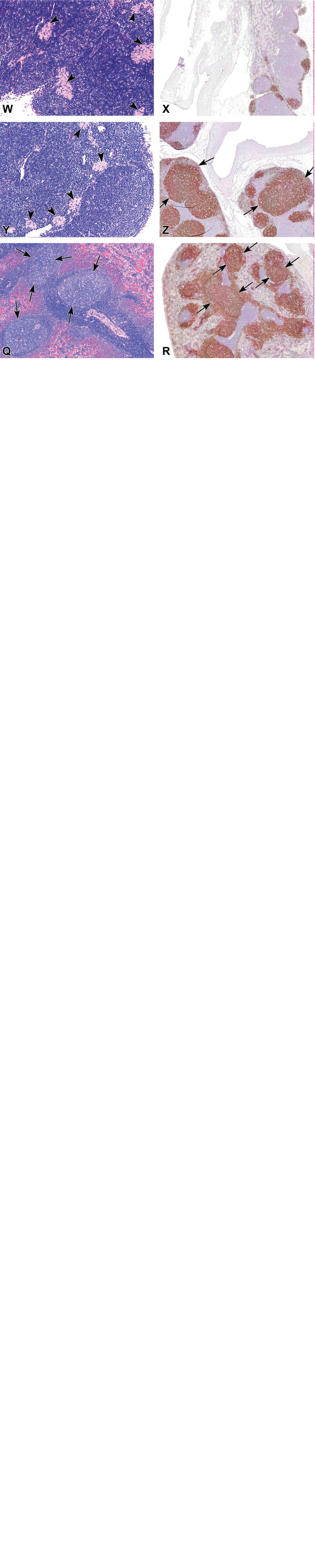
Coronavirus disease 2019 (COVID-19) has caused the ongoing COVID-19 pandemic and there is a growing demand for safe and effective vaccines. The thermophilic Thermothelomyces heterothallica filamentous fungal host, C1-cell, can be utilized as an expression platform for the rapid production of large quantities of antigens for developing vaccines. The aim of this study was to evaluate the local tolerance and the systemic toxicity of a C1-cell expressed receptor-binding domain (C1-RBD) vaccine, following repeated weekly intramuscular injections (total of 4 administrations), in New Zealand White rabbits. The animals were sacrificed either 3 days or 3 weeks following the last dose. No signs of toxicity were observed, including no injection site reactions. ELISA studies revealed severe acute respiratory syndrome coronavirus 2 (SARS-CoV-2)-specific immunoglobulin G antibodies in the sera of C1-RBD-treated animals starting from day 13 post injection, that were further elevated. Histopathology evaluation and immunohistochemical staining revealed follicular hyperplasia, consisting of B-cell type, in the spleen and inguinal lymph nodes of the treated animals that were sustained throughout the recovery phase. No local or systemic toxicity was observed. In conclusion, the SARS-CoV-2 C1-RBD vaccine candidate demonstrated an excellent safety profile and a lasting immunogenic response against receptor-binding domain, thus supporting its further development for use in humans.
Keywords: C1; COVID-19; RBD; SARS-CoV-2; Thermothelomyces heterothallica; rabbits; safety; toxicity; vaccine.
Conflict of interest statement
Figures
Similar articles
-
Thermophilic Filamentous Fungus C1-Cell-Cloned SARS-CoV-2-Spike-RBD-Subunit-Vaccine Adjuvanted with Aldydrogel®85 Protects K18-hACE2 Mice against Lethal Virus Challenge.Vaccines (Basel). 2022 Dec 11;10(12):2119. doi: 10.3390/vaccines10122119. Vaccines (Basel). 2022. PMID: 36560529 Free PMC article.
-
Preclinical immunogenicity and protective efficacy of a SARS-CoV-2 RBD-based vaccine produced with the thermophilic filamentous fungal expression system Thermothelomyces heterothallica C1.Front Immunol. 2023 Jun 9;14:1204834. doi: 10.3389/fimmu.2023.1204834. eCollection 2023. Front Immunol. 2023. PMID: 37359531 Free PMC article.
-
A recombinant SARS-CoV-2 receptor-binding domain expressed in an engineered fungal strain of Thermothelomyces heterothallica induces a functional immune response in mice.Vaccine. 2022 Feb 16;40(8):1162-1169. doi: 10.1016/j.vaccine.2022.01.007. Epub 2022 Jan 19. Vaccine. 2022. PMID: 35078661 Free PMC article.
-
A vaccine targeting the RBD of the S protein of SARS-CoV-2 induces protective immunity.Nature. 2020 Oct;586(7830):572-577. doi: 10.1038/s41586-020-2599-8. Epub 2020 Jul 29. Nature. 2020. PMID: 32726802
-
Potential for developing a SARS-CoV receptor-binding domain (RBD) recombinant protein as a heterologous human vaccine against coronavirus infectious disease (COVID)-19.Hum Vaccin Immunother. 2020 Jun 2;16(6):1239-1242. doi: 10.1080/21645515.2020.1740560. Epub 2020 Apr 16. Hum Vaccin Immunother. 2020. PMID: 32298218 Free PMC article. Review.
Cited by
-
Thermophilic Filamentous Fungus C1-Cell-Cloned SARS-CoV-2-Spike-RBD-Subunit-Vaccine Adjuvanted with Aldydrogel®85 Protects K18-hACE2 Mice against Lethal Virus Challenge.Vaccines (Basel). 2022 Dec 11;10(12):2119. doi: 10.3390/vaccines10122119. Vaccines (Basel). 2022. PMID: 36560529 Free PMC article.
-
Preclinical immunogenicity and protective efficacy of a SARS-CoV-2 RBD-based vaccine produced with the thermophilic filamentous fungal expression system Thermothelomyces heterothallica C1.Front Immunol. 2023 Jun 9;14:1204834. doi: 10.3389/fimmu.2023.1204834. eCollection 2023. Front Immunol. 2023. PMID: 37359531 Free PMC article.
-
A review of the scientific literature on experimental toxicity studies of COVID-19 vaccines, with special attention to publications in toxicology journals.Arch Toxicol. 2024 Nov;98(11):3603-3617. doi: 10.1007/s00204-024-03854-8. Epub 2024 Sep 3. Arch Toxicol. 2024. PMID: 39225797 Free PMC article. Review.
-
Advances in SARS-CoV-2 receptor-binding domain-based COVID-19 vaccines.Expert Rev Vaccines. 2023 Jan-Dec;22(1):422-439. doi: 10.1080/14760584.2023.2211153. Expert Rev Vaccines. 2023. PMID: 37161869 Free PMC article. Review.
-
Filamentous fungus-produced human monoclonal antibody provides protection against SARS-CoV-2 in hamster and non-human primate models.Nat Commun. 2024 Mar 14;15(1):2319. doi: 10.1038/s41467-024-46443-0. Nat Commun. 2024. PMID: 38485931 Free PMC article.
References
-
- WHO COVID-19 Dashboard [Internet]. Geneva: World Health Organization. Accessed October 13, 2021. https://covid19.who.int/.
Publication types
MeSH terms
Substances
LinkOut - more resources
Full Text Sources
Medical
Miscellaneous
