Periodontal disease affects oral cancer progression in a surrogate animal model for tobacco exposure
- PMID: 35514311
- PMCID: PMC9097773
- DOI: 10.3892/ijo.2022.5367
Periodontal disease affects oral cancer progression in a surrogate animal model for tobacco exposure
Abstract
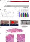
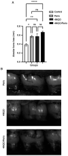
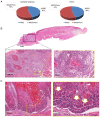
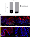
For decades, the link between poor oral hygiene and the increased prevalence of oral cancer has been suggested. Most recently, emerging evidence has suggested that chronic inflammatory diseases from the oral cavity (e.g., periodontal disease), to some extent, play a role in the development of oral squamous cell carcinoma (OSCC). The present study aimed to explore the direct impact of biofilm‑induced periodontitis in the carcinogenesis process using a tobacco surrogate animal model for oral cancer. A total of 42 Wistar rats were distributed into four experimental groups: Control group, periodontitis (Perio) group, 4‑nitroquinoline 1‑oxide (4‑NQO) group and 4NQO/Perio group. Periodontitis was stimulated by placing a ligature subgingivally, while oral carcinogenesis was induced by systemic administration of 4NQO in the drinking water for 20 weeks. It was observed that the Perio, 4NQO and 4NQO/Perio groups presented with significantly higher alveolar bone loss compared with that in the control group. Furthermore, all groups receiving 4NQO developed lesions on the dorsal surface of the tongue; however, the 4NQO/Perio group presented larger lesions compared with the 4NQO group. There was also a modest overall increase in the number of epithelial dysplasia and OSCC lesions in the 4NQO/Perio group. Notably, abnormal focal activation of cellular differentiation (cytokeratin 10‑positive cells) that extended near the basal cell layer of the mucosa was observed in rats receiving 4NQO alone, but was absent in rats receiving 4NQO and presenting with periodontal disease. Altogether, the presence of periodontitis combined with 4NQO administration augmented tumor size in the current rat model and tampered with the protective mechanisms of the cellular differentiation of epithelial cells.
Keywords: 4‑nitroquinoline‑1‑oxide; cell differentiation; head and neck cancer; ligature; periodontitis.
Conflict of interest statement
The authors declare that they have no competing interests.
Figures


Similar articles
-
Oral-specific ablation of Klf4 disrupts epithelial terminal differentiation and increases premalignant lesions and carcinomas upon chemical carcinogenesis.J Oral Pathol Med. 2015 Nov;44(10):801-9. doi: 10.1111/jop.12307. Epub 2015 Jan 21. J Oral Pathol Med. 2015. PMID: 25605610
-
Spontaneous alveolar bone loss after 4NQO exposure in Wistar rats.Arch Oral Biol. 2018 May;89:44-48. doi: 10.1016/j.archoralbio.2018.02.001. Epub 2018 Feb 7. Arch Oral Biol. 2018. PMID: 29448184 Clinical Trial.
-
4NQO induced carcinogenesis: A mouse model for oral squamous cell carcinoma.Methods Cell Biol. 2021;163:93-111. doi: 10.1016/bs.mcb.2021.01.001. Epub 2021 Mar 4. Methods Cell Biol. 2021. PMID: 33785171
-
Can propranolol act as a chemopreventive agent during oral carcinogenesis? An experimental animal study.Eur J Cancer Prev. 2021 Jul 1;30(4):315-321. doi: 10.1097/CEJ.0000000000000626. Eur J Cancer Prev. 2021. PMID: 33136608
-
4-nitroquinoline-1-oxide induced experimental oral carcinogenesis.Oral Oncol. 2006 Aug;42(7):655-67. doi: 10.1016/j.oraloncology.2005.10.013. Epub 2006 Jan 30. Oral Oncol. 2006. PMID: 16448841 Review.
Cited by
-
The immunomodulatory impact of naturally derived neem leaf glycoprotein on the initiation progression model of 4NQO induced murine oral carcinogenesis: a preclinical study.Front Immunol. 2024 Mar 22;15:1325161. doi: 10.3389/fimmu.2024.1325161. eCollection 2024. Front Immunol. 2024. PMID: 38585261 Free PMC article.
-
Network toxicology reveals collaborative mechanism of 4NQO and ethanol in esophageal squamous cell carcinogenesis.Biochem Biophys Rep. 2025 Aug 2;43:102187. doi: 10.1016/j.bbrep.2025.102187. eCollection 2025 Sep. Biochem Biophys Rep. 2025. PMID: 40800605 Free PMC article.
-
Alveolar bone loss is associated with oral cancer: a case-control study.Front Oral Health. 2025 May 9;6:1569491. doi: 10.3389/froh.2025.1569491. eCollection 2025. Front Oral Health. 2025. PMID: 40416513 Free PMC article.
-
Immunotherapeutic Strategies for Head and Neck Squamous Cell Carcinoma (HNSCC): Current Perspectives and Future Prospects.Vaccines (Basel). 2022 Aug 7;10(8):1272. doi: 10.3390/vaccines10081272. Vaccines (Basel). 2022. PMID: 36016159 Free PMC article. Review.
-
Periodontitis and Cancer: Beyond the Boundaries of Oral Cavity.Cancers (Basel). 2023 Mar 13;15(6):1736. doi: 10.3390/cancers15061736. Cancers (Basel). 2023. PMID: 36980622 Free PMC article.
References
MeSH terms
Substances
LinkOut - more resources
Full Text Sources
Medical
Research Materials
