. 2022 Apr 21:13:855959.
doi: 10.3389/fphys.2022.855959. eCollection 2022.

A Neural Crest-specific Overexpression Mouse Model Reveals the Transcriptional Regulatory Effects of Dlx2 During Maxillary Process Development

Affiliations

A Neural Crest-specific Overexpression Mouse Model Reveals the Transcriptional Regulatory Effects of Dlx2 During Maxillary Process Development

Jian Sun et al. Front Physiol. .

Abstract

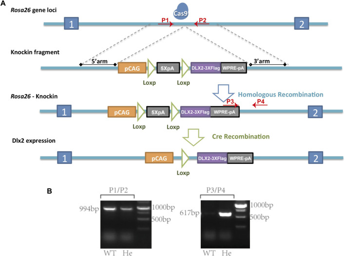
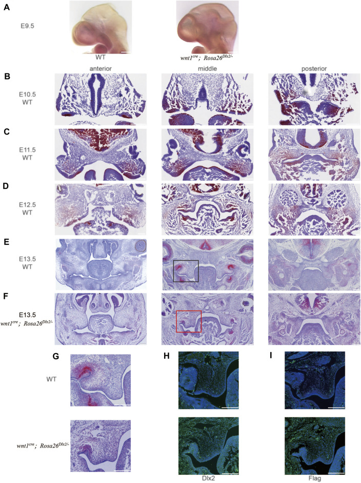
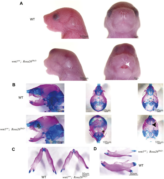
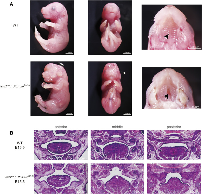
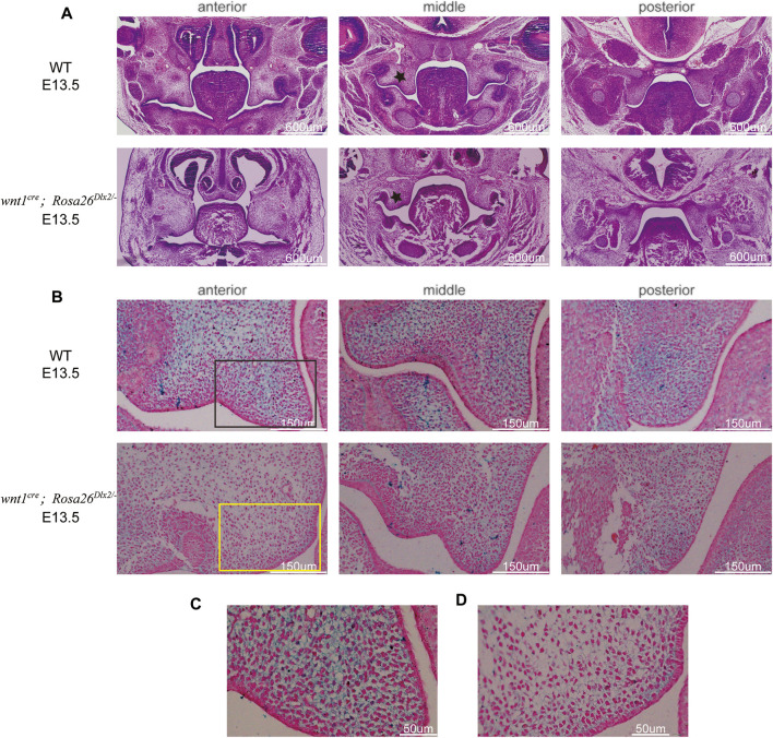
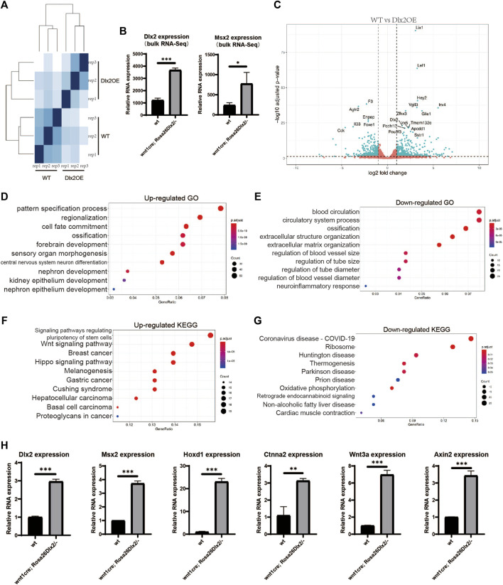
Craniofacial morphogenesis is a complex process that requires precise regulation of cell proliferation, migration, and differentiation. Perturbations of this process cause a series of craniofacial deformities. Dlx2 is a critical transcription factor that regulates the development of the first branchial arch. However, the transcriptional regulatory functions of Dlx2 during craniofacial development have been poorly understood due to the lack of animal models in which the Dlx2 level can be precisely modulated. In this study, we constructed a Rosa26 site-directed Dlx2 gene knock-in mouse model Rosa26 CAG-LSL-Dlx2-3xFlag for conditionally overexpressing Dlx2. By breeding with wnt1 cre mice, we obtained wnt1 cre ; Rosa26 Dlx2/- mice, in which Dlx2 is overexpressed in neural crest lineage at approximately three times the endogenous level. The wnt1 cre ; Rosa26 Dlx2/- mice exhibited consistent phenotypes that include cleft palate across generations and individual animals. Using this model, we demonstrated that Dlx2 caused cleft palate by affecting maxillary growth and uplift in the early-stage development of maxillary prominences. By performing bulk RNA-sequencing, we demonstrated that Dlx2 overexpression induced significant changes in many genes associated with critical developmental pathways. In summary, our novel mouse model provides a reliable and consistent system for investigating Dlx2 functions during development and for elucidating the gene regulatory networks underlying craniofacial development.

Keywords: Dlx2; RNA-seq; cleft palate; cranial neural crest cells; craniofacial development.


Conflict of interest statement

The authors declare that the research was conducted in the absence of any commercial or financial relationships that could be construed as a potential conflict of interest.

Figures

FIGURE 1
FIGURE 1
A Rosa26 CAG-LSL-Dlx2−3xFlag mouse model for conditional Dlx2 overexpression (A) Cartoons illustrate the design strategy for conditionally overexpressing Dlx2 by breeding Rosa26 CAG-LSL-Dlx2−3xFlag mice with Cre mice (B) In WT mice (P1, P2) generated a 994 bp band, whereas (P3, P4) did not generate a PCR product. In heterozygous mice (P1, P2) amplified a 994 bp PCR product, while (P3, P4) amplified a smaller PCR product of 617 bp. Cas9, CRISPR/Cas9 system; pCAG, CAG promoter; 5XpA, 5X PolyA (containing STOP cassette); DLX2-3XFlag, coding sequence of Dlx2 with 3X flag; WPRE-pA, woodchuck hepatitis virus post-transcriptional regulatory element (WPRE)-bGH polyA; WT, wild-type; He, heterozygote.
FIGURE 2
FIGURE 2
The temporal Dlx2 expression pattern during the early-stage development of maxillary processes from E9.5 to E13.5 (A) The whole in situ hybridization of E9.5 WT and wnt1 cre ; Rosa26 Dlx2/- mouse head (B-E) In situ hybridization on the head tissue sections of E10.5 (B), E11.5 (C), E12.5 (D), and E13.5 (E) WT embryos and the temporal Dlx2 expression pattern. Note that Dlx2 expression is largely diminished at E13.5 (F) In situ hybridization on the head tissue sections of E13.5 wnt1 cre ; Rosa26 Dlx2/- mouse embryo depicts Dlx2 overexpression in maxillary processes (G) Enlarged picture of the box area in E and F (H) Dlx2 immunofluorescence staining of E13.5 WT (up) and wnt1 cre ; Rosa26 Dlx2/- (down) mouse maxillary processes (I) Flag immunofluorescence staining of E13.5 WT (up) and wnt1 cre ; Rosa26 Dlx2/- (down) mouse maxillary processes. Bar, 200um.
FIGURE 3
FIGURE 3
Phenotype of wnt1 cre ; Rosa26 Dlx2/- mice at P0 (A) Stereo microscope images of P0 WT and wnt1 cre ; Rosa26 Dlx2/- mice. Wnt1 cre ; Rosa26 Dlx2/- mice exhibit a markedly shorter maxilla, a tiny cleft in the upper lip, abnormal eye development, and soft tissue bulges on the head (B-D) Skeletal staining of P0 WT and wnt1 cre ; Rosa26 Dlx2/- mice shows a range of evident craniofacial abnormality in wnt1 cre ; Rosa26 Dlx2/- mice (B) Wnt1 cre ; Rosa26 Dlx2/- mice exhibited a partial defect of the skull and parietal bones in addition to the short maxilla and cleft palate (C) Wnt1 cre ; Rosa26 Dlx2/- mice exhibit a shortened mandible (D) The temporomandibular joints of the wnt1 cre ; Rosa26 Dlx2/- mice are abnormally developed. The joint heads are smaller and the articular cartilage is insufficient. The dotted line shows the range of maxilla and palatine, excluding the premaxilla. Black arrows indicate cleft palate, the white arrow points to the tiny cleft in the upper lip, and the black star indicates the partial defect of the frontal bones.
FIGURE 4
FIGURE 4
Phenotype of wnt1 cre ; Rosa26 Dlx2/- mice at E15.5 (A) Stereo microscope images showing the cleft (black arrow) in the palate at E15.5 (B) HE staining of frontal sections showing the morphology of maxillary processes of WT and wnt1 cre ; Rosa26 Dlx2/- mouse embryos at E15.5. Wnt1 cre ; Rosa26 Dlx2/- embryos display a cleft in the palate. White star indicates the towering tongue.
FIGURE 5
FIGURE 5
Conditional Dlx2 overexpression in wnt1 cre ; Rosa26 Dlx2/- mice caused cleft palate by affecting maxillary growth and uplift (A) HE staining of frontal sections showing the morphology of maxillary processes of WT and wnt1 cre ; Rosa26 Dlx2/- mouse embryos at E13.5. The maxillary processes of the wnt1 cre ; Rosa26 Dlx2/- mice are smaller than those of WT. Black stars indicate the maxillary processes (B) HA staining of the right maxillary process at E13.5 showing HA expression in wnt1 cre ; Rosa26 Dlx2/- mice is decreased compared with WT mouse embryos (C,D) Locally enlarged palatal process sections display that HA fails to fill the ECM space and cell density is decreased in wnt1 cre ; Rosa26 Dlx2/- mouse embryos (C) Enlarged picture of the black box area in B (D) Enlarged picture of the yellow box area in B.
FIGURE 6
FIGURE 6
Bulk RNA-seq of the maxillary processes from E12.5 WT and wnt1 cre ; Rosa26 Dlx2/- embryos (A) Sample distances matrix showing the correlation between RNA-seq replicates (B) Bar graphs showing the Dlx2 and Msx2 expression levels in WT and wnt1 cre ; Rosa26 Dlx2/- samples according to bulk RNA-seq. (C) Volcano plots show differentially expressed genes between WT and wnt1 cre ; Rosa26 Dlx2/- samples (D,E) GO enrichment analysis of genes significantly upregulated or downregulated in wnt1 cre ; Rosa26 Dlx2/- (F,G) KEGG pathway enrichment results for genes significantly upregulated or downregulated in wnt1 cre ; Rosa26 Dlx2/- (H) Bar graphs show the Msx2, Hoxd1, Ctnna2, Wnt3a and Axin2 expression levels in WT and wnt1 cre ; Rosa26 Dlx2/- samples according to qPCR.

Similar articles

Cited by

References

    1. Al-Jaberi N., Lindsay S., Sarma S., Bayatti N., Clowry G. J. (2015). The Early Fetal Development of Human Neocortical GABAergic Interneurons. Cereb. Cortex 25 (3), 631–645. 10.1093/cercor/bht254 - DOI - PMC - PubMed
    1. Alzu'bi A., Clowry G. J. (2019). Expression of Ventral Telencephalon Transcription Factors ASCL1 and DLX2 in the Early Fetal Human Cerebral Cortex. J. Anat. 235 (3), 555–568. 10.1111/joa.12971 - DOI - PMC - PubMed
    1. Appukuttan B., Sood R., Ott S., Makalowska I., Patel R. J., Wang X., et al. (2000). Isolation and Characterization of the Human Homeobox Gene HOX D1. Mol. Biol. Rep. 27 (4), 195–201. 10.1023/a:1011048931477 - DOI - PubMed
    1. Carre A. L., James A. W., MacLeod L., Kong W., Kawai K., Longaker M. T., et al. (2010). Interaction of Wingless Protein (Wnt), Transforming Growth Factor-Β1, and Hyaluronan Production in Fetal and Postnatal Fibroblasts. Plast. Reconstr. Surg. 125 (1), 74–88. 10.1097/PRS.0b013e3181c495d1 - DOI - PubMed
    1. Chen J., Lan Y., Baek J.-A., Gao Y., Jiang R. (2009). Wnt/beta-catenin Signaling Plays an Essential Role in Activation of Odontogenic Mesenchyme during Early Tooth Development. Dev. Biol. 334 (1), 174–185. 10.1016/j.ydbio.2009.07.015 - DOI - PMC - PubMed