

A cellulose/β-cyclodextrin nanofiber patch as a wearable epidermal glucose sensor
- PMID: 35514507
- PMCID: PMC9067108
- DOI: 10.1039/c9ra03887f
A cellulose/β-cyclodextrin nanofiber patch as a wearable epidermal glucose sensor
Abstract
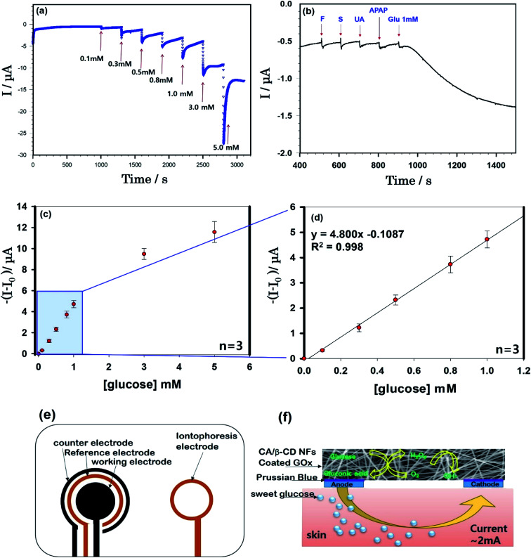
In this study, we aimed to develop a cellulose/β-cyclodextrin (β-CD) electrospun immobilized GOx enzyme patch with reverse iontophoresis for noninvasive monitoring of interstitial fluid (ISF) glucose levels (0.1-0.6 mM dm-3). In vitro analysis, performed using a sensor attached to flexible substrates, revealed that the high diffusion coefficient (9.0 × 10⁻⁵ cm2 s-1), the linear correlation coefficient (R 2 = 0.998), the detection limit (9.35 × 10⁻⁵ M), and the linear range sensitivity (0-1 mM) of the sensor (5.08 μA mM-1) remained unaffected by the presence of interfering substances (e.g., fructose, sucrose, uric acid, and acetaminophen) at physiological levels. The present results indicate that the new epidermal sensing strategy using nanofibers for continuous glucose monitoring has potential to be applied in diagnosis of diabetes.
This journal is © The Royal Society of Chemistry.
Conflict of interest statement
There are no conflicts to declare.
Figures
Similar articles
-
Novel glucose-responsive of the transparent nanofiber hydrogel patches as a wearable biosensor via electrospinning.Sci Rep. 2020 Nov 2;10(1):18858. doi: 10.1038/s41598-020-75906-9. Sci Rep. 2020. PMID: 33139822 Free PMC article.
-
A touch-actuated glucose sensor fully integrated with microneedle array and reverse iontophoresis for diabetes monitoring.Biosens Bioelectron. 2022 May 1;203:114026. doi: 10.1016/j.bios.2022.114026. Epub 2022 Jan 24. Biosens Bioelectron. 2022. PMID: 35114468
-
Fabrication of Amperometric Glucose Sensor Using Glucose Oxidase-Cellulose Nanofiber Aqueous Solution.Anal Sci. 2015;31(11):1111-4. doi: 10.2116/analsci.31.1111. Anal Sci. 2015. PMID: 26561252
-
Reverse iontophoresis with the development of flexible electronics: A review.Biosens Bioelectron. 2023 Mar 1;223:115036. doi: 10.1016/j.bios.2022.115036. Epub 2022 Dec 23. Biosens Bioelectron. 2023. PMID: 36580817 Review.
-
Flexible Electronics toward Wearable Sensing.Acc Chem Res. 2019 Mar 19;52(3):523-533. doi: 10.1021/acs.accounts.8b00500. Epub 2019 Feb 15. Acc Chem Res. 2019. PMID: 30767497 Review.
Cited by
-
Electrochemical Nanosensors for Sensitization of Sweat Metabolites: From Concept Mapping to Personalized Health Monitoring.Molecules. 2023 Jan 27;28(3):1259. doi: 10.3390/molecules28031259. Molecules. 2023. PMID: 36770925 Free PMC article. Review.
-
Optimization of Nanofiber Wearable Heart Rate Sensor Module for Human Motion Detection.Comput Math Methods Med. 2022 Jun 16;2022:1747822. doi: 10.1155/2022/1747822. eCollection 2022. Comput Math Methods Med. 2022. PMID: 35756404 Free PMC article.
-
A flexible glucose biosensor modified by reduced-swelling and conductive zwitterionic hydrogel enzyme membrane.Anal Bioanal Chem. 2024 Sep;416(22):4849-4860. doi: 10.1007/s00216-024-05429-z. Epub 2024 Jul 15. Anal Bioanal Chem. 2024. PMID: 39008068
-
Cyclodextrin Host-Guest Recognition in Glucose-Monitoring Sensors.ACS Omega. 2023 Sep 8;8(37):33202-33228. doi: 10.1021/acsomega.3c03746. eCollection 2023 Sep 19. ACS Omega. 2023. PMID: 37744789 Free PMC article. Review.
-
Production and Mechanical Characterisation of TEMPO-Oxidised Cellulose Nanofibrils/β-Cyclodextrin Films and Cryogels.Molecules. 2020 May 20;25(10):2381. doi: 10.3390/molecules25102381. Molecules. 2020. PMID: 32443918 Free PMC article.
References
LinkOut - more resources
Full Text Sources