. 2020 Jun 15;10(38):22622-22630.
doi: 10.1039/c9ra09058d. eCollection 2020 Jun 10.

Flaxseed orbitides, linusorbs, inhibit LPS-induced THP-1 macrophage inflammation

Affiliations

Flaxseed orbitides, linusorbs, inhibit LPS-induced THP-1 macrophage inflammation

Xian-Guo Zou et al. RSC Adv. .

Abstract

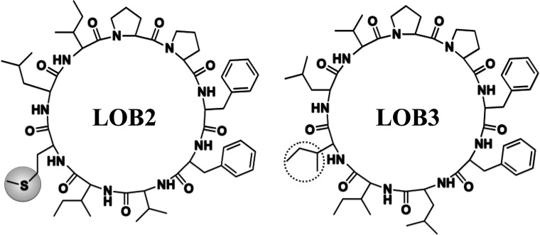
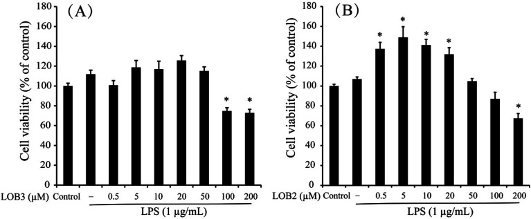
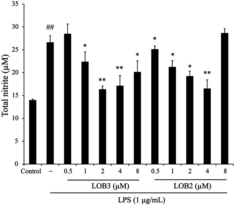
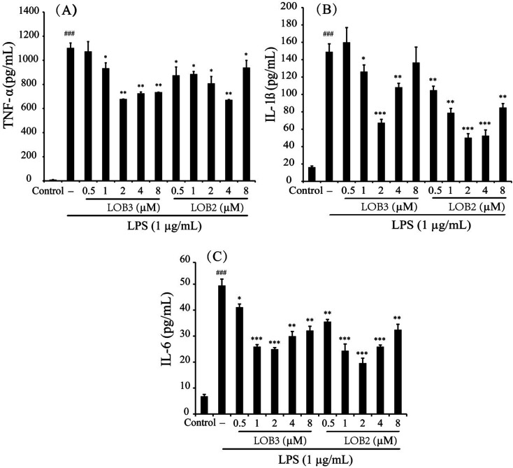
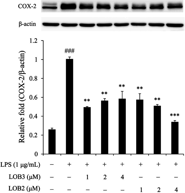
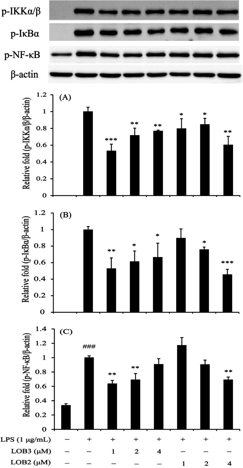
Linusorbs (flaxseed orbitides) are a family of naturally-occurring cyclic peptides. Previously, we reported that their anticancer effects were associated with their structures. In this study, we investigated the anti-inflammatory activities of 2 different linusorbs ([1-9-NαC]-linusorb B2 and [1-9-NαC]-linusorb B3) in lipopolysaccharide (LPS)-induced THP-1 macrophage activation as well as the underlying mechanism of this inflammatory response. Both molecules suppressed pro-inflammatory mediators (TNF-α, IL-1β, IL-6, NO and COX-2) and were involved in downregulating the NF-κB signaling pathway. The suppressive effects on pro-inflammatory mediators were comparable and the concentration range of action was similar (1-4 μM). However, the concentration of compound that induced downregulation of the NF-κB pathway was different for each compound. While [1-9-NαC]-linusorb B3 could inhibit the activation of the NF-κB pathway at concentrations of 1 and 2 μM, [1-9-NαC]-linusorb B2 induced a comparable inhibitory effect at a concentration of 4 μM.


Conflict of interest statement

There are no conflicts of interest to declare. Dr Martin J. T. Reaney is the founder of, and has an equity interest in, PTD (Saskatoon, SK, Canada: previous company name is Prairie Tide Chemicals Inc.). Dr Youn Young Shim is a Market Consultant for PTD. The terms of this arrangement have been reviewed and approved by the University of Saskatchewan (Saskatoon, SK, Canada) in accordance with its conflict of interest policies.

Figures

Fig. 1
Fig. 1. Chemical structures of LOB2 and LOB3.
Fig. 2
Fig. 2. Effects of LOB3 (A) and LOB2 (B) on the viability of THP-1 macrophages determined by MTT assay. THP-1 macrophages were pretreated with various concentrations (0, 0.5, 5, 10, 20, 50, 100 and 200 μM) of LOB3 or LOB2 for 1 h and then the cells were stimulated with LPS (1 μg mL−1) for 3 h. Significant differences compared with control group are indicated as *p < 0.05.
Fig. 3
Fig. 3. LOB3 and LOB2 prevented the NO production of LPS-induced THP-1 macrophage inflammation model. THP-1 macrophages were pretreated with various concentrations (0, 0.5, 1, 2, 4 and 8 μM) of LOB3 or LOB2 for 1 h and then the cells were stimulated with LPS (1 μg mL−1) for 3 h. After treatment, NO production was detected by the Griess reagent. Significant differences among LPS treatments are indicated as *p < 0.05, **p < 0.01. ##p < 0.01 versus the control group.
Fig. 4
Fig. 4. LOB3 and LOB2 inhibited the LPS-stimulated release of TNF-α (A), IL-1β (B) and IL-6 (C) in THP-1 macrophages. THP-1 macrophages were pretreated with different concentrations (0, 0.5, 1, 2, 4 and 8 μM) of LOB3 or LOB2 for 1 h and then the cells were stimulated with LPS (1 μg mL−1) for 3 h. After treatment, the production of cytokines was evaluated by corresponding human ELISA kits. Significant differences among LPS treatments are indicated as *p < 0.05, **p < 0.01, ***p < 0.001, ###p < 0.001 versus the control group.
Fig. 5
Fig. 5. LOB3 and LOB2 suppressed the protein expression of COX-2 in the LPS-stimulated THP-1 cells analyzed by western blot analysis. THP-1 macrophages were pretreated with different concentrations (0, 1, 2 and 4 μM) of LOB3 or LOB2 for 1 h and then the cells were stimulated with LPS (1 μg mL−1) for 3 h. COX-2 protein level was corrected to β-actin and was expressed relative to the LPS group (set as 1). ###p < 0.001 versus the control group. Significant differences compared with LPS group are indicated as **p < 0.01, ***p < 0.001.
Fig. 6
Fig. 6. LOB3 and LOB2 inhibited the phosphorylation of IKKα/β (A), IκBα (B) and p65-NF-κB (C) in the LPS-stimulated THP-1 cells evaluated by western blot analysis. THP-1 macrophages were pretreated with different concentrations (0, 1, 2 and 4 μM) of LOB3 or LOB2 for 1 h and then the cells were stimulated with LPS (1 μg mL−1) for 3 h. Protein levels of p-IKKα/β, p-IκBα and p-p65-NF-κB were corrected to β-actin and expressed relative to the LPS group (set as 1). ###p < 0.001 versus the control group. Significant differences compared with LPS group are indicated as *p < 0.05, **p < 0.01, ***p < 0.001.

Similar articles

Cited by

References

    1. Hansson G. K. N. Engl. J. Med. 2005;352:1685–1695. doi: 10.1056/NEJMra043430. - DOI - PubMed
    1. Pickup J. C. Chusney G. D. Thomas S. M. Burt D. Life Sci. 2000;67:291–300. doi: 10.1016/S0024-3205(00)00622-6. - DOI - PubMed
    1. Gregor M. F. Hotamisligil G. S. Annu. Rev. Immunol. 2011;29:415–445. doi: 10.1146/annurev-immunol-031210-101322. - DOI - PubMed
    1. Sartor R. B. Nat. Rev. Gastroenterol. Hepatol. 2006;3:390–407. doi: 10.1038/ncpgasthep0528. - DOI - PubMed
    1. Rakoff-Nahoum S. Yale J. Biol. Med. 2006;79:123–130. - PMC - PubMed