

miR-124 and miR-142 enhance cisplatin sensitivity of non-small cell lung cancer cells through repressing autophagy via directly targeting SIRT1
- PMID: 35514612
- PMCID: PMC9060797
- DOI: 10.1039/c8ra09914f
miR-124 and miR-142 enhance cisplatin sensitivity of non-small cell lung cancer cells through repressing autophagy via directly targeting SIRT1
Abstract
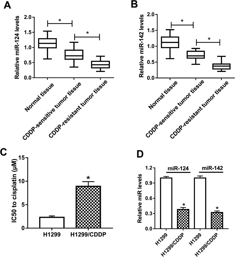
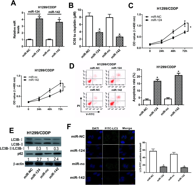
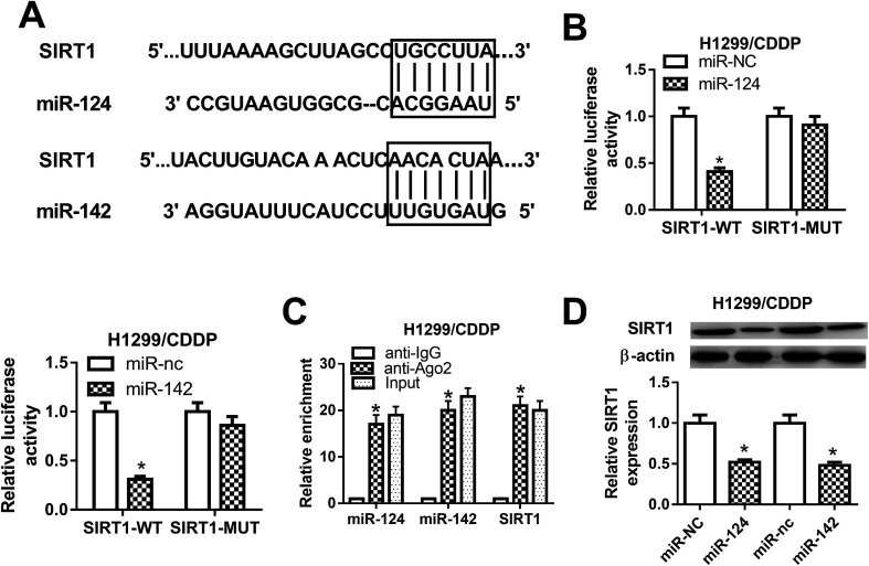
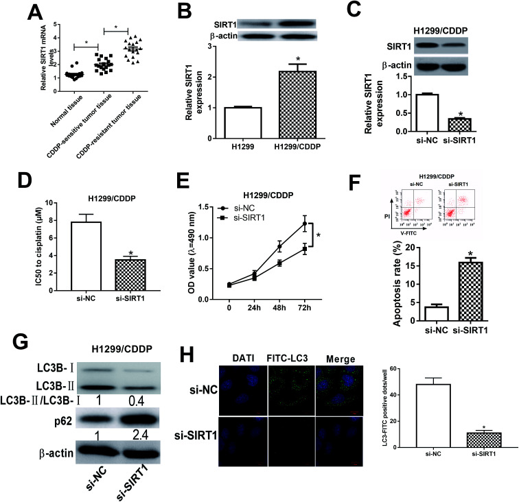
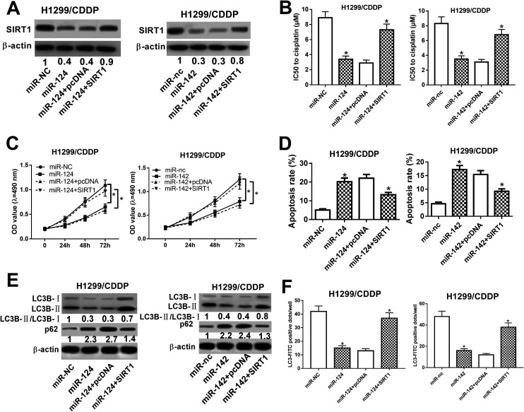
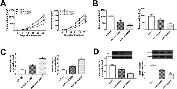
Background: Drug resistance is a major obstacle in the treatment of non-small cell lung cancer (NSCLC). Recently, miRNAs are reported to be involved in the drug resistance of NSCLC. The roles of miR-124 and miR-142 in the multidrug resistance of NSCLC cells have been reported. However, the underlying mechanism by which miR-124 and miR-142 regulate resistance to cisplatin (CDDP) remains unknown. Methods: The expressions of miR-124, miR-142 and sirtuin 1 (SIRT1) in CDDP-sensitive and CDDP-resistant NSCLC tissues and cells were detected by qRT-PCR and western blot. IC50 value and cell proliferation were determined by MTT assay. Apoptosis was assessed by flow cytometry analysis. Autophagy was evaluated by western blot analysis of the protein levels of LC3-I, LC3-II and p62, and FITC-LC3 punctate formation assay. The interaction between miR-124 or miR-142 and SIRT1 was determined by luciferase reporter, RNA immunoprecipitation (RIP) and western blot assays. A tumor xenograft was performed to further validate the role of miR-124 and miR-142 in the sensitivity of CDDP-resistant NSCLC to cisplatin. Results: miR-124 and miR-142 were downregulated, while SIRT1 was upregulated in CDDP-resistant NSCLC tissues and cells compared to CDDP-sensitive groups. Functionally, overexpression of miR-124 and miR-142 or SIRT1 silencing enhanced the CDDP sensitivity of H1299/CDDP cells via suppressing autophagy, as evidenced by the reduced LC3-II/LC3-I radio, elevated p62 protein, and suppressed FITC-LC3 punctate formation in H1299/CDDP cells. miR-124 and miR-142 were demonstrated to co-target SIRT1. Re-expression of SIRT1 overturned miR-124 and miR-142-mediated chemosensitivity in H1299/CDDP cells via triggering autophagy. Conclusion: miR-124 and miR-142 enhance the cytotoxic effect of CDDP through repressing autophagy via targeting SIRT1 in CDDP-resistant NSCLC cells.
This journal is © The Royal Society of Chemistry.
Conflict of interest statement
The authors declare none conflict of interest.
Figures
Similar articles
-
MicroRNAs as the critical regulators of autophagy-mediated cisplatin response in tumor cells.Cancer Cell Int. 2023 Apr 25;23(1):80. doi: 10.1186/s12935-023-02925-7. Cancer Cell Int. 2023. PMID: 37098542 Free PMC article. Review.
-
MicroRNA-1 overexpression increases chemosensitivity of non-small cell lung cancer cells by inhibiting autophagy related 3-mediated autophagy.Cell Biol Int. 2018 Sep;42(9):1240-1249. doi: 10.1002/cbin.10995. Epub 2018 Jun 15. Cell Biol Int. 2018. PMID: 29851226
-
CircPTK2 inhibits cell cisplatin (CDDP) resistance by targeting miR-942/TRIM16 axis in non-small cell lung cancer (NSCLC).Bioengineered. 2022 Feb;13(2):3651-3664. doi: 10.1080/21655979.2021.2024321. Bioengineered. 2022. PMID: 35230201 Free PMC article.
-
MicroRNA-217 inhibits the proliferation and invasion, and promotes apoptosis of non-small cell lung cancer cells by targeting sirtuin 1.Oncol Lett. 2021 May;21(5):386. doi: 10.3892/ol.2021.12647. Epub 2021 Mar 17. Oncol Lett. 2021. PMID: 33777209 Free PMC article.
-
miR-30 decreases multidrug resistance in human gastric cancer cells by modulating cell autophagy.Exp Ther Med. 2018 Jan;15(1):599-605. doi: 10.3892/etm.2017.5354. Epub 2017 Oct 23. Exp Ther Med. 2018. PMID: 29375703 Free PMC article.
Cited by
-
Emerging role of sirtuins in non‑small cell lung cancer (Review).Oncol Rep. 2024 Oct;52(4):127. doi: 10.3892/or.2024.8786. Epub 2024 Aug 2. Oncol Rep. 2024. PMID: 39092574 Free PMC article. Review.
-
MicroRNAs as the critical regulators of autophagy-mediated cisplatin response in tumor cells.Cancer Cell Int. 2023 Apr 25;23(1):80. doi: 10.1186/s12935-023-02925-7. Cancer Cell Int. 2023. PMID: 37098542 Free PMC article. Review.
-
The sirtuin family in health and disease.Signal Transduct Target Ther. 2022 Dec 29;7(1):402. doi: 10.1038/s41392-022-01257-8. Signal Transduct Target Ther. 2022. PMID: 36581622 Free PMC article. Review.
-
Molecular targets and therapies associated with poor prognosis of triple‑negative breast cancer (Review).Int J Oncol. 2025 Jun;66(6):52. doi: 10.3892/ijo.2025.5758. Epub 2025 May 30. Int J Oncol. 2025. PMID: 40444482 Free PMC article. Review.
-
MiRNA-Based Therapies for Lung Cancer: Opportunities and Challenges?Biomolecules. 2023 May 23;13(6):877. doi: 10.3390/biom13060877. Biomolecules. 2023. PMID: 37371458 Free PMC article. Review.
References
-
- Raez L. E. Lilenbaum R. Clin. Adv. Hematol. Oncol. 2004;2:173–178. - PubMed
LinkOut - more resources
Full Text Sources
Miscellaneous