

Radio-Histological Correlation of Lung Features in Severe COVID-19 Through CT-Scan and Lung Ultrasound Evaluation
- PMID: 35514757
- PMCID: PMC9063463
- DOI: 10.3389/fmed.2022.820661
Radio-Histological Correlation of Lung Features in Severe COVID-19 Through CT-Scan and Lung Ultrasound Evaluation
Abstract
Background: Patients with coronavirus disease 2019 (COVID-19) can develop severe bilateral pneumonia leading to respiratory failure. Lung histological samples were scarce due to the high risk of contamination during autopsies. We aimed to correlate histological COVID-19 features with radiological findings through lung ultrasound (LU)-guided postmortem core needle biopsies (CNBs) and computerized tomography (CT) scans.
Methodology: We performed an observational prospective study, including 30 consecutive patients with severe COVID-19. The thorax was divided into 12 explorations regions to correlate LU and CT-scan features. Histological findings were also related to radiological features through CNBs.
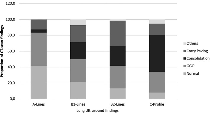
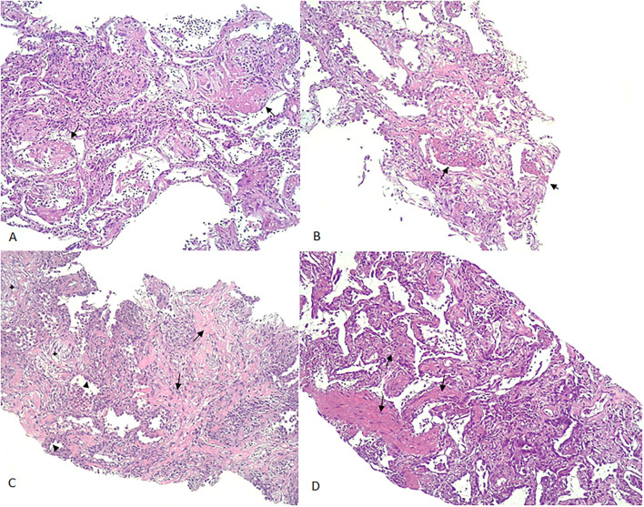
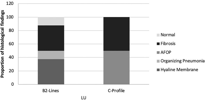
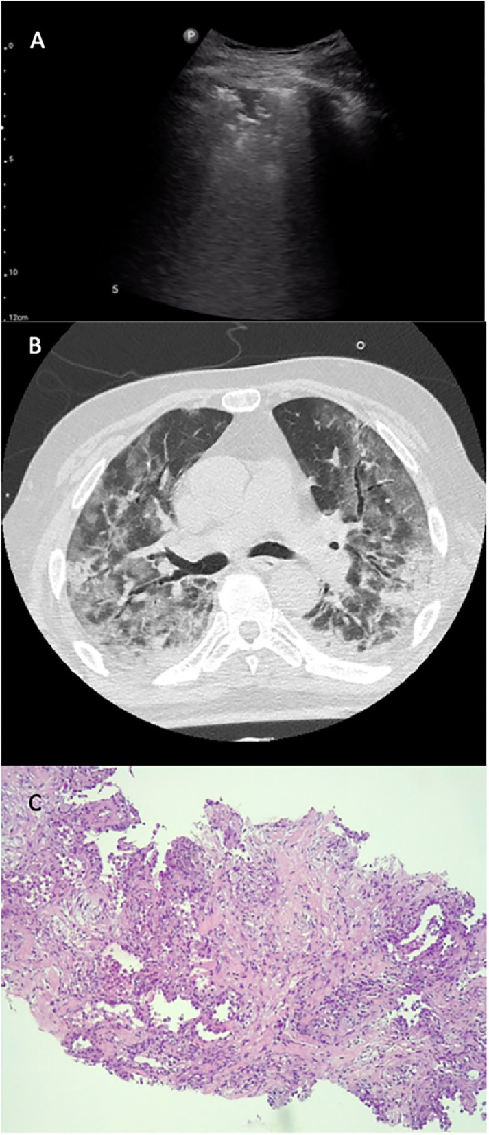
Results: Mean age was 62.56 ± 13.27 years old, with 96.7% male patients. Postmortem LU-guided CNBs were performed in 13 patients. Thirty patients were evaluated with both thoracic LU and chest CT scan, representing a total of 279 thoracic regions explored. The most frequent LU finding was B2-lines (49.1%). The most CT-scan finding was ground-glass opacity (GGO, 29%). Pathological CT-scan findings were commonly observed when B2-lines or C-lines were identified through LU (positive predictive value, PPV, 87.1%). Twenty-five postmortem echo-guided histological samples were obtained from 12 patients. Histological samples showed diffuse alveolar damage (DAD) (75%) and chronic interstitial inflammation (25%). The observed DAD was heterogeneous, showing multiple evolving patterns of damage, including exudative (33.3%), fibrotic (33.3%), and organizing (8.3%) phases. In those patients with acute or exudative pattern, two lesions were distinguished: classic hyaline membrane; fibrin "plug" in alveolar space (acute fibrinous organizing pneumonia, AFOP). C-profile was described in 33.3% and presented histological signs of DAD and lung fibrosis. The predominant findings were collagen deposition (50%) and AFOP (50%). B2-lines were identified in 66.7%; the presence of hyaline membrane was the predominant finding (37.5%), then organizing pneumonia (12.5%) and fibrosis (37.5%). No A-lines or B1-lines were observed in these patients.
Conclusion: LU B2-lines and C-profile are predominantly identified in patients with severe COVID-19 with respiratory worsening, which correspond to different CT patterns and histological findings of DAD and lung fibrosis.
Keywords: COVID-19; biopsy; lung ultrasonography (LU); pathology; radiology.
Copyright © 2022 Trias-Sabrià, Dorca Duch, Molina-Molina, Aso, Díez-Ferrer, Marín Muñiz, Bordas-Martínez, Sabater, Luburich, del Rio, Solanich, Dorca, Santos and Suárez-Cuartin.
Conflict of interest statement
The authors declare that the research was conducted in the absence of any commercial or financial relationships that could be construed as a potential conflict of interest.
Figures
Similar articles
-
Acute fibrinous and organizing pneumonia: a histological pattern of lung injury and possible variant of diffuse alveolar damage.Arch Pathol Lab Med. 2002 Sep;126(9):1064-70. doi: 10.5858/2002-126-1064-AFAOP. Arch Pathol Lab Med. 2002. PMID: 12204055
-
Postmortem chest computed tomography in COVID-19: A minimally invasive autopsy method.Eur J Radiol Open. 2024 Jan 13;12:100546. doi: 10.1016/j.ejro.2024.100546. eCollection 2024 Jun. Eur J Radiol Open. 2024. PMID: 38293283 Free PMC article.
-
COVID-19: Histopathological correlates of imaging patterns on chest computed tomography.Respirology. 2021 Sep;26(9):869-877. doi: 10.1111/resp.14101. Epub 2021 Jun 22. Respirology. 2021. PMID: 34159661 Free PMC article.
-
Acute fibrinous and organizing pfneumonia: two case reports and literature review.Diagn Pathol. 2021 Oct 10;16(1):90. doi: 10.1186/s13000-021-01155-7. Diagn Pathol. 2021. PMID: 34629105 Free PMC article. Review.
-
Similarities and Differences of Early Pulmonary CT Features of Pneumonia Caused by SARS-CoV-2, SARS-CoV and MERS-CoV: Comparison Based on a Systemic Review.Chin Med Sci J. 2020 Sep 30;35(3):254-261. doi: 10.24920/003727. Chin Med Sci J. 2020. PMID: 32972503 Free PMC article.
Cited by
-
Identification of Histopathological Biomarkers in Fatal Cases of Coronavirus Disease: A Study on Lung Tissue.Diagnostics (Basel). 2023 Jun 12;13(12):2039. doi: 10.3390/diagnostics13122039. Diagnostics (Basel). 2023. PMID: 37370934 Free PMC article.
-
The Role of POCUS to Face COVID-19: A Narrative Review.J Clin Med. 2024 May 7;13(10):2756. doi: 10.3390/jcm13102756. J Clin Med. 2024. PMID: 38792298 Free PMC article. Review.
-
Association between glucocorticoid administration and outcomes in patients with ARDS based on the MIMIC-III database.Medicine (Baltimore). 2024 Aug 9;103(32):e39239. doi: 10.1097/MD.0000000000039239. Medicine (Baltimore). 2024. PMID: 39121259 Free PMC article.
References
-
- WHO Coronavirus Disease (COVID-19) Dashboard. Available online at: https://covid19.who.int/ (accessed May 11, 2020).
LinkOut - more resources
Full Text Sources
Miscellaneous