

Critical role for the lung endothelial nonmuscle myosin light-chain kinase isoform in the severity of inflammatory murine lung injury
- PMID: 35514774
- PMCID: PMC9063969
- DOI: 10.1002/pul2.12061
Critical role for the lung endothelial nonmuscle myosin light-chain kinase isoform in the severity of inflammatory murine lung injury
Abstract
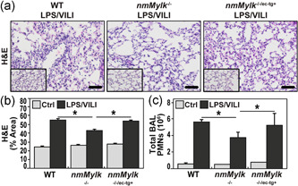
Global knockout of the nonmuscle isoform of myosin light-chain kinase (nmMLCK), a primary cellular regulator of cytoskeletal machinery, is strongly protective in preclinical murine models of inflammatory lung injury. The current study was designed to assess the specific contribution of endothelial cell (EC) nmMLCK to the severity of murine inflammatory lung injury produced by lipopolysaccharide (LPS) and mechanical ventilation ventilator-induced lung injury or ventilation (VILI). Responses to combined LPS/VILI exposure were assessed in: (i) wild-type (WT) C57BL/6J mice; (ii) transgenic mice with global deletion of nmMLCK (nmMylk -/-); (iii) transgenic nmMylk -/- mice with overexpression of nmMLCK restricted to the endothelium (nmMylk -/-/ec-tg+). Lung inflammation indices included lung histology, bronchoalveolar lavage (BAL) polymorphonuclear leukocytes (PMNs), lung protein biochemistry, tissue albumin levels, Evans blue dye (EBD) lung extravasation, and plasma cytokines (interleukin-6 [IL-6], keratinocyte chemoattractant [KC]/IL-8, IL-1bβ, extracellular nicotinamide phosphoribosyltransferase, tumor necrosis factor-α). Compared to WT C57BL/6J mice, the severity of LPS/VILI-induced lung injury was markedly reduced in mice with global nmMLCK deletion reflected by reductions in histologic inflammatory lung injury, BAL PMN counts, mitogen-activated protein kinase, and NF-kB pathway activation in lung homogenates, plasma cytokine levels, and parameters of lung permeability (increased BAL protein, tissue albumin levels, EBD lung extravasation). In contrast, mice with restricted overexpression of nmMLCK in EC (nmMylk -/-/ec-tg+) showed significant persistence of LPS/VILI-induced lung injury severity compared to WT mice. In conclusion, these studies strongly endorse the role of EC nmMLCK in driving the severity of preclinical inflammatory lung injury. Precise targeting of EC nmMLCK may represent an attractive therapeutic strategy to reduce lung inflammation and both lung and systemic vascular permeability.
Keywords: ARDS; MLCK.
© 2022 The Authors. Pulmonary Circulation published by Wiley Periodicals LLC on behalf of the Pulmonary Vascular Research Institute.
Conflict of interest statement
Joe G. N. Garcia is CEO and founder of Aqualung Therapeutics Corporation. The remaining authors declare no conflicts of interest.
Figures


Similar articles
-
Genetic and epigenetic regulation of the non-muscle myosin light chain kinase isoform by lung inflammatory factors and mechanical stress.Clin Sci (Lond). 2021 Apr 16;135(7):963-977. doi: 10.1042/CS20201448. Clin Sci (Lond). 2021. PMID: 33792658 Free PMC article.
-
Non-muscle myosin light chain kinase isoform is a viable molecular target in acute inflammatory lung injury.Am J Respir Cell Mol Biol. 2011 Jan;44(1):40-52. doi: 10.1165/rcmb.2009-0197OC. Epub 2010 Feb 5. Am J Respir Cell Mol Biol. 2011. PMID: 20139351 Free PMC article.
-
Nonmuscle myosin light chain kinase regulates murine asthmatic inflammation.Am J Respir Cell Mol Biol. 2014 Jun;50(6):1129-35. doi: 10.1165/rcmb.2013-0434OC. Am J Respir Cell Mol Biol. 2014. PMID: 24428690 Free PMC article.
-
Sp1-mediated nonmuscle myosin light chain kinase expression and enhanced activity in vascular endothelial growth factor-induced vascular permeability.Pulm Circ. 2015 Dec;5(4):707-15. doi: 10.1086/684124. Pulm Circ. 2015. PMID: 26697178 Free PMC article.
-
Interaction in endothelium of non-muscular myosin light-chain kinase and the NF-κB pathway is critical to lipopolysaccharide-induced vascular hyporeactivity.Clin Sci (Lond). 2015 Oct 1;129(8):687-98. doi: 10.1042/CS20140625. Epub 2015 Jun 15. Clin Sci (Lond). 2015. PMID: 26201020
Cited by
-
Edema and lymphatic clearance: molecular mechanisms and ongoing challenges.Clin Sci (Lond). 2023 Sep 27;137(18):1451-1476. doi: 10.1042/CS20220314. Clin Sci (Lond). 2023. PMID: 37732545 Free PMC article. Review.
-
Dexamethasone restores TNFα-induced epithelial barrier dysfunction in primary rat alveolar epithelial cells.PLoS One. 2023 Dec 27;18(12):e0295684. doi: 10.1371/journal.pone.0295684. eCollection 2023. PLoS One. 2023. PMID: 38150443 Free PMC article.
-
Unraveling the deadly dance: endothelial cells and neutrophils in sepsis-induced acute lung injury/acute respiratory distress syndrome.Front Cell Dev Biol. 2025 May 22;13:1551138. doi: 10.3389/fcell.2025.1551138. eCollection 2025. Front Cell Dev Biol. 2025. PMID: 40476000 Free PMC article. Review.
-
A Razor's Edge: Vascular Responses to Acute Inflammatory Lung Injury/Acute Respiratory Distress Syndrome.Annu Rev Physiol. 2024 Feb 12;86:505-529. doi: 10.1146/annurev-physiol-042222-030731. Annu Rev Physiol. 2024. PMID: 38345908 Free PMC article. Review.
-
TLR4 activation induces inflammatory vascular permeability via Dock1 targeting and NOX4 upregulation.Biochim Biophys Acta Mol Basis Dis. 2022 Dec 1;1868(12):166562. doi: 10.1016/j.bbadis.2022.166562. Epub 2022 Sep 27. Biochim Biophys Acta Mol Basis Dis. 2022. PMID: 36179995 Free PMC article.
References
-
- Garcia JG, Lazar V, Gilbert‐McClain LI, Gallagher PJ, Verin AD. Myosin light chain kinase in endothelium: molecular cloning and regulation. Am J Respir Cell Mol Biol. 1997;16:489–94. - PubMed
Grants and funding
LinkOut - more resources
Full Text Sources
Miscellaneous
