Case Reports
. 2022 Apr 23:26:101556.
doi: 10.1016/j.ajoc.2022.101556. eCollection 2022 Jun.

Multilayered macular hemorrhages as an unusual complication of transorbital neuroendoscopic surgery

Affiliations
Case Reports

Multilayered macular hemorrhages as an unusual complication of transorbital neuroendoscopic surgery

William Foulsham et al. Am J Ophthalmol Case Rep. .

Abstract

Purpose: To report a case of multilayered intraocular hemorrhage at the posterior pole as a complication of transorbital neuroendoscopic surgery.

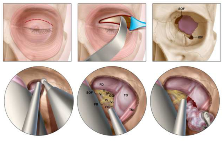
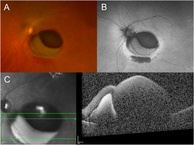
Observations: Our patient underwent an uncomplicated endoscopic transorbital resection of a left sphenoid wing meningioma. In the immediate post-operative period, the patient reported blurred vision of her left eye, and dilated fundus examination demonstrated multilayered hemorrhages at the posterior pole. No intracranial hemorrhage was identified on post-operative imaging. Due to persistent subnormal visual acuity and non-clearing hemorrhage over several weeks of follow-up, a pars plana vitrectomy with peeling of the internal limiting membrane was performed to clear the hemorrhagic component obscuring the macula.

Conclusions and importance: We report the first case of multilayered intraocular hemorrhages at the posterior pole, mimicking Terson syndrome, in the absence of intracranial hemorrhage or elevated intracranial pressure as a complication of transorbital surgery.

Keywords: Meningioma; Neuroendoscopic; Spheno-orbital; Sphenoid wing; Surgery; Terson syndrome; Transorbital.


Conflict of interest statement

The following authors have no financial disclosures: WF, VSN, BWB, MJD, FH, KJG, DJD, THS, AO.

Figures

Fig. 1
Fig. 1
Pre-operative magnetic resonance imaging T1 post-contrast MRI (left) and T2 FLAIR (middle) showing dural-based mass and surrounding edema (arrows). Preoperative non-contrast computerized tomography scan showing left sphenoid bone hyperostosis underlying the tumor (arrows).
Fig. 2
Fig. 2
Illustration demonstrating the endoscopic transorbital eyelid approach for the resection of spheno-orbital meningiomas The upper eyelid is incised, and the orbital contents are retracted medially (upper left and central panels). The sphenoid bone between the superior orbital fissure and inferior orbital fissures is exposed (upper right). Decompression of the hyperostotic bone is performed (bottom left). Key landmarks are identified including the superior orbital fissure, the foramen rotundum, the foramen ovale, the geniculate ganglion as well as the dura overlying the frontal and temporal lobes (bottom center). The dura is incised, and the tumor decompressed (bottom right). FD = frontal dura; TD = temporal dura; SOF = superior orbital fissure; FR = foramen rotundum; FO = foramen ovale; III = CN III (oculomotor nerve); IV = CN IV (trochlear nerve); GG = gasserian ganglion; V1 = ophthalmic branch of CN V (trigeminal nerve); V2 = maxillary branch of CN V; V3 = mandibular branch of CN V; TM = temporalis muscle.
Fig. 3
Fig. 3
Post-operative magnetic resonance imaging 3-month post-operative T1 post-contrast MRI (left) and T2 FLAIR (middle) showing resection of meningioma and resolution of surrounding edema. Postoperative day 1 non-contrast computerized tomography scan showing resection of hyperostotic component of sphenoid bone (right).
Fig. 4
Fig. 4
Multimodal imaging performed 10 days after neuroendoscopic surgery Multilayered hemorrhages of the posterior pole are shown on Optos imaging and autofluorescence (A and B, respectively), as well as spectral-domain optical coherence tomography (C).
Fig. 5
Fig. 5
Multimodal imaging performed 1 week following pars plana vitrectomy with peeling of the internal limiting membrane The component of the hemorrhage obscuring the macula is cleared after peeling the internal limiting membrane. Optos imaging and autofluorescence are shown (A and B, respectively), in addition to spectral-domain optical coherence tomography (C).

References

    1. Fountas K.N., Kapsalaki E.Z., Lee G.P., et al. Terson hemorrhage in patients suffering aneurysmal subarachnoid hemorrhage: predisposing factors and prognostic significance. J Neurosurg. 2008;109(3):439–444. - PubMed
    1. Naseri A., Blumenkranz M.S., Horton J.C. Terson's syndrome following epidural saline injection. Neurology. 2001;57(2):364. - PubMed
    1. Clark C.J., Whitwell J. Intraocular haemorrhage after epidural injection. Br Med J. 1961;2(5267):1612. - PMC - PubMed
    1. Pagani-Estévez G.L., Chen J.J., Watson J.C., Leavitt J.A. Acute vision loss secondary to epidural blood patch: Terson syndrome. Reg Anesth Pain Med. 2016;41(2):164–168. - PubMed
    1. Matsuoka T., Matsuda S., Harino S., et al. Subarachnoid hemorrhage-negative Terson syndrome after intracranial artery treatment with a flow diverter device. Am J Ophthalmol Case Reports. 2020;20 - PMC - PubMed

Publication types

LinkOut - more resources