. 2022 Apr 19:13:882721.
doi: 10.3389/fimmu.2022.882721. eCollection 2022.

Ablation of BATF Alleviates Transplant Rejection via Abrogating the Effector Differentiation and Memory Responses of CD8+ T Cells

Affiliations

Ablation of BATF Alleviates Transplant Rejection via Abrogating the Effector Differentiation and Memory Responses of CD8+ T Cells

Shuang Li et al. Front Immunol. .

Abstract

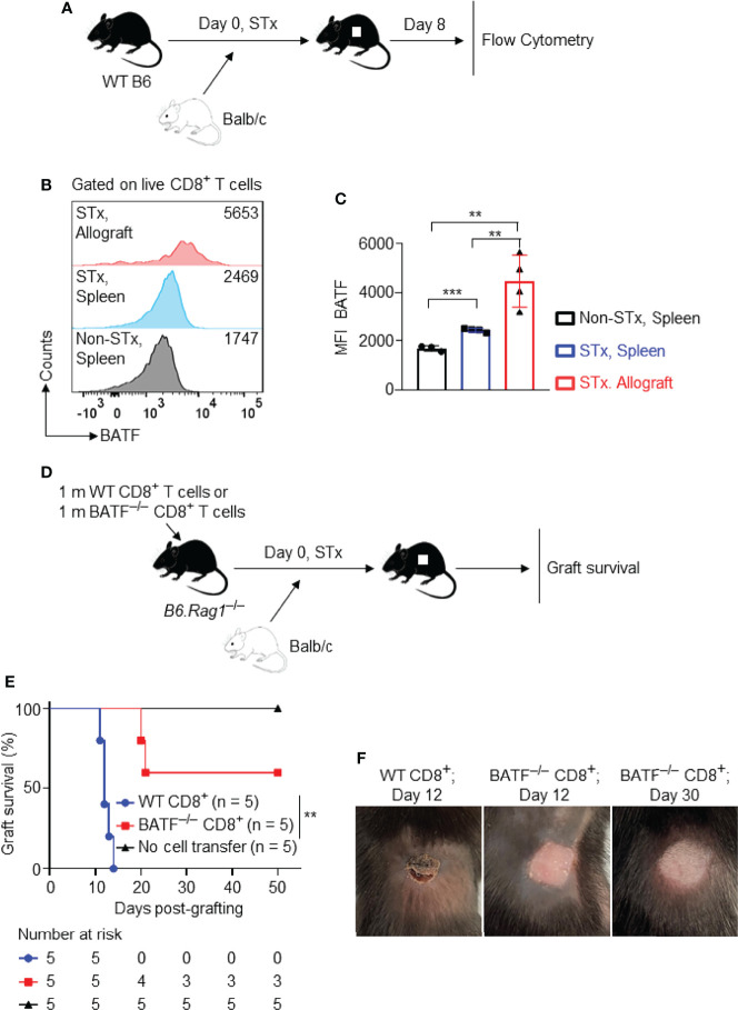
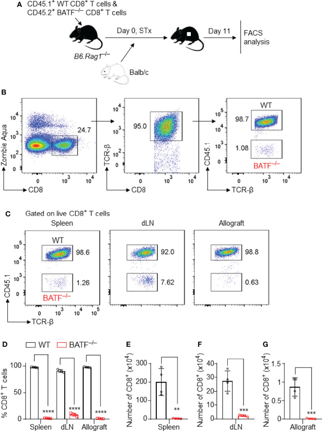
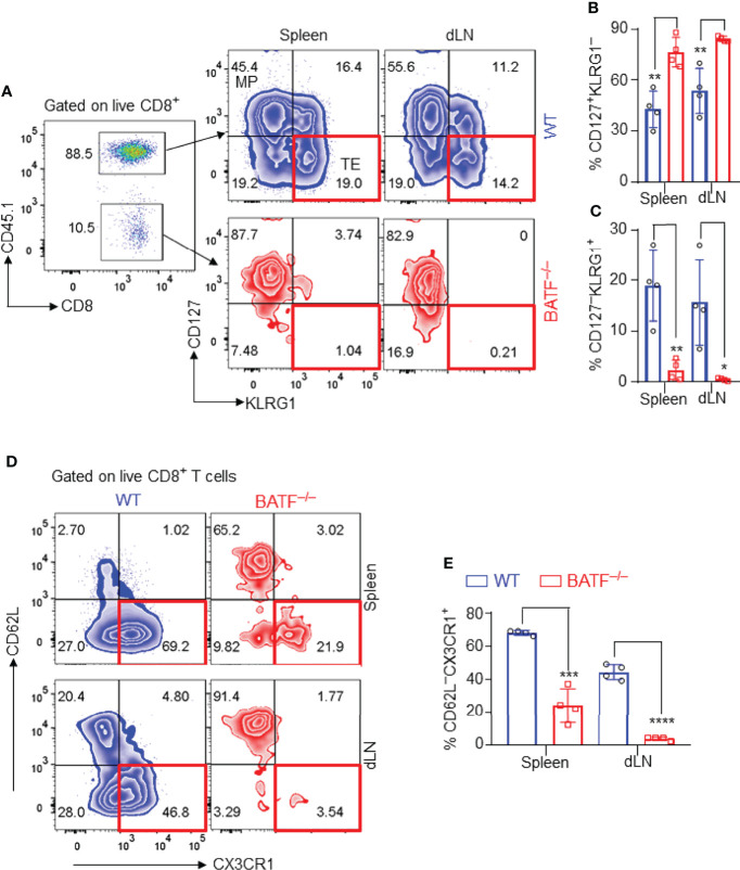
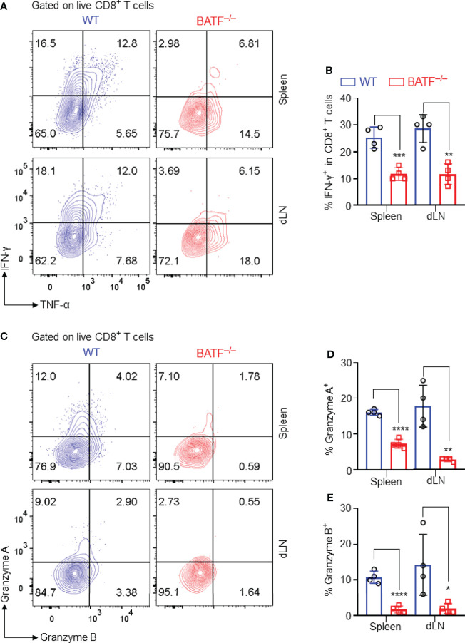
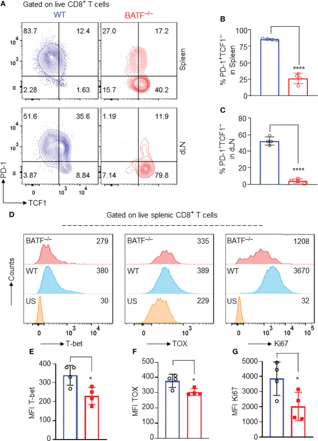
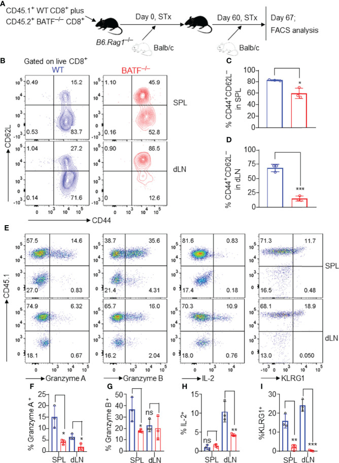
Allogeneic CD8+ T cells are prominently involved in allograft rejection, but how their effector differentiation and function are regulated at a transcriptional level is not fully understood. Herein, we identified the basic leucine zipper ATF-like transcription factor (BATF) as a key transcription factor that drives the effector program of allogeneic CD8+ T cells. We found that BATF is highly expressed in graft-infiltrating CD8+ T cells, and its ablation in CD8+ T cells significantly prolonged skin allograft survival in a fully MHC-mismatched transplantation model. To investigate how BATF dictates allogeneic CD8+ T cell response, BATF-/- and wild-type (WT) CD8+ T cells were mixed in a 1:1 ratio and adoptively transferred into B6.Rag1-/- mice 1 day prior to skin transplantation. Compared with WT CD8+ T cells at the peak of rejection response, BATF-/- CD8+ T cells displayed a dysfunctional phenotype, evident by their failure to differentiate into CD127-KLRG1+ terminal effectors, impaired proliferative capacity and production of pro-inflammatory cytokines/cytotoxic molecules, and diminished capacity to infiltrate allografts. In association with the failure of effector differentiation, BATF-/- CD8+ T cells largely retained TCF1 expression and expressed significantly low levels of T-bet, TOX, and Ki67. At the memory phase, BATF-deficient CD8+ T cells displayed impaired effector differentiation upon allogeneic antigen re-stimulation. Therefore, BATF is a critical transcriptional determinant that governs the terminal differentiation and memory responses of allogeneic CD8+ T cells in the transplantation setting. Targeting BATF in CD8+ T cells may be an attractive therapeutic approach to promote transplant acceptance.

Keywords: BATF; CD8+ T cells; allograft rejection; effector differentiation; memory; transplantation.


Conflict of interest statement

The authors declare that the research was conducted in the absence of any commercial or financial relationships that could be construed as a potential conflict of interest.

Figures

Figure 1
Figure 1
Graft-infiltrating CD8+ T cells highly express BATF, and BATF deficiency in CD8+ T cells prolongs skin allograft survival. Wild-type B6 recipients were transplanted with Balb/c skin allografts or left untransplanted on day 0. The expression levels of BATF in CD8+ T cells in these recipients were detected by flow cytometry on day 8 post skin grafting. All plots were gated on live CD8+ T cells. (A) Schematic of the experimental design. (B, C) Representative plots and bar graphs display the BATF expression levels in CD8+ T cells from spleens and skin allografts. Data represent the mean ± SD (n = 3-4). **p < 0.01, ***p < 0.001; (unpaired Student’s t-test). B6.Rag1 –/– hosts were adoptive transferred with either 1 x 10⁶ WT CD8+ T cells or 1 x 10⁶ BATF–/– CD8+ T cells, followed by Balb/c skin transplantation. (D) Schematic of the experimental design. (E) % skin allograft survival (n = 5). **p < 0.01; (Mann-Whitney test). (F) Representative images of the skin allografts on B6.Rag1 –/– recipients at indicated days post skin grafting.
Figure 2
Figure 2
BATF-sufficient CD8+ T cells outcompete their BATF-deficient counterparts in infiltration into skin allografts. B6.Rag1 –/– recipients were adoptively transferred with a mixture of 0.5 x 10⁶ CD45.1+ WT CD8+ T cells plus 0.5 x 10⁶ CD45.2+ BATF–/– CD8+ T cells, followed by Balb/c skin transplantation. The frequencies of the transferred CD8+ T cells were determined on day 11 post skin grafting. (A) Schematic of the experimental design. (B) Gating strategy for detecting live WT and BATF–/– CD8+ T cells. (C, D) Representative plots and bar graphs show the percentages of the CD45.1+ WT and CD45.2+ BATF–/– cells among live CD8+ T cells. (E–G) Bar graphs display the absolute cell numbers of WT and BATF–/– CD8+ T cells. Data represent the mean ± SD (n = 4). **p < 0.01, ***p < 0.001, ****p < 0.0001; (unpaired Student’s t-test).
Figure 3
Figure 3
BATF-deficient CD8+ T cells fail to differentiate into terminal effector cells in recipients. The effector phenotypes of the transferred CD8+ T cells in B6.Rag1 –/– recipients were analyzed on day 11 post skin transplantation. (A–C) Representative plots and bar graphs show % CD127KLRG1+ terminal effector cells and % CD127+KLRG1 cells. (D, E) Representative plots and bar graphs show % CD62LCX3CR1+ effector cells. Data represent the mean ± SD (n = 4). *p < 0.05, **p < 0.01, ***p < 0.001, ****p < 0.0001; (unpaired Student’s t-test).
Figure 4
Figure 4
BATF deficiency in CD8+ T cells impairs their expression of proinflammatory cytokines and cytotoxic molecules. The production of the effector cytokines and cytotoxic molecules of the adoptively transferred CD8+ T cells in recipients were analyzed on day 11 post skin grafting. All plots were gated on live CD8+ T cells. (A, B) Representative plots and bar graph display % IFN-γ+ cells in spleens and dLNs. (C–E) Representative plots and bar graphs display % Granzyme A+ cells and % Granzyme B+ cells. Data represent the mean ± SD (n = 4). *p < 0.05, **p < 0.01, ***p < 0.001, ****p < 0.0001; (unpaired Student’s t-test).
Figure 5
Figure 5
BATF deficiency may perturb the transcriptional networks that control the effector programs of CD8+ T cells. B6.Rag1 –/– mice were reconstituted with a mixture of WT and BATF–/– CD8+ T cells, and were transplanted with Balb/c skins 1 day later. The expression levels of several transcription factors were determined on day 11 after skin transplantation. All plots were gated on live splenic CD8+ T cells. (A–C) Representative plots and bar graphs show the expression levels of TCF1 and PD-1 of the transferred WT and BATF–/– CD8+ T cells. (D–G) Representative histograms and bar graphs display the expression levels of T-bet, TOX and Ki67 in the transferred CD8+ T cells. Data represent the mean ± SD (n = 4). *p < 0.05, ****p < 0.0001; (unpaired Student’s t-test).
Figure 6
Figure 6
BATF-deficient CD8+ T cells display impaired memory responses upon allogeneic antigen re-stimulation. B6.Rag1 –/– mice were reconstituted with a mixture of WT and BATF CD8+ T cells–/– (in a 1:1 ratio) on day -1, transplanted with Balb/c skins on day 0, and re-transplanted with Balb/c allografts on day 60 post-initial transplantation. On day 7 post-secondary transplantation, the cell states of the transferred CD8+ T cells were determined by flow cytometry. All the representative plots are gated on live CD8+ T cells. (A) Schematic of the experimental design. (B–D) Representative plots and bar graphs show % CD44+CD62L cells. (E–I) Representative plots and bar graphs display the expression of Granzyme A, Granzyme B, IL-2, and KLRG1. Data represent the mean ± SD (n = 3). ns, p > 0.05, *p < 0.05, **p < 0.01, ***p < 0.001; (unpaired Student’s t-test).

Similar articles

Cited by

References

    1. Rocha PN, Plumb TJ, Crowley SD, Coffman TM. Effector Mechanisms in Transplant Rejection. Immunol Rev (2003) 196:51–64. doi: 10.1046/j.1600-065x.2003.00090.x - DOI - PubMed
    1. Jacquemont L, Tilly G, Yap M, Doan-Ngoc TM, Danger R, Guerif P, et al. . Terminally Differentiated Effector Memory CD8+ T Cells Identify Kidney Transplant Recipients at High Risk of Graft Failure. J Am Soc Nephrol (2020) 31(4):876–91. doi: 10.1681/Asn.2019080847 - DOI - PMC - PubMed
    1. Schowengerdt KO, Fricker FJ, Bahjat KS, Kuntz ST. Increased Expression of the Lymphocyte Early Activation Marker CD69 in Peripheral Blood Correlates With Histologic Evidence of Cardiac Allograft Rejection. Transplantation (2000) 69(10):2102–7. doi: 10.1097/00007890-200005270-00023 - DOI - PubMed
    1. Yap M, Brouard S, Pecqueur C, Degauque N. Targeting CD8 T-Cell Metabolism in Transplantation. Front Immunol (2015) 6:547. doi: 10.3389/fimmu.2015.00547 - DOI - PMC - PubMed
    1. Youssef AR, Otley C, Mathieson PW, Smith RM. Role of CD4+ and CD8+ T Cells in Murine Skin and Heart Allograft Rejection Across Different Antigenic Desparities. Transpl Immunol (2004) 13(4):297–304. doi: 10.1016/j.trim.2004.10.005 - DOI - PubMed

Publication types

Substances