. 2022 Apr 20:13:853094.
doi: 10.3389/fimmu.2022.853094. eCollection 2022.

miR-582 Suppresses the Proliferation of B-Cell Precursor Acute Lymphoblastic Leukemia (BCP-ALL) Cells and Protects Them From Natural Killer Cell-Mediated Cytotoxicity

Affiliations

miR-582 Suppresses the Proliferation of B-Cell Precursor Acute Lymphoblastic Leukemia (BCP-ALL) Cells and Protects Them From Natural Killer Cell-Mediated Cytotoxicity

Xinxin Li et al. Front Immunol. .

Abstract

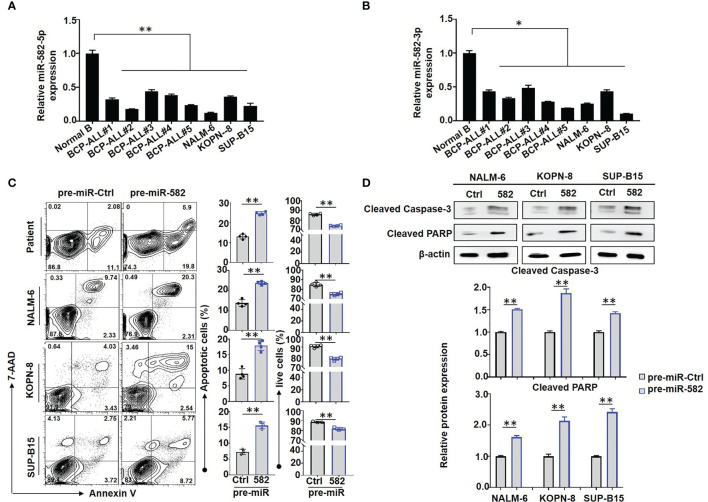
B-cell precursor acute lymphoblastic leukemia (BCP-ALL) is a malignancy characterized by the aberrant accumulation of immature B-cell precursors in bone marrow and other lymphoid organs. Although several intrinsic regulatory signals participating in BCP-ALL have been clarified, detailed intrinsic and extrinsic mechanisms that regulate BCP-ALL progression have not been fully understood. In the current study, we report that miR-582 is downregulated in BCP-ALL cells compared with normal B cells. Forced overexpression of miR-582 attenuated BCP-ALL cell proliferation and survival. We found that miR-582 overexpression disturbed the mitochondrial metabolism of BCP-ALL cells, leading to less ATP but more ROS production. Mechanistically, we identified PPTC7 as a direct target of miR-582. MiR-582 overexpression inhibited the activity of CoQ10, which is downstream of PPTC7 and played an important positive regulatory role in mitochondrial electron transportation. Finally, we found that overexpression of miR-582 upregulated the expression of immune checkpoint molecule CD276 and reduced NK cell-mediated cytotoxicity against BCP-ALL cells. CD276 blockade significantly increased NK cell-mediated cytotoxicity against miR-582-overexpressing BCP-ALL cells. Together, our research demonstrates that miR-582 acts as a negative regulator of BCP-ALL cells by reducing proliferation and survival, but protects BCP-ALL cells from NK cell-mediated cytotoxicity, suggesting that miR-582 may be a new therapeutic biomarker for BCP-ALL with CD276 blocker.

Keywords: BCP-ALL; CD276; NK cells; PPTC7; immune checkpoint; metabolism; miR-582; mitochondria.


Conflict of interest statement

The authors declare that the research was conducted in the absence of any commercial or financial relationships that could be construed as a potential conflict of interest.

Figures

Figure 1
Figure 1
Overexpression of miR-582 inhibits the survival of BCP-ALL cells. (A, B) Relative expression of human miR-582-5p and miR-582-3p in normal B cells, BCP-ALL patients B cells, NALM-6, KOPN-8 and SUP-B15 cells were determined by qRT-PCR. (C) BCP-ALL patients B cells, NALM-6, KOPN-8 and SUP-B15 cells were infected with pre-miR-582 lentivirus or their control lentivirus, and cultured for 72 h. Apoptosis was evaluated by using flow cytometry after staining of Annexin V and 7-AAD. The percentages of infected live (Annexin V-/7-AAD-) BCP-ALL cells, infected early and late apoptotic (Annexin V+/7-AAD-/+) BCP-ALL cells were quantitatively compared (n = 3-4). (D) BCP-ALL cells were infected with pre-miR-582 lentivirus or their control lentivirus for 72 h, and Cleaved Caspase-3, Cleaved PARP proteins were determined by Western blotting, with β-actin as an internal control (n = 3). Bars represent means ± SD, *P < 0.05, **P < 0.01.
Figure 2
Figure 2
Overexpression of miR-582 inhibits the proliferation of BCP-ALL cells. (A) NALM-6, KOPN-8 and SUP-B15 cells were infected with pre-miR-582 lentivirus or their control lentivirus. Cell proliferation of infected cells was determined using the CFSE labeling assay (n = 3-6). (B) NALM-6, KOPN-8 and SUP-B15 cells were infected with pre-miR-582 lentivirus or their control lentivirus, and cultured for 72 h. Cell viability was detected by MTT method (n = 6). (C, D) Experimental timeline for in vivo study. luciferase (luci)-labeled NALM-6 were infected with pre-miR-582 lentivirus or their control lentivirus, then, an orthotopic xenograft BCP-ALL mouse model was established by i.v injecting 1 × 10⁶ luci-labeling pre-miR-582 NALM-6 cells or pre-miR-Ctrl NALM-6 cells into NCG mice on day 0. Tumor burden was determined on day 14 and 21. (E, F) On Day 21, single cell suspensions of BM and spleen from BCP-ALL mouse model were analyzed by flow cytometry (E), and the percentage of human CD19+ B cells were quantitatively compared (F) (n = 3). (G) Survival of NCG mice which injected with miR-582-overexpressing NALM-6 cells and control NALM-6 cells. Bars represent means ± SD, *P < 0.05, **P < 0.01.
Figure 3
Figure 3
Overexpression of miR-582 inhibits mitochondrial energy metabolism of BCP-ALL cells. (A) After infection with pre-miR-582 lentivirus or their control lentivirus for 72 h, the contents of ATP in NALM-6, KOPN-8 and SUP-B15 cells were measured by using a luciferase reporter assay system. The luminescence level was normalized to pre-miR-Ctrl group in each individual (n = 3). (B) NALM-6, KOPN-8 and SUP-B15 cells were infected with pre-miR-582 lentivirus or their control lentivirus for 72 h, the mitochondrial metabolic pressure was detected by using XF Extracellular Flux Analyzer (n = 3). (C, D) NALM-6, KOPN-8 and SUP-B15 cells were infected with pre-miR-582 lentivirus and their control lentivirus for 72 h, the total intracellular ROS and mitochondria ROS were determined by flow cytometry, total intracellular ROS was labeled by DCFH-DA, the mitochondria ROS was labeled by MitoTacker and DCFH-DA (n = 3-5). (E) NALM-6, KOPN-8 and SUP-B15 cells were infected with pre-miR-582 lentivirus or their control lentivirus for 72 h, 2-NBDG was added to glucose-free medium for 30 min and intracellular 2-NBDG was analyzed by flow cytometry and quantitatively by statistics the proportion of 2-NBDG+ cells (n = 3-5). Bars represent means ± SD, **P < 0.01.
Figure 4
Figure 4
miR-582 downregulates PPTC7 in BCP-ALL cells. (A) Significantly different expressed genes (differentially upregulated genes and differentially downregulated genes) in pre-miR-582 NALM-6 cells/pre-miR-Ctrl NALM-6 cells. (B) 94 genes which related to metabolism are significantly downregulated in pre-miR-582 NALM-6 cells. (C) 7 significantly downregulated metabolism-related genes were predicted target genes of human miR-582-5p by combined with RNA-seq data and TargetScan 6.2 database. (D, E) The mRNA expression of PPTC7 in pre-miR-Ctrl NALM-6 cells and pre-miR-582 NALM-6 cells were showed by volcano plot and heatmap. (F) NALM-6, KOPN-8 and SUP-B15 cells were infected with pre-miR-582 lentivirus or their control lentivirus for 72 h, and PPTC7 mRNA were determined by qRT-PCR, with β-actin as an internal control (n = 3). (G) Alignment of the seed sequence of human miR-582-5p with human PPTC7 3’UTR. Complementary bases are marked with blue color. (H) The sequences of PPTC7 3’UTR-wt and PPTC7 3’UTR-mut used for schematic of the reporter constructs. (I) HEK293T cells were transfected with miR-582-5p and different 3’UTR reporters of human PPTC7 for 72 h. Luciferase activity in cell lysates were determined by the dual luciferase reporter assay (n = 3). (J, K) NALM-6, KOPN-8 and SUP-B15 cells were infected with pre-miR-582 lentivirus or their controls for 72 h, and PPTC7 protein were determined by Western blotting, with β-actin as an internal control (n = 3). Bars represent means ± SD, *P < 0.05,**P < 0.01.
Figure 5
Figure 5
miR-582 attenuates BCP-ALL cells mitochondrial energy metabolism by inhibiting PPTC7/CoQ10 signaling. (A) NALM-6, KOPN-8 and SUP-B15 cells were infected with pre-miR-582 lentivirus or their control lentivirus for 72 h, CoQ10 contents in cell lysates was evaluated and compared (n = 3). (B) NALM-6, KOPN-8 and SUP-B15 cells were infected with PPTC7 overexpression lentivirus (LV-PPTC7), with/without cultured with pre-miR-582 lentivirus for 72 h. CoQ10 contents in cell lysates was evaluated and compared (n = 3). (C) NALM-6, KOPN-8 and SUP-B15 cells were infected with LV-PPTC7, with/without cultured with pre-miR-582 lentivirus for 72 h, relative ATP level in cell lysates was evaluated and compared. (D, E) NALM-6, KOPN-8 and SUP-B15 cells were infected with LV-PPTC7, with/without cultured with pre-miR-582 lentivirus for 72 h, the total ROS and Mitochondria ROS were determined by flow cytometry, total intracellular ROS was labeled by DCFH-DA, the mitochondria ROS was labeled by MitoTacker and DCFH-DA. Bars represent means ± SD, *P < 0.05, **P < 0.01.
Figure 6
Figure 6
Overexpression of miR-582 protects BCP-ALL cells from NK cell-mediated cytotoxicity. (A) NALM-6 cells were infected with pre-miR-582 lentivirus or its control lentivirus for 72 h, and CD276 mRNA were determined by RNA-seq. (B, C) NALM-6, KOPN-8 and SUP-B15 cells were infected with pre-miR-582 lentivirus or its control lentivirus for 72 h, and CD276 mRNA were determined by qRT-PCR (B); the proportion of CD276+ cells were determined by flow cytometry (C). (D, E) NALM-6, KOPN-8 and SUP-B15 cells were infected with pre-miR-582 lentivirus and their control lentivirus, cultured with or without anti-CD276 antibody, and co-cultured with NK cells for 4 h, the proportion of CD107a+ NK and GZMB+ NK cells were determined by flow cytometry. (F) The signaling pathway of miR-582 regulate the progression of BCP-ALL and protect BCP-ALL cells from natural killer cell-mediated cytotoxicity, figure created with BioRender.com. Bars represent means ± SD, *P < 0.05, **P < 0.01.

Similar articles

Cited by

References

    1. Geron I, Savino AM, Tal N, Brown J, Turati V, James C, et al. . An Instructive Role for IL7RA in the Development of Human B-Cell Precursor Leukemia. Nat Commun (2022) 13(1):659. doi: 10.1038/s41467-022-28218-7 - DOI - PMC - PubMed
    1. Wang W, Wu F, Ma P, Gan S, Li X, Chen L, et al. . LncRNA CRNDE Promotes the Progression of B-Cell Precursor Acute Lymphoblastic Leukemia by Targeting the miR-345-5p/CREB Axi. Mol Cells (2020) 43(8):718–27. doi: 10.14348/molcells.2020.0065 - DOI - PMC - PubMed
    1. Neri LM, Cani A, Martelli A, Simioni C, Junghanss C, Tabellini G, et al. . Targeting the PI3K/Akt/mTOR Signaling Pathway in B-Precursor Acute Lymphoblastic Leukemia and Its Therapeutic Potential. Leukemia (2014) 28(4):739–48. doi: 10.1038/leu.2013.226 - DOI - PubMed
    1. Buske C, Becker D, Feuring-Buske M, Hannig H, Wulf G, Schäfer C, et al. . TGF-β Inhibits Growth and Induces Apoptosis in Leukemic B Cell Precursors. Leukemia (1997) 11(3):386–92. doi: 10.1038/sj.leu.2400586 - DOI - PubMed
    1. Heisterkamp N, ji Joo E, Yang L, Groffen J, Fei F. Cell-Intrinsic and Extrinsic Effects of Galectin-1 and Galectin-3 in B-Cell Precursor Acute Lymphoblastic Leukemia. bioRxiv (2021). doi: 10.1101/2021.09.22.461145 - DOI

Publication types