. 2019 Apr 5;9(19):10766-10775.
doi: 10.1039/c9ra00770a. eCollection 2019 Apr 3.

Comparative effects of mercury chloride and methylmercury exposure on early neurodevelopment in zebrafish larvae

Affiliations

Comparative effects of mercury chloride and methylmercury exposure on early neurodevelopment in zebrafish larvae

Jun Zhu et al. RSC Adv. .

Abstract

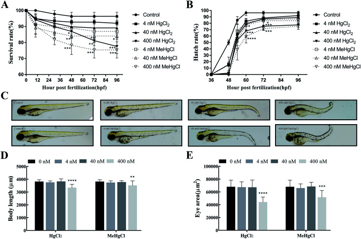
Mercury (Hg) is a ubiquitous environmental toxicant with important public health implications. Hg causes neurotoxicity through astrocytes, Ca2+, neurotransmitters, mitochondrial damage, elevations of reactive oxygen species and post-translational modifications. However, the similarities and differences between the neurotoxic mechanisms caused by different chemical forms of Hg remain unclear. Zebrafish embryos were exposed to methylmercury (MeHgCl) or mercury chloride (HgCl2) (0, 4, 40, 400 nM) up for 96 h. HgCl2 exposure could significantly decrease survival rate, body length and eye size, delay the hatching period, induce tail bending and reduce the locomotor activity, and these effects were aggravated in the MeHgCl group. The compounds could increase the number of apoptotic cells in the brain and downregulate the expression of Shha, Ngn1 and Nrd, which contribute to early nervous development. The underlying mechanisms were investigated by metabolomics data. Galactose metabolism, tyrosine metabolism and starch and sucrose metabolism pathways were disturbed after HgCl2 or MeHgCl exposure. In addition, the levels of three neurotransmitters including tyrosine, dopamine and tryptophan were reduced after HgCl2 or MeHgCl exposure. Oxidative stress is related to metabolite changes, such as changes in the putrescine, niacinamide and uric acid contents in the HgCl2 group, and squalene in the MeHgCl group. These data indicated that downregulation of these genes and abnormal metabolic profile and pathways contribute to the neurotoxicity of HgCl2 and MeHgCl.


Conflict of interest statement

The authors declare that there are no conflicts of interest.

Figures

Fig. 1
Fig. 1. Effects of mercury chloride (HgCl2) and methylmercury chloride (MeHgCl) on the embryonic development. Zebrafish embryos were exposed to MeHgCl (control, 4, 40, 400 nM) and HgCl2 (control, 4, 40, 400 nM) for up to 96 hpf. (A) Survival rates, (B) hatching rates, (C) morphological abnormalities, (D) body length, (E) eye size of embryos were investigated. Data represent the mean ± SD of three experiments. *P < 0.05; **P < 0.01, ***P < 0.001, ****P < 0.0001; compared with control group.
Fig. 2
Fig. 2. Effects of mercury chloride (HgCl2) and methylmercury chloride (MeHgCl) on the locomotor activity. The embryos/larvae were exposed to MeHgCl (control, 4, 40, 400 nM) or HgCl2 (control, 4, 40, 400 nM), and their locomotor behavior were analyzed by the dark-light-dark photoperiod stimulation test at 96 hpf. (A) Average moving distance, (B) locomotor traces chart, (C) locomotor traces. *P < 0.05; **P < 0.01; compared with control group.
Fig. 3
Fig. 3. Effects of mercury chloride (HgCl2) and methylmercury chloride (MeHgCl) on brain cell apoptosis and the expression of neural development related genes. The embryos/larvae were exposed to MeHgCl (control, 4, 40, 400 nM) or HgCl2 (control, 4, 40, 400 nM), and the apoptotic cells in the brain were analyzed by acridine orange (AO) staining at 96 hpf. (A) Representative images of AO staining and (B) the number of AO positive cells in brain. n = 12 embryos per group. The expression of (C) Shha, (D) Ngn1 and (E) Nrd in the zebrafish treated with HgCl2 and MeHgCl for 30 hpf. Data represent the mean ± SD. **P < 0.01, ***P < 0.001, ****P < 0.0001; compared with control group.
Fig. 4
Fig. 4. Effects of mercury chloride (HgCl2) and methylmercury chloride (MeHgCl) on the metabolic profile. Heat map and hierarchical clustering analyses of zebrafish exposed to 40 nM of HgCl2 (A) and MeHgCl (B). Red colour represents an upregulation of the metabolite while blue color represents a downregulation. Relevant pathways disturbed by HgCl2 (C) and MeHgCl (D). Bubble area is proportional to the impact of each pathway, with color denoting the significance from highest in red to lowest in white. Peak area of different metabolites across the different exposure groups at 30 hpf. The effect of HgCl2 and MeHgCl on (E) tyrosine, (F) dopamine, (G) tryptophan. Bars represent mean values (n = 8) and error bars represent standard deviations. *P < 0.05, **P < 0.01; compared with control group.

Similar articles

Cited by

References

    1. Shi Q. Sun N. Kou H. Wang H. Zhao H. Ecotoxicol. Environ. Saf. 2018;164:500–509. doi: 10.1016/j.ecoenv.2018.08.058. - DOI - PubMed
    1. Reif J. S. Schaefer A. M. Bossart G. D. Vet. Sci. 2015;2:407–422. doi: 10.3390/vetsci2040407. - DOI - PMC - PubMed
    1. The L. Lancet. 2017;390:822. - PubMed
    1. Liang S. Wang Y. Cinnirella S. Pirrone N. Environ. Sci. Technol. 2015;49:3566–3574. doi: 10.1021/es503977y. - DOI - PubMed
    1. Dong A. Zhai S. Louchouarn P. Izon G. Zhang H. Jiang X. Environ. Sci. Pollut. Res. Int. 2019;26:4667–4679. doi: 10.1007/s11356-018-3880-3. - DOI - PubMed