

Targeting Inhibition of Notch1 Signaling Pathway on the Study of Human Gastric Cancer Stem Cells with Chemosensitization
- PMID: 35515501
- PMCID: PMC9064537
- DOI: 10.1155/2022/1098394
Targeting Inhibition of Notch1 Signaling Pathway on the Study of Human Gastric Cancer Stem Cells with Chemosensitization
Retraction in
-
Retracted: Targeting Inhibition of Notch1 Signaling Pathway on the Study of Human Gastric Cancer Stem Cells with Chemosensitization.Comput Intell Neurosci. 2022 Dec 20;2022:9846409. doi: 10.1155/2022/9846409. eCollection 2022. Comput Intell Neurosci. 2022. PMID: 36582407 Free PMC article.
Abstract
Background: Gastric cancer is the second most frequent cause of cancer death worldwide, although much geographical variation in incidence exists. Prevention and personalized treatment are regarded as the best options to reduce gastric cancer mortality rates (Hartgrink et al., 2009). Numerous studies have suggested that Notch1 and its ligands are overexpressed in gastric cancer, and its knockdown can inhibit the proliferation and survival of gastric cancer cells.
Objective: To investigate the effect of Notch1 on the stemness and drug sensitivity of human gastric cancer SGC-7901 cells.
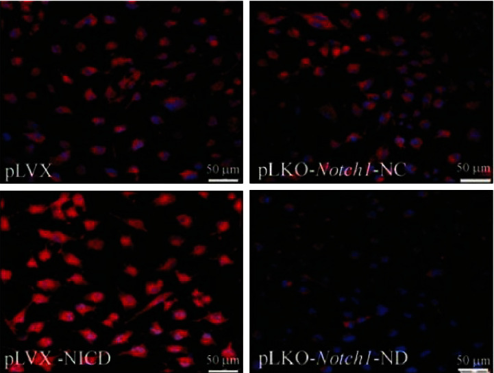
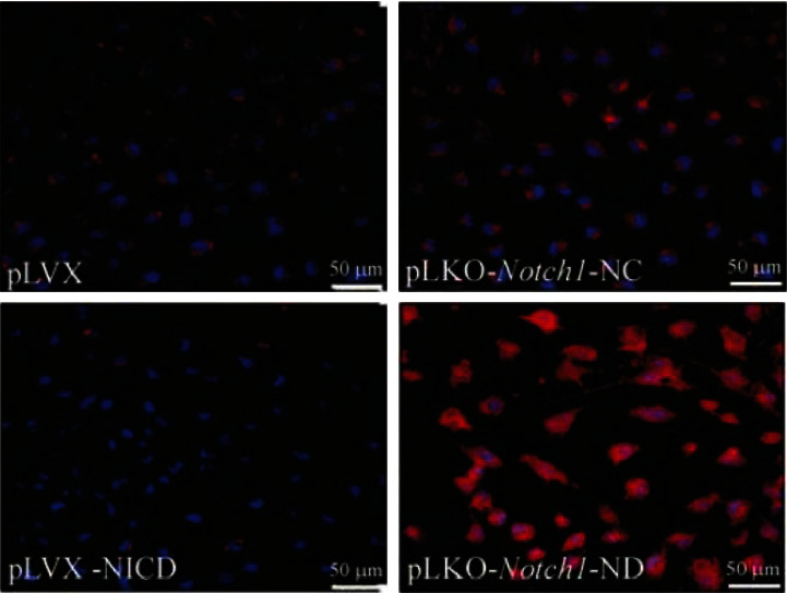
Methods: Highly expressed Notch1 intracellular domain (NICD1) and Notch1-shRNA lentiviral expression vector were used to infect human gastric cancer SGC-7901 cells cultured in vitro, and western blot and immunofluorescence staining were used to identify highly expressed NICD and Notch1 silenced cells. The percentage of CD133+ cells was analyzed by flow cytometry, the expression of nestin and CFAP by immunofluorescence staining, the formation rate of tumor cell spheres and the tumorigenicity of SCID mice in vivo, and the regulation of cell stemness by Notch1. The sensitivity of each group of cells to the chemotherapeutic drugs teniposide (VM-26) and carmustine (BCNU) was also detected by the MTT method.
Results: The stemness phenotype of tumor cells with the increased NICD expression was enhanced, such as an increased proportion of CD133+ cells, enhanced nestin expression, decreased GFAP expression, increased tumor cell sphere formation rate and tumorigenic rate of SCID mice implantation, and decreased sensitivity to VM-26 and BCNU. In contrast, the stemness phenotype of tumor cells with downregulated Notch1 gene expression was significantly suppressed, while the sensitivity to VM-26 and BCNU was increased.
Conclusion: High Notch1 expression increased the stemness of SGC-7901 cells and decreased the sensitivity of SGC-7901 cells to chemotherapeutic drugs.
Copyright © 2022 Yan Dou and Jinghong Wang.
Conflict of interest statement
The authors declare no potential conflicts of interest with respect to the research, authorship, and/or publication of this article.
Figures


Similar articles
-
Intracellular Notch1 Signaling in Cancer-Associated Fibroblasts Dictates the Plasticity and Stemness of Melanoma Stem/Initiating Cells.Stem Cells. 2019 Jul;37(7):865-875. doi: 10.1002/stem.3013. Epub 2019 Apr 19. Stem Cells. 2019. PMID: 30941836 Free PMC article.
-
RECK inhibits stemness gene expression and tumorigenicity of gastric cancer cells by suppressing ADAM-mediated Notch1 activation.J Cell Physiol. 2014 Feb;229(2):191-201. doi: 10.1002/jcp.24434. J Cell Physiol. 2014. PMID: 23881612
-
Notch1 directly induced CD133 expression in human diffuse type gastric cancers.Oncotarget. 2016 Aug 30;7(35):56598-56607. doi: 10.18632/oncotarget.10967. Oncotarget. 2016. PMID: 27489358 Free PMC article.
-
DLL4 overexpression increases gastric cancer stem/progenitor cell self-renewal ability and correlates with poor clinical outcome via Notch-1 signaling pathway activation.Cancer Med. 2017 Jan;6(1):245-257. doi: 10.1002/cam4.962. Epub 2016 Nov 28. Cancer Med. 2017. PMID: 27891816 Free PMC article.
-
Xiaotan Sanjie decoction attenuates tumor angiogenesis by manipulating Notch-1-regulated proliferation of gastric cancer stem-like cells.World J Gastroenterol. 2014 Sep 28;20(36):13105-18. doi: 10.3748/wjg.v20.i36.13105. World J Gastroenterol. 2014. PMID: 25278704 Free PMC article.
Cited by
-
RAB4A is a master regulator of cancer cell stemness upstream of NUMB-NOTCH signaling.Cell Death Dis. 2024 Oct 27;15(10):778. doi: 10.1038/s41419-024-07172-w. Cell Death Dis. 2024. PMID: 39463384 Free PMC article.
-
Retracted: Targeting Inhibition of Notch1 Signaling Pathway on the Study of Human Gastric Cancer Stem Cells with Chemosensitization.Comput Intell Neurosci. 2022 Dec 20;2022:9846409. doi: 10.1155/2022/9846409. eCollection 2022. Comput Intell Neurosci. 2022. PMID: 36582407 Free PMC article.
-
Targeting Gastric Cancer Stem Cells to Enhance Treatment Response.Cells. 2022 Sep 10;11(18):2828. doi: 10.3390/cells11182828. Cells. 2022. PMID: 36139403 Free PMC article. Review.
References
-
- Merz V., Zecchetto C., Simionato F., et al. A phase II trial of the FGFR inhibitor pemigatinib in patients with metastatic esophageal-gastric junction/gastric cancer trastuzumab resistant: the FiGhTeR trial. Therapeutic advances in medical oncology . 2020;12 doi: 10.1177/1758835920937889.1758835920937889 - DOI - PMC - PubMed
Publication types
MeSH terms
Substances
LinkOut - more resources
Full Text Sources
Medical
Research Materials
Miscellaneous
