Review
. 2020 Jul 21;10(45):27064-27080.
doi: 10.1039/d0ra03194a. eCollection 2020 Jul 15.

Systematic overview of soft materials as a novel frontier for MRI contrast agents

Affiliations
Review

Systematic overview of soft materials as a novel frontier for MRI contrast agents

Enrico Gallo et al. RSC Adv. .

Abstract

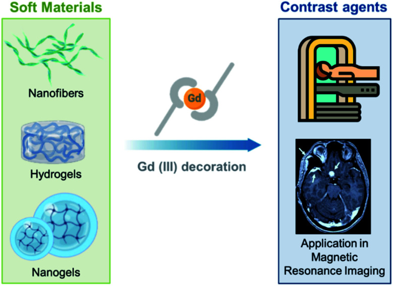
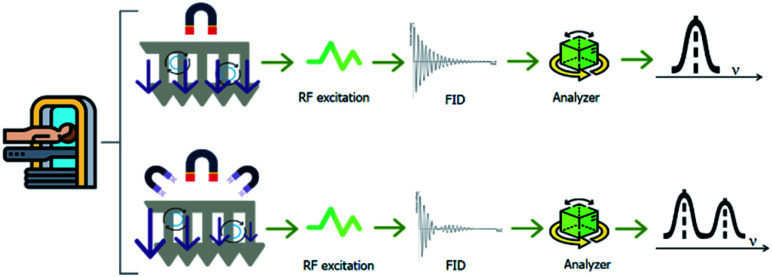
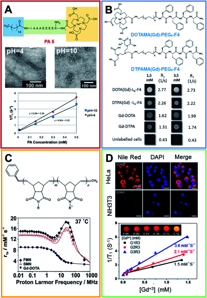
Magnetic resonance imaging (MRI) is a well-known diagnostic technique used to obtain high quality images in a non-invasive manner. In order to increase the contrast between normal and pathological regions in the human body, positive (T1) or negative (T2) contrast agents (CAs) are commonly intravenously administered. The most efficient class of T1-CAs are based on kinetically stable and thermodynamically inert gadolinium complexes. In the last two decades many novel macro- and supramolecular CAs have been proposed. These approaches have been optimized to increase the performance of the CAs in terms of the relaxivity values and to reduce the administered dose, decreasing the toxicity and giving better safety and pharmacokinetic profiles. The improved performances may also allow further information to be gained on the pathological and physiological state of the human body. The goal of this review is to report a systematic overview of the nanostructurated CAs obtained and developed by manipulating soft materials at the nanometer scale. Specifically, our attention is centered on recent examples of fibers, hydrogels and nanogel formulations, that seem particularly promising for overcoming the problematic issues that have recently pushed the European Medicines Agency (EMA) to withdraw linear CAs from the market.


Conflict of interest statement

There are no conflicts to declare.

Figures

Fig. 1
Fig. 1. Techniques: MRI (A), fluorescence (B), PET (C) and SPECT (D).
Fig. 2
Fig. 2. The class of soft materials includes nanofibers, hydrogels and nanogels. Using appropriate strategies for decoration, these supramolecular aggregates can be functionalized with Gd(iii) ions and complexes in order to became suitable for application in MRI as T1-positive contrast agents.
Fig. 3
Fig. 3. To obtain an MRI image, the body is located in a uniform magnetic field. Hydrogen nuclei align with the magnetic field and create a net magnetic moment parallel to it. When a radio-frequency pulse (RF excitation) is applied perpendicularly, the net magnetic moment of the nuclei tilts away from the magnetic field. When the RF pulse stops, the nuclei return to the equilibrium initial state (relaxation step). During the relaxation, the nuclei lose energy and a measurable signal, indicated as the free-induction decay (FID), is detected. 3D MRI images can be generated, encoding the FID in each dimension. An additional magnetic field in the gradient changes the FID as a function of the proton 3D position.
Fig. 4
Fig. 4. (A) Chemical formula for the PA 5 compound, the peptide sequence of PA 5 is reported according to the one code letter. TEM images (0.5 mM solution of PA 5) at pH 4.0 (on the left) and 10.0 (on the right). T1 relaxivity graph of PA 5 under basic and acidic conditions (adapted with permission 2012-American Chemical Society). (B) Chemical structure of DOTAMA(Gd)-PEG6-F4 and DTPAMA(Gd)-PEG6-F4. T1-weighted MR-images of pellets of the J774A.1 cell line labelled with 1.5 and 3.5 × 10⁻³ mol L−1 of the two Gd-complexes. The relative observed relaxation rates are reported too (reproduced with permission from ref. 41). (C) Chemical structure of Gd3+-containing an amphiphilic block copolymer. R and R′ can be a phenyl group or a modified Gd-DOTA complex (norbornenyl-Gd-DOTA monoamide). 1H NMRD profiles for Gd-DOTA, spherical and fibrillar micellar nanoparticles (reproduced with permission, published by The Royal Society of Chemistry). (D) Laser scanning microscopy images of HeLa and NIH3T3 cells lines treated with Nile red-loaded porous networks of Gd3+-G3R3 (1 h after incubation): left, Nile red (red); middle, nuclei stained with DAPI (blue); and right, merged images. T1 relaxivity plots of Gd3+-chelating nanostructures (0.2 mM) as a function of the Gd3+ concentration ([Gd3+]) (4.7 T, 25 °C) and (inset) T1-weighted MR images of Gd3+-G3R3 (adapted with permission, copyright 2017-American Chemical Society).
Fig. 5
Fig. 5. (A) On the left, schematic illustration of the formation of pH-sensitive injectable hydrogel composed by a modified PEG polymer (blue ribbon) and a DTPA-decorated chitosan (purple ribbon). The hydrogel self-healing feature is reported too. On the right, MRI intensity of the visible hydrogel as a function of the Gd(iii) concentration (from ref. 57. Reproduced by permission of The Royal Society of Chemistry). (B) Chemical structure for the bifunctional hydrogelator containing ureidopyrimidinone (UPy). 10 wt% UP-PEG hydrogels T1-weighted 1.5 T MRI scans reported in pseudo-color of the released solution and for the gels. Hydrogels contain either (1) 1 × 10⁻³ mol L−1 Gadoteridol or (2) 1 × 10⁻³ mol L−1 UPy-Gd(iii) modified monomers. The relative release curves are reported too (arranged with permission). (C) On the left, Cryo-TEM of PAs conjugates in 10 mmol L−1 in Tris buffer after a process of thermal annealing (80 °C for 30 min) and their relative molecular graphical representation. The scale bar represents 200 nm and Gd macrocycles are represented as green elements. On the left, the NMRD profiles for all PAs at three different conditions recorded at 37 °C and a PAs concentration of 2 mmol L−1 (adapted with permission, copyright 2014-American Chemical Society).
Fig. 6
Fig. 6. (A) (1) Optical fluorescence microscopy photo of flow-focusing pattern used for obtaining the nanogel formulation using a microfluidic approach. (2) Schematic crosslinking reaction involving HA hydroxyl groups with DVS. FE-SEM images of crosslinked HA nanoparticles (cHANPs) in aqueous solution in (3) 0.8% v/v of DVS added in the middle channel and (4) 4% v/v of DVS added in the side channels. (5) In vitro relaxation time distribution reported for: Gd-DTPA in water solution at (purple line) 10 μmol L−1, (light blue line) 60 μmol L−1 and (blue) 100 μmol L−1; un-loaded cHANPs (black line). Also reported are the loaded HA nanoparticles at standard conditions obtained using (red line) 4% v/v DVS in the side channels, at pH 12.3, reported at 12 μmol L−1 of Gd-DTPA, (green line) 0.8% v/v DVS and Cspan80 tensioactive 0.5% v/v in the middle channel, reported at Gd-DTPA of 10 μM (reproduced with permission). (B) Upper row, nanogel formulation obtained by self-assembling of a cholesterol and acryloyl-modified polysaccharide pullulan (CHPOA). The photoinduced crosslinkage of the acryloyl groups on the nanogel surface of the DOTA-Gd modified chelating agent (GdCHPOA) allows the creation of T1 MRI contrast agents (adapted with permission, copyright 2015-American Chemical Society). Lower row: chemical structure of two macrocyclic DOTA-based crosslinkers. Polymers were converted in polyacrylamide-based nanogels using an inverse emulsion process via ammonium persulfate (APS) as the initiator and by adding tetramethylethylenediamine (TMEDA) to control the radical polymerization rate (arranged from ref. 88. Reproduced by permission of The Royal Society of Chemistry). (C) Graphical representation of the functional metal–organic coordinated nanogels (GdNGs) obtained using the branched poly(ethyleneimine) (PEI) polymer via the colloidal reverse microemulsion method. On the right, in vivo NIRF images of the SCC7 tumor-bearing mouse before and after tail vein injection (1 d and 1 week) of the GdNGs and the biodistribution of the GdNGs, obtained by NIRF signals from the dissected organs and tumors before (control) and after injection (reproduced with permission).
Scheme 1
Scheme 1. Scheme describing the systematic literature review performed according to PRISMA (Preferred Reporting Items for Systematic Reviews and Meta-Analyses) criteria.

Similar articles

Cited by

References

    1. Kherlopian A. R. Song T. Duan Q. Neimark M. A. Po M. J. Gohagan J. K. Laine A. F. BMC Syst. Biol. 2008;2:74–92. doi: 10.1186/1752-0509-2-74. - DOI - PMC - PubMed
    1. Salzer R., Biomedical imaging: principles and applications, John Wiley & Sons, Inc., Hoboken, NJ USA, 2012
    1. Lin E. C. Mayo Clin. Proc. 2010;85:1142–1146. doi: 10.4065/mcp.2010.0260. - DOI - PMC - PubMed
    1. Seibert J. A. Morin R. L. Pediatr. Radiol. 2011;41:573–581. doi: 10.1007/s00247-010-1954-6. - DOI - PMC - PubMed
    1. Gallagher F. A. Clin. Radiol. 2010;65:557–566. doi: 10.1016/j.crad.2010.04.006. - DOI - PubMed