Review
. 2020 Jul 21;10(45):27194-27214.
doi: 10.1039/d0ra04736h. eCollection 2020 Jul 15.

Cerium oxide nanoparticles: properties, biosynthesis and biomedical application

Affiliations
Review

Cerium oxide nanoparticles: properties, biosynthesis and biomedical application

Kshitij Rb Singh et al. RSC Adv. .

Abstract

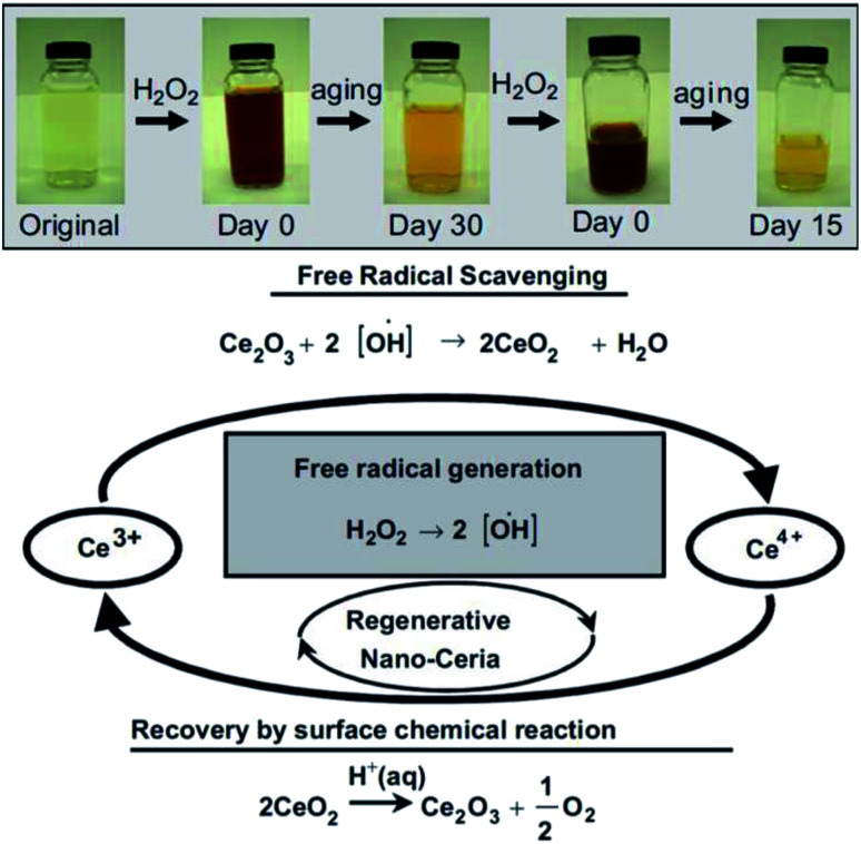
Nanotechnology is the branch of science which deals with particles ranging between 1-100 nm. These particles are called nanoparticles, and they exhibit unique electronic, optical, magnetic, and mechanical properties, which make them different from the bulk material. These properties of nanomaterials help them to find a variety of applications in the biomedical, agricultural, and environmental domains. Cerium oxide nanoparticles have gained a lot of attention as a potential future candidate for ending various kinds of problems by exhibiting redox activity, free radical scavenging property, biofilm inhibition, etc. Synthesis of these nanoparticles can be performed very easily by utilizing chemical or biological methods. But in this review, the focus is laid on the biosynthesis of these nanoparticles; as the biosynthesis method makes the cerium oxide nanoparticle less toxic and compatible with the living tissues, which helps them to find their path as an anticancer, anti-inflammatory and antibacterial agents. The pre-existing reviews have only focused on details relating to properties/applications/synthesis; whereas this review draws attention towards all the aspects in single review covering all the details in depth such as biosynthesis methods and its effect on the living tissues, along with properties, biomedical applications (diagnostic and therapeutic) and future outlook of the cerium oxide nanoparticle.


Conflict of interest statement

Author's declare no conflict of interest for this work.

Figures

Fig. 1
Fig. 1. General structure: face centered cubic structure of cerium oxide (CeO2) nanoparticles.
Fig. 2
Fig. 2. Structurally analyzed ceria crystals Ce4O8 (unit cell); in (a) and (b) yellow color represents eight-folds of cerium atoms and red represents four-fold oxygen atoms in ceria crystal structure; (c) is the basic fcc fluorite lattice structure of Ce4O8 (reproduced from K. Reed et al., Environ. Sci.: Nano, The Royal Society of Chemistry, 2014 (ref. 22)).
Fig. 3
Fig. 3. (a) Represents the unit cell structure of cerium oxide nanoparticles and (b) represents single oxygen vacancy of cerium oxide unit cell, where absence of one oxygen atom in left side (uppermost) and forward octant position can be seen (reproduced from K. Reed et al., Environ. Sci.: Nano, The Royal Society of Chemistry, 2014 (ref. 22)).
Fig. 4
Fig. 4. Oxygen vacancy created by CeO2 particle while oxidizing CO to CO2, and two Ce4+ atoms were reduced simultaneously (adapted from K. Reed et al., Environ. Sci.: Nano, The Royal Society of Chemistry, 2014 (ref. 22)).
Fig. 5
Fig. 5. Schematic representation of SOD reaction mechanism by cerium oxide nanoparticle (reproduced from I. Celardo et al., Nanoscale, The Royal Society of Chemistry, 2011 (ref. 24)).
Fig. 6
Fig. 6. Top: Observation of change in color when, cerium oxide nanoparticle coated with dextran was treated with H2O2 at different time intervals. Bottom: Schema of chemical reaction showing auto-regenerative properties cerium oxide nanoparticles and possible mode of mechanism data of the cerium oxide nanoparticle autocatalytic behavior and free-radical scavenging property (reproduced from M. Das et al., Biomaterials, Elsevier, 2007 (ref. 105)).
Fig. 7
Fig. 7. Top: Color change in solutions of cerium oxide nanoparticle coated with dextran at basic and acidic pH environment on addition of H2O2. Bottom: Detailed probable revised mode of mechanism data cerium oxide nanoparticle auto-regenerative attribute and free-radical scavenging property (reproduced from J. M. Perez et al., Small, John Wiley and Sons, 2008 (ref. 106)).
Fig. 8
Fig. 8. Method of nanoparticle synthesis; (A) top-down approach and (B) bottom-up approach of nanoparticle synthesis with examples.
Fig. 9
Fig. 9. Potential applications of cerium oxide nanoparticles in the biomedical, environmental, and agricultural domains.
Fig. 10
Fig. 10. Schematic illustration of cerium oxide nanoparticles loaded doxorubicin mode of mechanism interaction data of stromal-tumor for cancer treatment by combinational approach (reproduced from P. Brenneisen and A. Reichert, Antioxidants, MDPI, 2018 (ref. 150)).
Fig. 11
Fig. 11. Combination therapy for non-small-cell lung cancer (NSCLC) by using NC (cerium oxide nanoparticles), which results as an excellent tool for targeted drug delivery system. In this illustration, FNC is folate decorated cerium oxide nanoparticles, GT is ganetespib, Dox is doxorubicin, and nanoceria is cerium oxide nanoparticles (reproduced from S. Sulthana et al., Mol. Pharm., American Chemical Society, 2017 (ref. 172)).
Fig. 12
Fig. 12. Immunofluorescence monitoring of in vitro culture of adult murine resident cardiac stem cell incubated by different concentration of cerium oxide nanoparticle-poly(d,l-lactic-co-glycolic acid) composite material for 6 days: (a) 5, (b) 10, and (c) 20% wt%, together with (d) poly(d,l-lactic-co-glycolic acid) (PLGA) as a control (reproduced from C. Mandoli et al., Adv. Funct. Mater., John Wiley and Sons, 2010 (ref. 178)).
None
Kshitij RB Singh
None
Vanya Nayak
None
Tanushri Sarkar
None
Ravindra Pratap Singh

Similar articles

Cited by

References

    1. Rico C. M. Majumdar S. Duarte-Gardea M. Peralta-Videa J. R. Gardea-Torresdey J. L. J. Agric. Food Chem. 2011;59:3485–3498. doi: 10.1021/jf104517j. doi: 10.1021/jf104517j. - DOI - DOI - PMC - PubMed
    1. Razzaque S. Hussain S. Z. Hussain I. Tan B. Polymers. 2016;8:156. doi: 10.3390/polym8040156. doi: 10.3390/polym8040156. - DOI - DOI - PMC - PubMed
    1. Singh R. P. Int. J. Electrochem. 2011;2011:1–30. doi: 10.4061/2011/125487. doi: 10.4061/2011/125487. - DOI - DOI
    1. Singh R. P. Oh B. K. Koo K. K. Jyoung J. Y. Jeong S. Choi J. W. BioChip J. 2009;2:223–234.
    1. Singh R. P. J. Bioanal. Biomed. 2016;8:e143. doi: 10.4172/1948-593x.1000e143. - DOI