. 2019 Feb 14;9(10):5674-5681.
doi: 10.1039/c8ra09157a. eCollection 2019 Feb 11.

Three-dimensional paper-based microfluidic electrochemical integrated devices (3D-PMED) for wearable electrochemical glucose detection

Affiliations

Three-dimensional paper-based microfluidic electrochemical integrated devices (3D-PMED) for wearable electrochemical glucose detection

Qingpeng Cao et al. RSC Adv. .

Abstract

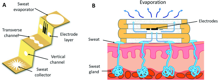
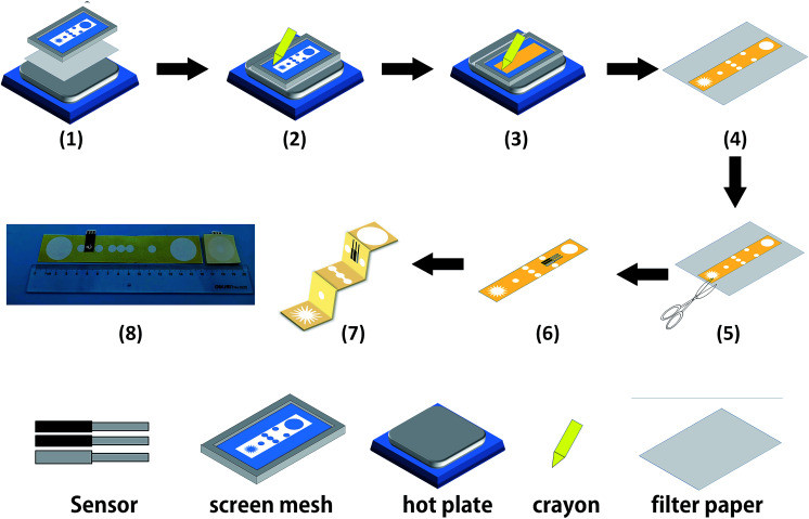
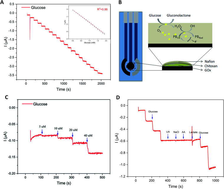
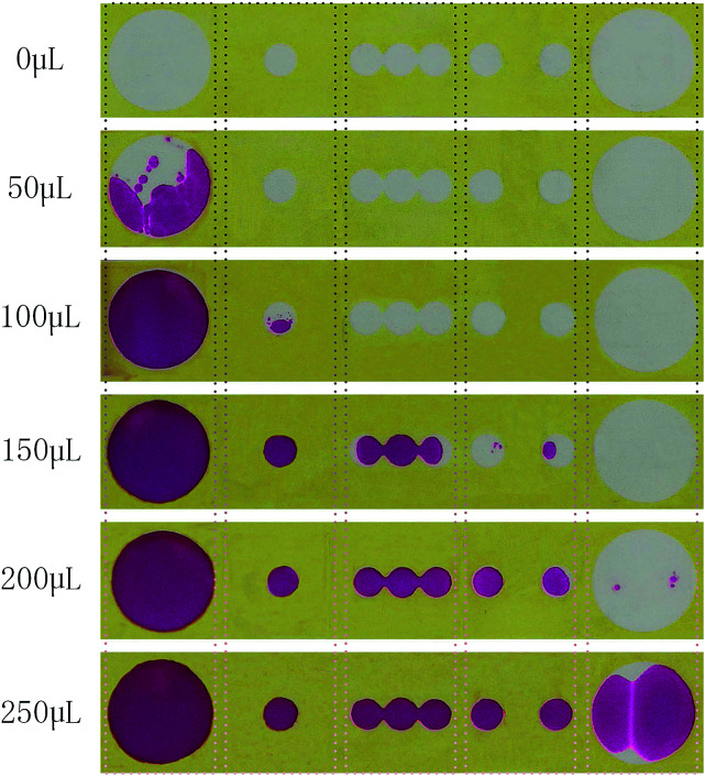
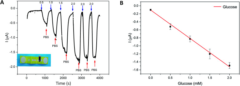
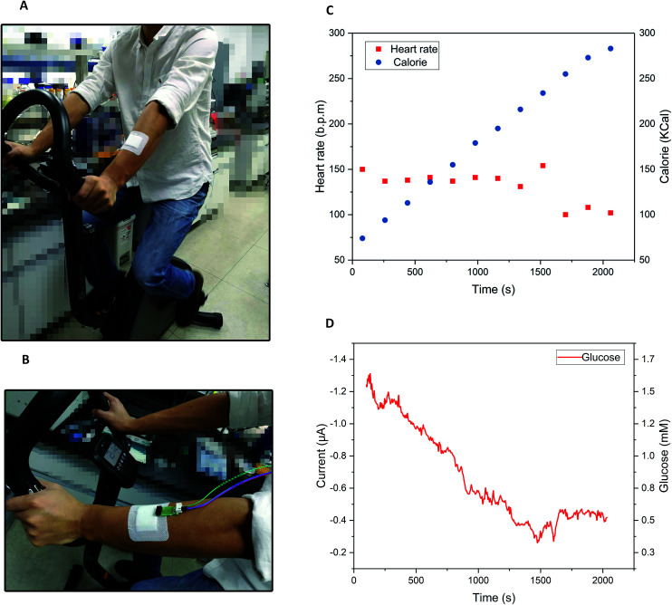
Wearable electrochemical sensors have attracted tremendous attention in recent years. Here, a three-dimensional paper-based microfluidic electrochemical integrated device (3D-PMED) was demonstrated for real-time monitoring of sweat metabolites. The 3D-PMED was fabricated by wax screen-printing patterns on cellulose paper and then folding the pre-patterned paper four times to form five stacked layers: the sweat collector, vertical channel, transverse channel, electrode layer and sweat evaporator. A sweat monitoring device was realized by integrating a screen-printed glucose sensor on polyethylene terephthalate (PET) substrate with the fabricated 3D-PMED. The sweat flow process in 3D-PMED was modelled with red ink to demonstrate the capability of collecting, analyzing and evaporating sweat, due to the capillary action of filter paper and hydrophobicity of wax. The glucose sensor was designed with a high sensitivity (35.7 μA mM-1 cm-2) and low detection limit (5 μM), considering the low concentration of glucose in sweat. An on-body experiment was carried out to validate the practicability of the three-dimensional sweat monitoring device. Such a 3D-PMED can be readily expanded for the simultaneous monitoring of alternative sweat electrolytes and metabolites.


Conflict of interest statement

There are no conflicts to declare.

Figures

Fig. 1
Fig. 1. Schematic diagram of the three-dimensional paper-based microfluidic electrochemical integrated device (3D-PMED). (A) The layered structure of the device. 3D-PMED includes: sweat collector, vertical channel, transverse channel, electrode layer and sweat evaporator. The yellow areas or the white areas on the device were hydrophobic areas formed by wax screen-printing or hydrophilic areas of filter paper, respectively. A three-electrode electrochemical sensor by screen-printing was affixed onto the electrode layer of the device by double-side adhesive tape. (B) The schematic diagram of the 3D-PMED applied on the skin of human. A 3D-flow channel was formed by folding the device with the tiered structure, to help the sweat flow from skin into the device and thus refresh the sweat under the electrodes.
Fig. 2
Fig. 2. Schematic diagram of the fabrication process for 3D-PMED. (1) A filter paper was pasted on the back of screen mesh to avoid the paper slide. (2 and 3) The screen mesh was rubbed with heating (50–55 °C). (4) Distinct patterns were formed on the paper. (5) The resulting paper was cut along the edge of the patterns. (6) A prepared three-electrode sensor was pasted onto the electrode layer with double-side adhesive tape. (7) The device was folded up to form a 3D-PMED. (8) The photograph of the 3D-PMED. The patterned filter paper was cut into rectangle shape (3 cm × 15 cm) and then folded into an origami structure with five equal parts (3 cm × 3 cm).
Fig. 3
Fig. 3. Characterization of glucose sensor. (A) Amperometric current response at −0.1 V (vs. Ag/AgCl reference electrode) with successive addition of 0.1 mM glucose in concentration ranging from 0 mM to 1.9 mM in 0.01 M PBS solution; inset: the calibration plot. (B) Schematic diagram of glucose sensor, along with the recognition and transduction events. (C) Detection limit curve of amperometric current response with successive addition of 5 μM, 10 μM, 20 μM, 40 μM. (D) Selectivity experiment using amperometric current responses of the biosensor with successive addition of 0.1 mM glucose (threefold), followed by electroactive interfering species of 2 μM UA, 0.1 mM NaCl, 2 μM AA, and 5 mM lactate, and finally 0.1 mM, 0.3 mM glucose in 0.01 M PBS at an applied potential of 0.1 V.
Fig. 4
Fig. 4. The simulation of the flow of sweat in 3D-PMED. The red ink was applied to simulate the sweat flow in 3D-PMED drove by capillary effect. Different volumes (0, 50 μL, 100 μL, 150 μL, 200 μL, 250 μL) red ink were injected into the 3D-PMED. After the flowing process for around 5 seconds, the 3D-PMED was unfolded for recording the results. The photographs displayed that the red ink first spread laterally, then penetrated into the next layer with increase of ink. The 250 μL red ink almost spread through the whole chamber of 3D-PMED.
Fig. 5
Fig. 5. Amperometric detection of glucose in the flow injection analysis system with 3D-PMED. (A) Amperometric current response at –0.1 V (vs. Ag/AgCl reference electrode) with alternant injecting of glucose (successive addition of 0.5 mM, 1.0 mM, 1.5 mM, 2.0 mM glucose) and PBS. (B) The calibration plot with the error bar ranging from 0 mM to 2.0 mM.
Fig. 6
Fig. 6. On-body measurement of glucose in sweat with 3D-PMED. (A and B) The photograph of the 3D-PMED was applied on the forearm of a subject, who was asked to perform the cycling exercise. (C) The subject heart rate and the calorie subject consumed were recorded by the multifunctional cycle ergometer. (D) The current response of glucose in sweat was measured by an electrochemical workstation.

Similar articles

Cited by

References

    1. Khan Y. Ostfeld A. E. Lochner C. M. Pierre A. Arias A. C. Adv. Mater. 2016;28:4373–4395. doi: 10.1002/adma.201504366. - DOI - PubMed
    1. Tran Quang T. Lee N.-E. Adv. Mater. 2016;28:4338–4372. doi: 10.1002/adma.201504244. - DOI - PubMed
    1. Kim D.-H. Lu N. Ma R. Kim Y.-S. Kim R.-H. Wang S. Wu J. Won S. M. Tao H. Islam A. Yu K. J. Kim T.-i. Chowdhury R. Ying M. Xu L. Li M. Chung H.-J. Keum H. McCormick M. Liu P. Zhang Y.-W. Omenetto F. G. Huang Y. Coleman T. Rogers J. A. Science. 2011;333:838–843. doi: 10.1126/science.1206157. - DOI - PubMed
    1. Bariya M. Nyein H. Y. Y. Javey A. Nature Electronics. 2018;1:160–171. doi: 10.1038/s41928-018-0043-y. - DOI
    1. Mukhopadhyay S. C. IEEE Sens. J. 2015;15:1321–1330.