

Biomimetic composite scaffold from an in situ hydroxyapatite coating on cellulose nanocrystals
- PMID: 35515933
- PMCID: PMC9060865
- DOI: 10.1039/c8ra09523j
Biomimetic composite scaffold from an in situ hydroxyapatite coating on cellulose nanocrystals
Abstract
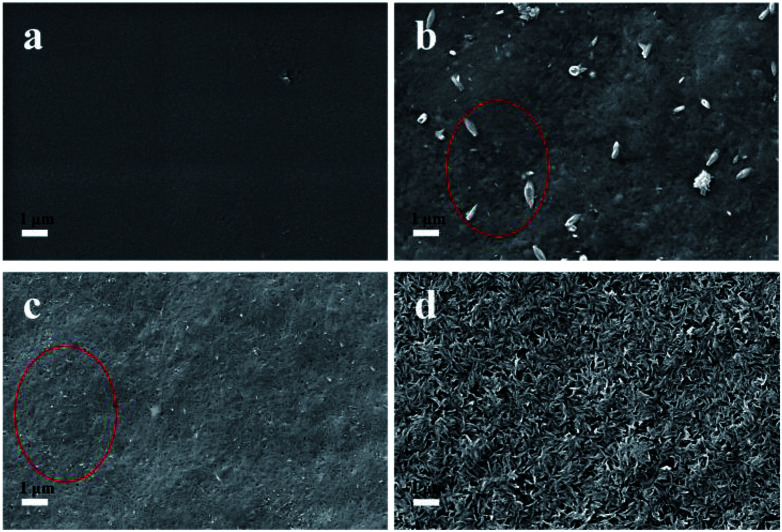
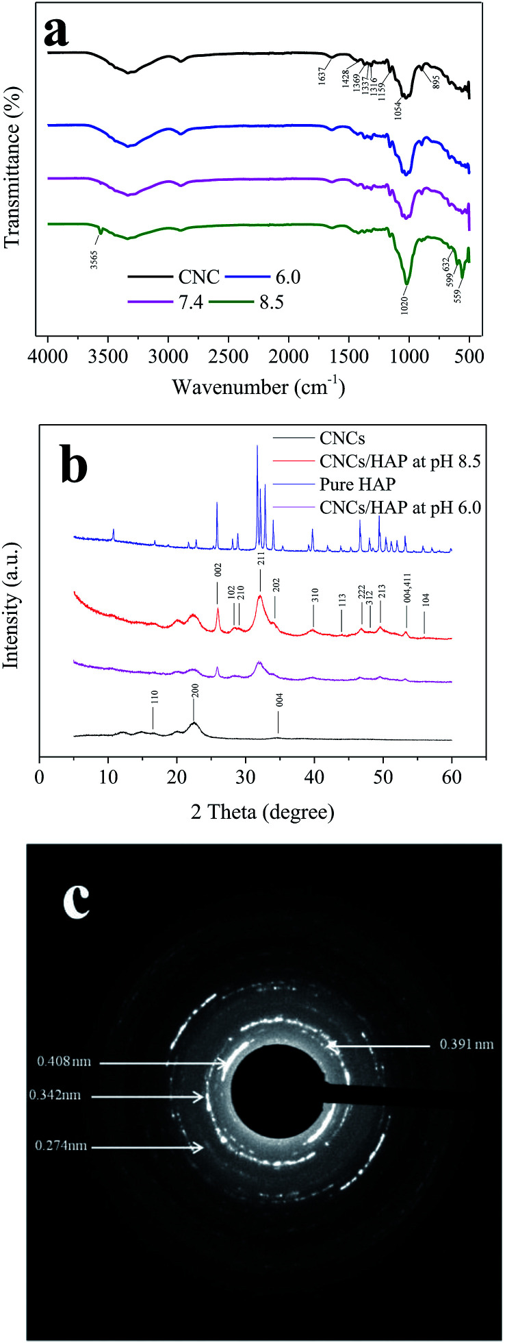
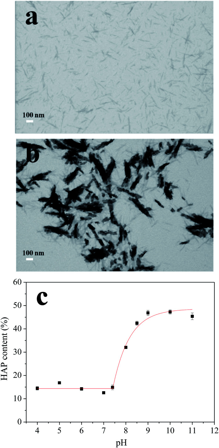
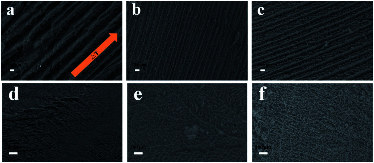
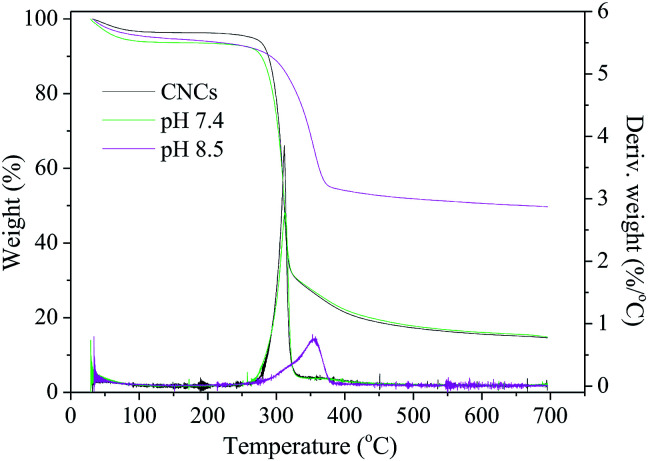
A novel nanocomposite scaffold was developed by homogeneous deposition of hydroxyapatite (HAP) on a cellulose nanocrystals (CNCs) matrix suspended in a simulated body fluid (SBF). By adjusting the pH of the SBF, the HAP content in the nanocomposite could be controlled between 15 wt% and 47 wt%. Physical and chemical characteristics of the nanocomposites were analyzed by SEM, FTIR, XRD, SAED, and TEM, which confirmed the successful incorporation of HAP onto the CNCs. The nanocomposites were then freeze-casted into porous scaffolds by different solidification technologies (i.e., directional freezing (DF), plunging in liquid N2 (PL) or in a -20 °C freezer (FZ)) followed by lyophilization. Compression testing of the HAP/CNCs foams indicated that DF caused significant improvement in mechanical properties due to the specific orientation and anisotropic porous structure compared to conventional freezing methods such as PL and FZ. Moreover, the scaffold with high HAP content exhibited improved mechanical and thermal properties, which holds potential for application in bone tissue engineering.
This journal is © The Royal Society of Chemistry.
Conflict of interest statement
The authors declare no competing financial interests.
Figures
References
-
- Olszta M. J. Cheng X. Jee S. S. Kumar R. Kim Y. Y. Kaufman M. J. Douglas E. P. Gower L. B. Mater. Sci. Eng., R. 2007;58:77–116. doi: 10.1016/j.mser.2007.05.001. - DOI
LinkOut - more resources
Full Text Sources
Miscellaneous