. 2020 Dec 22;10(73):45199-45206.
doi: 10.1039/d0ra09112j. eCollection 2020 Dec 17.

Design, synthesis and bioevaluation of novel 6-substituted aminoindazole derivatives as anticancer agents

Affiliations

Design, synthesis and bioevaluation of novel 6-substituted aminoindazole derivatives as anticancer agents

Ngo Xuan Hoang et al. RSC Adv. .

Abstract

In the present study, a series of 6-substituted aminoindazole derivatives were designed, synthesized, and evaluated for bio-activities. The compounds were initially designed as indoleamine 2,3-dioxygenase 1 (IDO1) inhibitors based on the structural feature of five IDO1 inhibitors, which are currently on clinical trials, and the important anticancer activity of the indazole scaffold. One of them, compound N-(4-fluorobenzyl)-1,3-dimethyl-1H-indazol-6-amine (36), exhibited a potent anti-proliferative activity with an IC50 value of 0.4 ± 0.3 μM in human colorectal cancer cells (HCT116). This compound also remarkably suppressed the IDO1 protein expression. In the cell-cycle studies, the suppressive activity of compound 36 in HCT116 cells was related to the G2/M cell cycle arrest. Altogether, the current findings demonstrate that compound 36 would be promising for further development as a potential anticancer agent.


Conflict of interest statement

There are no conflicts to declare.

Figures

Fig. 1
Fig. 1. (A) Clinical IDO1 inhibitors; (B) some IDO1 inhibitors bearing the indazole structure; (C) novel designed indazole derivatives.
Scheme 1
Scheme 1. Reagents and conditions: (a) CH3I (2.0 equiv.), excess K2CO3, DMF, 60 °C, 4 h, 95–98%; (b) H2, Pd/C (10 mol%), r.t., 4 h, 85–96%; (c) ketone or aldehyde (1.0 equiv.), NaBH3CN (5.0 equiv.), AcOH, MeOH, 40 °C, 4 h, 73–96%; (d) excess Ac2O, TEA, DMC, r.t., 2 h, 89–92%.
Fig. 2
Fig. 2. Effect of 6-substituted aminoindazole derivatives on the IDO1 expression in human colorectal cancer cells. HCT116 were treated with 10 μM of indicative compounds for 24 h. The immunoblotting analysis was performed to determine IDO1 and β-actin expression.
Fig. 3
Fig. 3. HCT116 cells were treated with compound 36 at various concentrations. The immunoblotting analysis was performed to determine IDO1 and β-actin protein levels.
Fig. 4
Fig. 4. Docking model of IDO1 with 5PK and compounds. (A) The docking models of 5PK, compound 35, and compound 36 are shown in yellow green, light yellow, and pink models, respectively. (B) A close-up view of docked compounds in the active site cavity of IDO1. (C) The interaction mode of IDO1 with compound 36. The IDO1 residue recognizing ligands are shown in the green stick model. The heme ion is shown in a grey stick. Hydrogen bonds and π–π interactions are shown in red and black dots, respectively.
Fig. 5
Fig. 5. Anti-proliferative effect of compound 36 on HCT116 cells. HCT116 cells were treated with compound 36 at various concentrations for 72 h, and cell proliferation was determined by the SRB assay. The data were presented as mean ± SD. The experiment was performed in triplicate (n = 3).
Fig. 6
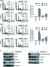
Fig. 6. Effect of compound 36 on the cell cycle distribution in human colorectal cancer cells. (A and B) HCT116 cells were treated with compound 36 for 24 h or 48 h. The cell cycle distribution was analyzed via flow cytometry. (C) Effects of 36 on the expression of cell cycle-related proteins in HCT116 cells. HCT116 cells were treated with 36 for 48 h. Subsequently, the protein expression levels of cell cycle-related proteins were analyzed by the immunoblotting assay.

References

    1. Bach D. H. Kim D. Bae S. Y. Kim W. K. Hong J. Y. Lee H. J. Rajasekaran N. Kwon S. Fan Y. Luu T. T. Shin Y. K. Lee J. Lee S. K. Mol. Ther.--Nucleic Acids. 2018;11:455–467. doi: 10.1016/j.omtn.2018.03.011. - DOI - PMC - PubMed
    1. Bach D. H. Luu T. T. Kim D. An Y. J. Park S. Park H. J. Lee S. K. Mol. Ther.--Nucleic Acids. 2018;12:817–828. doi: 10.1016/j.omtn.2018.07.016. - DOI - PMC - PubMed
    1. Kim D. Bach D. H. Fan Y. H. Luu T. T. Hong J. Y. Park H. J. Lee S. K. Cell Death Dis. 2019;10:361. doi: 10.1038/s41419-019-1601-6. - DOI - PMC - PubMed
    1. Luu T. T. Bach D. H. Kim D. Hu R. Park H. J. Lee S. K. Anticancer Res. 2020;40:1855–1866. doi: 10.21873/anticanres.14139. - DOI - PubMed
    1. Huong P. T. Nguyen L. T. Nguyen X. B. Lee S. K. Bach D. H. Cancers. 2019;11:240. doi: 10.3390/cancers11020240. - DOI - PMC - PubMed