

Alpha-hemolysin nanopore allows discrimination of the microcystins variants
- PMID: 35516306
- PMCID: PMC9064141
- DOI: 10.1039/c8ra10384d
Alpha-hemolysin nanopore allows discrimination of the microcystins variants
Abstract
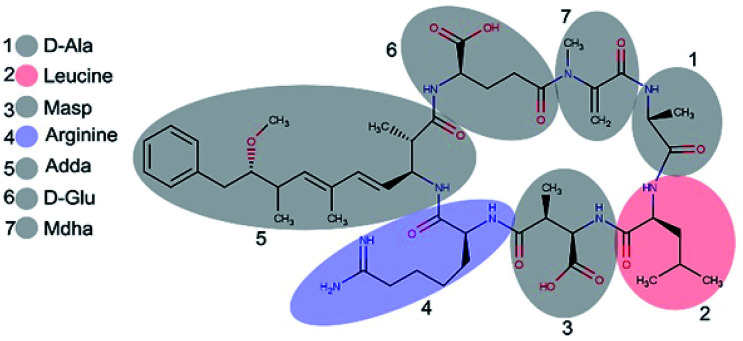
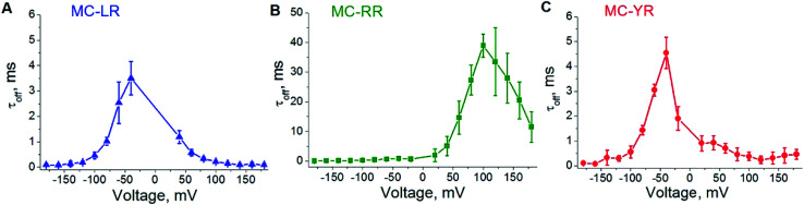
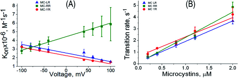
Microcystins (MCs) are a class of cyclic heptapeptides with more than 100 variants produced by cyanobacteria present in surface waters. MCs are potent hepatotoxic agents responsible for fatal poisoning in animals and humans. Several techniques are employed in the detection of MCs, however, there is a shortage of methods capable of discriminating variants of MCs. In this work we demonstrate that the α-hemolysin (αHL) nanopore can detect and discriminate the variants (LR, YR and RR) of MCs in aqueous solution. The discrimination process is based on the analysis of the residence times of each variant of MCs within the unitary nanopore, as well as, on the amplitudes of the blockages in the ionic current flowing through it. Simulations of molecular dynamics and calculation of the electrostatic potential revealed that the variants of MCs present different charge distribution and correlated with the three patterns on the amplitudes of the blockages in the ionic current. Additionally, molecular docking analysis indicates different patterns of interaction of the variants of MCs with two specific regions of the nanopore. We conclude that αHL nanopore can discriminate variants of microcystins by a mechanism based mainly on electrostatic interaction. Finally, we propose the use of nanopore-based technology as a promising method for analyzing microcystins in aqueous solutions.
This journal is © The Royal Society of Chemistry.
Conflict of interest statement
There are no conflicts to declare.
Figures


Similar articles
-
Ecotoxicity assessment of microcystins from freshwater samples using a bioluminescent cyanobacterial bioassay.Chemosphere. 2020 Feb;240:124966. doi: 10.1016/j.chemosphere.2019.124966. Epub 2019 Sep 25. Chemosphere. 2020. PMID: 31726608
-
Single-molecule Study on the Interactions between Cyclic Nonribosomal Peptides and Protein Nanopore.ACS Appl Bio Mater. 2020 Jan 21;3(1):554-560. doi: 10.1021/acsabm.9b00961. Epub 2019 Dec 16. ACS Appl Bio Mater. 2020. PMID: 34169233 Free PMC article.
-
Analysis of microcystins in cyanobacteria blooms and surface water samples from Meiliang Bay, Taihu Lake, China.Environ Int. 2003 Aug;29(5):641-7. doi: 10.1016/S0160-4120(03)00047-3. Environ Int. 2003. PMID: 12742407
-
Exposure routes and health effects of microcystins on animals and humans: A mini-review.Toxicon. 2018 Sep 1;151:156-162. doi: 10.1016/j.toxicon.2018.07.010. Epub 2018 Jul 10. Toxicon. 2018. PMID: 30003917 Review.
-
[Application of ELISA for microcystins detection].Wei Sheng Yan Jiu. 2007 May;36(3):388-90. Wei Sheng Yan Jiu. 2007. PMID: 17712969 Review. Chinese.
Cited by
-
Pioneering Role of Nanopore Single-Molecule Sensing in Environmental and Food Surveillance.Biosensors (Basel). 2025 Jan 13;15(1):41. doi: 10.3390/bios15010041. Biosensors (Basel). 2025. PMID: 39852092 Free PMC article. Review.
-
Long-term memory in Staphylococcus aureus α-hemolysin ion channel kinetics.Eur Biophys J. 2023 Nov;52(8):661-671. doi: 10.1007/s00249-023-01675-8. Epub 2023 Aug 5. Eur Biophys J. 2023. PMID: 37542583
-
Recent advances in biological nanopores for nanopore sequencing, sensing and comparison of functional variations in MspA mutants.RSC Adv. 2021 Aug 31;11(46):28996-29014. doi: 10.1039/d1ra02364k. eCollection 2021 Aug 23. RSC Adv. 2021. PMID: 35478559 Free PMC article. Review.
-
Alphatoxin Nanopore Detection of Aflatoxin, Ochratoxin and Fumonisin in Aqueous Solution.Toxins (Basel). 2023 Feb 28;15(3):183. doi: 10.3390/toxins15030183. Toxins (Basel). 2023. PMID: 36977074 Free PMC article.
-
Nanopore Environmental Analysis.JACS Au. 2025 Apr 1;5(4):1570-1590. doi: 10.1021/jacsau.5c00114. eCollection 2025 Apr 28. JACS Au. 2025. PMID: 40313842 Free PMC article. Review.
References
-
- Puddick J. Prinsep M. R. Wood S. A. Kaufononga S. A. F. Cary S. C. Hamilton D. P. High levels of structural diversity observed in microcystins from microcystis CAWBG11 and characterization of six new microcystin congeners. Mar. Drugs. 2014;12(11):5372–5395. doi: 10.3390/md12115372. - DOI - PMC - PubMed
LinkOut - more resources
Full Text Sources