

Photoinduced electron transfer in novel CdSe-Cu2Se type II core-shell quantum dots
- PMID: 35516312
- PMCID: PMC9064209
- DOI: 10.1039/c9ra02027f
Photoinduced electron transfer in novel CdSe-Cu2Se type II core-shell quantum dots
Abstract
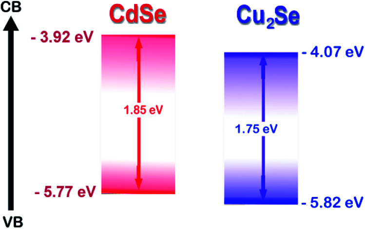
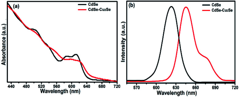
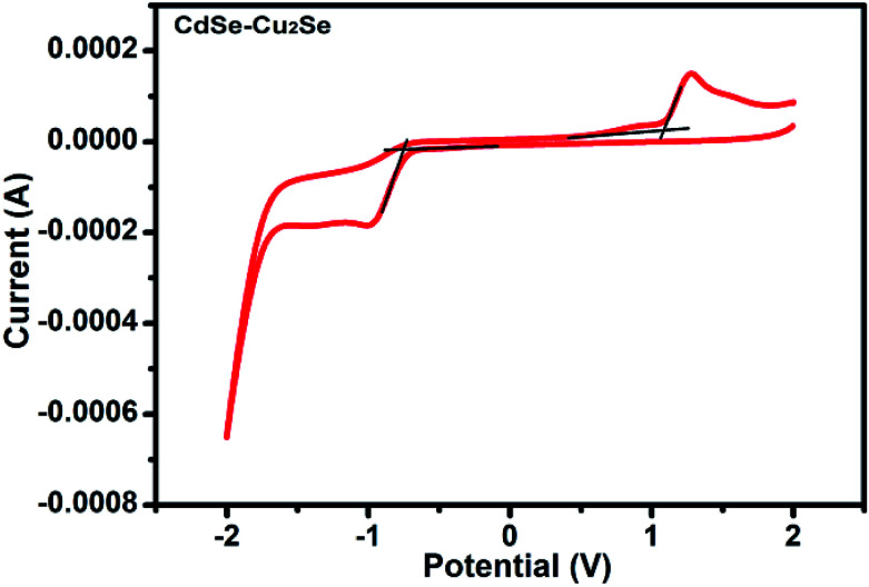
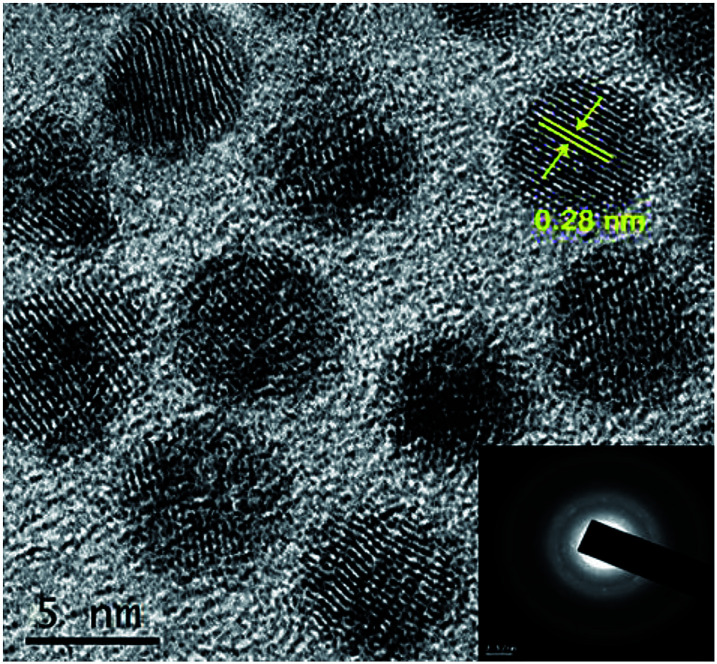
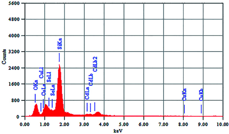
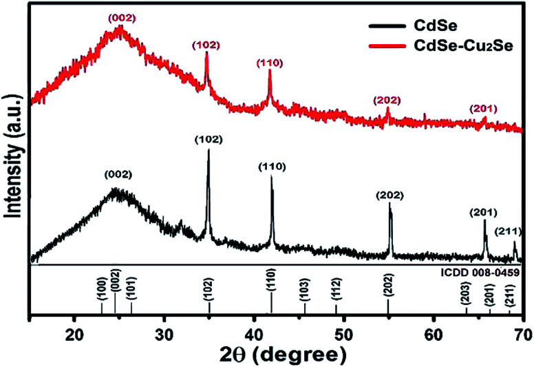
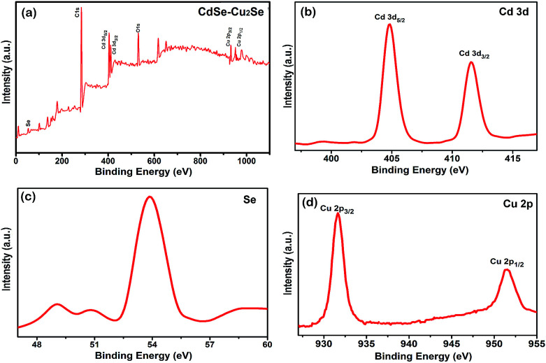
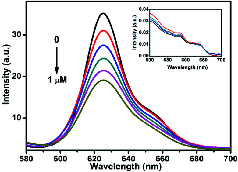
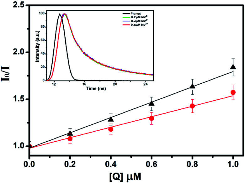
Herein we report the synthesis, characterisation and electron transfer studies of CdSe-Cu2Se QDs, a novel type II core-shell system. The synthesis was achieved by a high temperature organometallic method with oleylamine as ligand. Structural and optical properties of the nanostructures were investigated using X-ray diffraction, high resolution transmission electron microscopy, selected area electron diffraction, energy dispersive X-ray spectroscopy, inductive coupled plasma optical emission spectroscopy, cyclic voltammetry, X-ray photoelectron spectroscopy and absorption spectroscopy. The electron transfer dynamics were investigated by observing the variations in steady state and time resolved emission spectra in the presence of an electron acceptor-methyl viologen. Localization of electrons in the shells was evident from the studies performed indicating efficient charge separation.
This journal is © The Royal Society of Chemistry.
Conflict of interest statement
There are no conflicts to declare.
Figures
Similar articles
-
Surface-state-mediated charge-transfer dynamics in CdTe/CdSe core-shell quantum dots.Chemphyschem. 2011 Jun 20;12(9):1729-35. doi: 10.1002/cphc.201100105. Epub 2011 May 12. Chemphyschem. 2011. PMID: 21567706
-
CuInS2-In2Se3 quantum dots - a novel material via a green synthesis approach.RSC Adv. 2018 Nov 5;8(65):37146-37150. doi: 10.1039/c8ra07389a. eCollection 2018 Nov 1. RSC Adv. 2018. PMID: 35557798 Free PMC article.
-
CuZn2InTe4 quantum dots-a novel nanostructure employing a green synthesis route.RSC Adv. 2020 May 18;10(32):18560-18564. doi: 10.1039/d0ra02980g. eCollection 2020 May 14. RSC Adv. 2020. PMID: 35518340 Free PMC article.
-
Concurrent Ultrafast Electron- and Hole-Transfer Dynamics in CsPbBr3 Perovskite and Quantum Dots.ACS Omega. 2018 Mar 7;3(3):2706-2714. doi: 10.1021/acsomega.8b00276. eCollection 2018 Mar 31. ACS Omega. 2018. PMID: 31458549 Free PMC article.
-
Spectroscopy and femtosecond dynamics of type-II CdTe/CdSe core-shell quantum dots.Chemphyschem. 2006 Jan 16;7(1):222-8. doi: 10.1002/cphc.200500307. Chemphyschem. 2006. PMID: 16404768
Cited by
-
Composite formation in CdSe:Cu2Se nanocrystal films, charge transport characteristics and heterojunction performance.RSC Adv. 2020 Mar 2;10(15):8842-8852. doi: 10.1039/c9ra10251e. eCollection 2020 Feb 27. RSC Adv. 2020. PMID: 35496572 Free PMC article.
References
-
- van Embden J. Jasieniak J. Gómez D. E. Mulvaney P. Giersig M. Aust. J. Chem. 2007;60:457–471. doi: 10.1071/CH07046. - DOI
LinkOut - more resources
Full Text Sources