. 2022 Apr 18:13:856471.
doi: 10.3389/fmicb.2022.856471. eCollection 2022.

Interactome and Ubiquitinome Analyses Identify Functional Targets of Herpes Simplex Virus 1 Infected Cell Protein 0

Affiliations

Interactome and Ubiquitinome Analyses Identify Functional Targets of Herpes Simplex Virus 1 Infected Cell Protein 0

Fujun Hou et al. Front Microbiol. .

Abstract

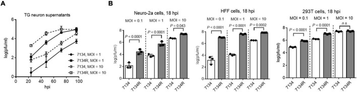
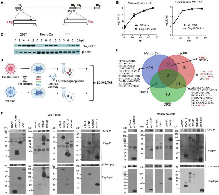
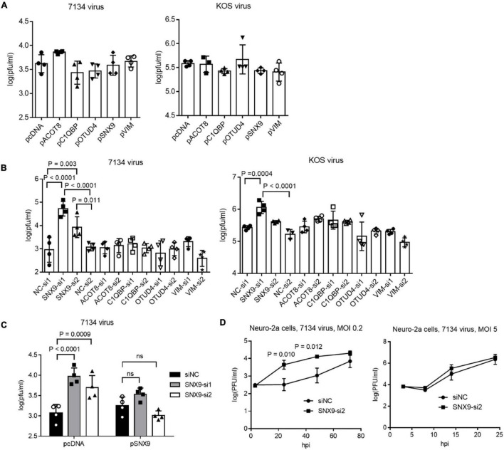
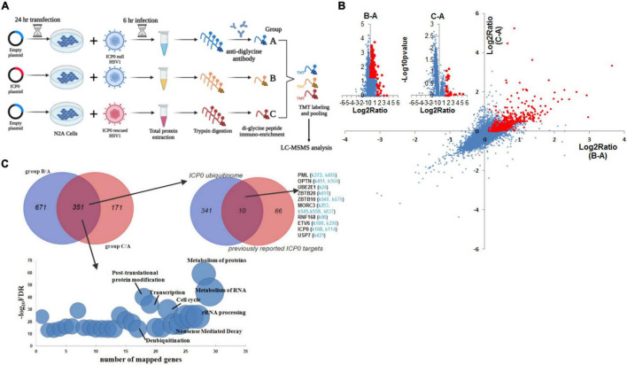
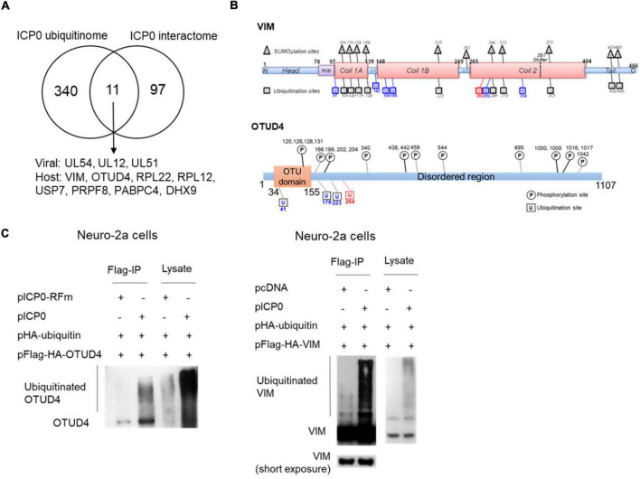
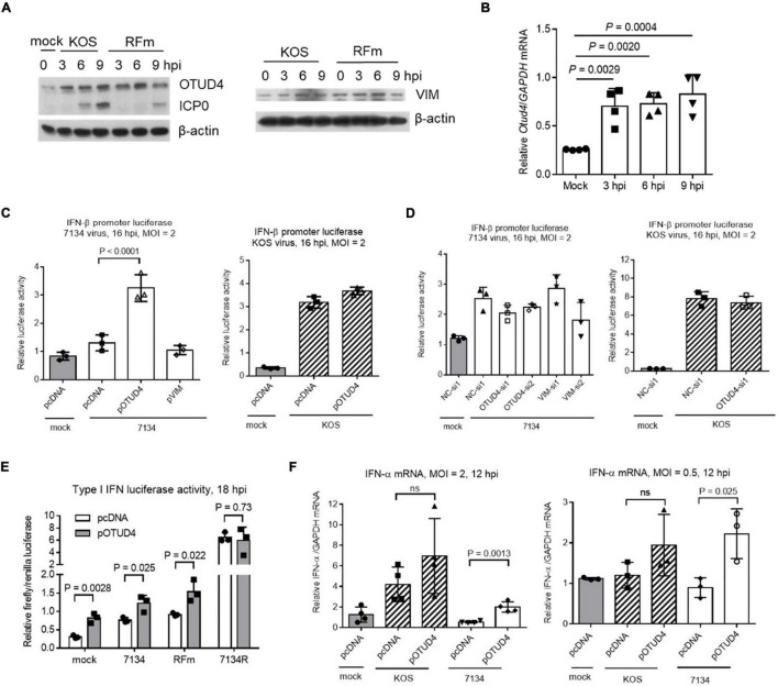
Herpes simplex virus 1 (HSV-1) can productively infect multiple cell types and establish latent infection in neurons. Infected cell protein 0 (ICP0) is an HSV-1 E3 ubiquitin ligase crucial for productive infection and reactivation from latency. However, our knowledge about its targets especially in neuronal cells is limited. We confirmed that, like in non-neuronal cells, ICP0-null virus exhibited major replication defects in primary mouse neurons and Neuro-2a cells. We identified many ICP0-interacting proteins in Neuro-2a cells, 293T cells, and human foreskin fibroblasts by mass spectrometry-based interactome analysis. Co-immunoprecipitation assays validated ICP0 interactions with acyl-coenzyme A thioesterase 8 (ACOT8), complement C1q binding protein (C1QBP), ovarian tumour domain-containing protein 4 (OTUD4), sorting nexin 9 (SNX9), and vimentin (VIM) in both Neuro-2a and 293T cells. Overexpression and knockdown experiments showed that SNX9 restricted replication of an ICP0-null but not wild-type virus in Neuro-2a cells. Ubiquitinome analysis by immunoprecipitating the trypsin-digested ubiquitin reminant followed by mass spectrometry identified numerous candidate ubiquitination substrates of ICP0 in infected Neuro-2a cells, among which OTUD4 and VIM were novel substrates confirmed to be ubiquitinated by transfected ICP0 in Neuro-2a cells despite no evidence of their degradation by ICP0. Expression of OTUD4 was induced independently of ICP0 during HSV-1 infection. Overexpressed OTUD4 enhanced type I interferon expression during infection with the ICP0-null but not wild-type virus. In summary, by combining two proteomic approaches followed by confirmatory and functional experiments, we identified and validated multiple novel targets of ICP0 and revealed potential restrictive activities of SNX9 and OTUD4 in neuronal cells.

Keywords: ICP0; herpes simplex virus; interactome; proteomics; ubiquitinome.


Conflict of interest statement

The authors declare that the research was conducted in the absence of any commercial or financial relationships that could be construed as a potential conflict of interest.

Figures

FIGURE 1
FIGURE 1
ICP0 is important for HSV-1 replication in neuronal cells. (A) Primary mouse TG neurons were infected with 7134 or 7134R at an MOI of 1 or 10 as indicated. At the indicated times post-infection, supernatants were collected for virus titration. (B) Neuro-2a (left), HFF (middle), and 293T (right) cells were infected with 7134 or 7134R at the indicated MOI and harvested at 18 hpi for virus titration. Data were analyzed by one-way ANOVA with Sidak’s multiple comparisons tests and are presented as mean values ± standard deviations (SD).
FIGURE 2
FIGURE 2
ICP0-interactome analysis. (A) Diagram of recombinant Flag-ICP0 virus. The horizontal line at the top represents HSV-1 genome and the boxes labeled with TRL, IRL, TRS, and IRS represent the repeated sequences. At the bottom are expanded ICP0 gene regions with the inserted N-terminal Flag tags indicated. (B) Virus growth curves of WT and Flag-ICP0 virus in Vero (left) and Neuro-2a (right) cells after infection at an MOI of 0.01 and 0.1, respectively. Data are presented as mean values ± SD. (C) Neuro-2a, HFF, and 293T cells were infected with Flag-ICP0 virus at an MOI of 10 and cells were lysed at the indicated time points for examination of Flag-ICP0 expression by Western blots with anti-Flag and anti-β-actin antibodies. (D) Schematic diagram of the procedures for ICP0 interactome analysis. Neuro-2a, 293T, and HFF cells were infected with WT (control) or Flag-ICP0 virus for 6 h at an MOI of 20 with three biological replicates. Proteins were immunoprecipitated by the anti-Flag antibody and examined by liquid chromatography with tandem mass spectrometry (LC-MS/MS). (E) Venn diagram showing candidate ICP0-interacting proteins identified by the interactome analysis in the three cell types. Previously reported ICP0-interacting proteins are marked in red. (F) 293T (left) or Neuro-2a (right) cells in each 100-mm plate were co-transfected with 7.5 μg of an empty vector (pcDNA) or a plasmid expressing the indicated protein linked to a Flag-tag and 7.5 μg of a plasmid expressing untagged ICP0 for 48 h. After the cells were lysed, the indicated proteins were immunoprecipitated by an anti-Flag antibody and analyzed by Western blots for ICP0 and the corresponding proteins using an ICP0 and Flag antibody, respectively. Results from immunoprecipitated samples (IP) were shown in the upper panels and those from the lysates (Input) were shown in the lower panels. Note that prolyl 4-hydroxylase subunit alpha 2 (P4HA2) and OTUD4 expression from the plasmids were not detected in the input samples due to low expression but were clearly detected in the IP samples.
FIGURE 3
FIGURE 3
Effects of ICP0-interacting proteins on HSV-1 replication in Neuro-2a cells. (A) Neuro-2a cells were transfected with the indicated plasmids (400 ng/ml) for 40 h and then infected with 7134 (left) or KOS (right) virus for 48 h at an MOI of 0.2 before the cells were harvested for virus titration by plaque assays. (B) Neuro-2a cells were transfected with the indicated siRNAs (80 nM) for 40 h and then infected with 7134 (left) or KOS (right) virus for 48 h at an MOI of 0.2 before the cells were harvested for virus titration. NC, negative control. (C) Neuro-2a cells were transfected with the indicated siRNAs (80 nM) for 17 h and then transfected with the indicated plasmids (600 ng/ml) for 33 h before infection with 7134 virus at an MOI of 0.2 for 44 h. The cells were then harvested for viral titration. (D) Neuro-2a cells were transfected with a negative control siRNA or an siRNA against SNX9 for 40 h before infection with 7134 virus at an MOI of 0.2 (left) or 5 (right). The cells were harvested at the indicated times for virus titration. Data were analyzed by one-way ANOVA with Dunnett’s multiple comparisons tests (A,B), two-way ANOVA with Sidak’s multiple comparisons tests (C), or two-tailed unpaired t-tests (D). Data are presented as mean values ± SD.
FIGURE 4
FIGURE 4
ICP0 ubiquitinome analysis in Neuro-2a cells. (A) Schematic diagram of the experimental design for ICP0 ubiquitinome analysis. (B) Potential ICP0 target sites (red) showing higher ubiquitination levels in group B than A (x-axis) or those showing higher ubiquitination levels in group C than A (y-axis) according to quantitative mass spectrometry. Individual volcano diagrams showing B-A and C-A comparisons were also displayed in the upper left corner. (C) Potential ICP0 ubiquitination target proteins identified by comparing groups B and A as well as comparing groups C and A. The overlap of the two comparisons represents 351 candidate targets for subsequent analysis. The pathways enriched in these proteins are shown in the bottom graph. The upper right Venn diagram shows the overlap of our ICP0 ubiquitinome results and previously reported ICP0 substrates or interacting proteins. For the targets in our data that were also previously reported, potential ubiquitination sites on lysine differentially represented between ICP0-positive and negative cells in our data are indicated in light blue.
FIGURE 5
FIGURE 5
ICP0-mediated ubiquitination of OTUD4 and VIM. (A) Venn diagram showing 12 proteins identified by both ICP0 interactome and ubiquitinome analyses. (B) Summary of ubiquitination sites on VIM (upper) and OTUD4 (lower). Schematic representation of functional domains of VIM and OTUD4 decorated with previously documented SUMOylation (for VIM, in triangles) or phosphorylation (for OTUD4, in circles) sites and newly identified ubiquitination sites (in squares). L, linker region; PCD, pre-coiled domain. Blue indicates sites with ubiquitination but not SUMOylation. Red indicates the ubiquitination site potentially targeted by ICP0. (C) Neuro-2a cells in each 100-mm plate were co-transfected with 5 μg of a plasmid expressing Flag-HA-tagged OTUD4 (left) or VIM (right), 2 μg of a plasmid expressing HA-tagged ubiquitin, and 3 μg of a plasmid expressing ICP0 or its RING finger mutant (RFm) or an empty vector (pcDNA) for 48 h. Cell lysates were immunoprecipitated by an anti-Flag antibody. The lysates and immunoprecipitated samples (IP) were analyzed by Western blots for ubiquitination levels by an anti-HA antibody.
FIGURE 6
FIGURE 6
Functions of OTUD4 and VIM in Neuro-2a cells. (A) Cells were infected with KOS or ICP0 RING finger mutant virus (RFm) at an MOI of 20 for the indicated times before the cells were harvested for Western blot analysis for the indicated proteins. (B) After infection of cells with KOS at an MOI of 20, the cells were harvested at the indicated times for qRT-PCR analysis of Otud4 mRNA levels normalized to glyceraldehyde-3 phosphate dehydrogenase (GAPDH) mRNA levels. (C) Cells were co-transfected with the indicated plasmid (400 ng/ml), pIFN-β (80 ng/ml) (an IFN-β promoter luciferase plasmid), and pRL-TK (40 ng/ml; a control plasmid expressing renilla luciferase) for 48 h and then infected with 7134 (left) or KOS (right) at an MOI of 2 for 16 h before measurement of luciferase activities. (D) Neuro-2a cells were co-transfected with the indicated siRNA (80 nM), pIFN-β (80 ng/ml), and pRL-TK (40 ng/ml) for 48 h and then infected with 7134 (left) or KOS (right) at an MOI of 2 for 16 h before measurement of luciferase activities. (E) The assay was performed as in (C) except that different viruses were used. (F) Cells were transfected with the indicated plasmid (400 ng/ml) and then infected with the indicated virus at an MOI of 2 (left) or 0.5 (right) for 12 h before qRT-PCR quantification of IFN-α mRNA normalized to GAPDH mRNA. Data were analyzed by one-way ANOVA with Dunnett’s multiple comparisons tests (B–D,F) or two-tailed unpaired t-tests (E). Data are presented as mean values ± SD.

Similar articles

Cited by

References

    1. Alandijany T., Roberts A. P. E., Conn K. L., Loney C., Mcfarlane S., Orr A., et al. (2018). Distinct temporal roles for the promyelocytic leukaemia (PML) protein in the sequential regulation of intracellular host immunity to HSV-1 infection. PLoS Pathog. 14:e1006769. 10.1371/journal.ppat.1006769 - DOI - PMC - PubMed
    1. Bendris N., Schmid S. L. (2017). Endocytosis, metastasis and beyond: multiple facets of SNX9. Trends Cell Biol. 27 189–200. 10.1016/j.tcb.2016.11.001 - DOI - PMC - PubMed
    1. Blum M., Chang H. Y., Chuguransky S., Grego T., Kandasaamy S., Mitchell A., et al. (2020). The InterPro protein families and domains database: 20 years on. Nucleic Acids Res. 49 D344–D354. 10.1093/nar/gkaa977 - DOI - PMC - PubMed
    1. Boutell C., Everett R. D. (2003). The herpes simplex virus type 1 (HSV-1) regulatory protein ICP0 interacts with and Ubiquitinates p53. J. Biol. Chem. 278 36596–36602. 10.1074/jbc.M300776200 - DOI - PubMed
    1. Boutell C., Canning M., Orr A., Everett R. D. (2005). Reciprocal activities between herpes simplex virus type 1 regulatory protein ICP0, a ubiquitin E3 ligase, and ubiquitin-specific protease USP7. J. Virol. 79 12342–12354. 10.1128/JVI.79.19.12342-12354.2005 - DOI - PMC - PubMed