. 2020 Sep 1;10(54):32393-32399.
doi: 10.1039/d0ra05117a.

Reporter-recruiting bifunctional aptasensor for bioluminescent analytical assays

Affiliations

Reporter-recruiting bifunctional aptasensor for bioluminescent analytical assays

Anna Davydova et al. RSC Adv. .

Abstract

We report a novel bioluminescent aptasensor, which consists of 2'-F-RNA aptamer modules joined into a bi-specific aptamer construct. One aptamer module binds the analyte, then after structural rearrangement the second module recruits non-covalently Ca2+-dependent photoprotein obelin from the solution, thus providing a bioluminescent signal. This concept allows using free protein as a reporter, which brings such advantages as no need for aptamer-protein conjugation, a possibility of thermal re-folding of aptamer component with no harm to a protein, and simpler detection protocol. We developed the new 2'-F-RNA aptamer for obelin, and proposed the strategy for engineering structure-switching bi-modular aptamer constructs which bind the analyte and the obelin in a sequential manner. With the use of hemoglobin as a model analyte, we showed the feasibility of utilizing the aptasensor in a fast and straightforward bioluminescent microplate assay. With a proper design of a secondary structure, this strategy of aptasensor engineering might be further extended to bi-specific aptamer-based bioluminescent sensors for other analytes of interest.


Conflict of interest statement

There are no conflicts to declare.

Figures

Fig. 1
Fig. 1. Obelin-binding aptamer O79 and its truncated versions. (A) Predicted secondary structures for O79, O79t, O79t1. (B) Example of binding of O79t to close related photoproteins obelin, aequorin, and clytin examined by the DRaCALA assay.
Fig. 2
Fig. 2. A general scheme of microplate bioluminescent detection of an analyte with bi-specific aptamers.
Fig. 3
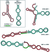
Fig. 3. Secondary structures of obelin-binding (red) and hemoglobin-binding (blue) aptamer modules and bi-specific aptamers on their basis (obtained by Vienna RNA fold). Dashed lines connect G residues involved in quadruplex formation. Hexanucleotide linker (green) between the modules is derived from the 3′-terminal fragment of O79t1.
Fig. 4
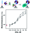
Fig. 4. Bioluminescent detection of human hemoglobin with bispecific aptamers Apt79t1-H9t11 (–□–), H9t11-L-Apt79t1 (–○–) or Apt79t1-L-H9t11 (–Δ–) at simultaneous addition of obelin (100 nM) and hemoglobin (1.56–100 nM). Red symbols show the signals from hemoglobin-free (control) samples. Biotinylated aptamers were immobilized in streptavidin-coated microplate wells. Each point is the average ± standard deviation, n = 3.

References

    1. Poolsup S. Kim C. Y. Curr. Opin. Biotechnol. 2017;48:180–186. doi: 10.1016/j.copbio.2017.05.004. - DOI - PubMed
    1. Zhang H. Zhou L. Zhu Z. Yang C. Chem.–Eur. J. 2016;22:9886–9900. doi: 10.1002/chem.201503543. - DOI - PubMed
    1. Ni S. Yao H. Wang L. Lu J. Jiang F. Lu A. Zhang G. Int. J. Mol. Sci. 2017;18:1683. doi: 10.3390/ijms18081683. - DOI - PMC - PubMed
    1. Wang G. Wang Y. Chen L. Choo J. Biosens. Bioelectron. 2010;25:1859–1868. doi: 10.1016/j.bios.2009.11.012. - DOI - PubMed
    1. Fallmann J. Will S. Engelhardt J. Grüning B. Backofen R. Stadler P. F. J. Biotechnol. 2017;261:97–104. doi: 10.1016/j.jbiotec.2017.07.007. - DOI - PubMed