

Testicular alterations in cryptorchid/orchiopexic rats chronically exposed to acrylamide or di-butyl-phthalate
- PMID: 35516837
- PMCID: PMC9018398
- DOI: 10.1293/tox.2021-0045
Testicular alterations in cryptorchid/orchiopexic rats chronically exposed to acrylamide or di-butyl-phthalate
Abstract
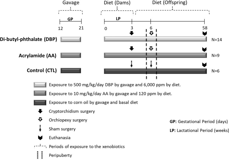
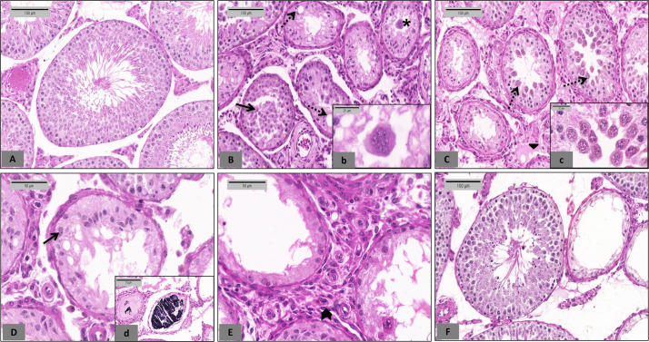
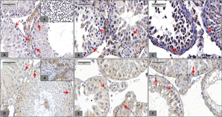
Exposure of Sprague-Dawley (SD) rats to acrylamide (AA) or di-butyl-phthalate (DBP) from the 12th gestational day to the 16th postnatal week (PNW) has been shown to reduce the effectiveness of orchiopexy in recovering the testicular alterations associated with experimental cryptorchidism established at weaning. Herein, we provide information about the long-term effects of AA or DBP on the testes of cryptorchid/orchiopexic rats. Male offspring exposed in utero to 10 mg/kg/day AA or 500 mg/kg/day DBP underwent bilateral surgical cryptorchidism at the 3rd PNW and orchiopexy at the 6th week, with continuous exposure to the chemicals through diet until the 58th week. Regardless of the test chemical, there were severe qualitative/quantitative alterations in the seminiferous tubules and increased numbers of Leydig cells. There was an increase and decrease in the number of tubules with c-Kit- and placental alkaline phosphatase-labeled germ cells, respectively, as compared to those in the control group, suggesting an imbalance between apoptosis and cell proliferation processes. The histological scores of the testicular lesions at the end of this one-year study were higher than those in the previous 16-week study, indicating that exposure of rats to the toxicants AA or DBP enhanced the testicular alterations induced by the chemicals beginning at the intra-uterine life, and impaired the effectiveness of orchiopexy in restoring the testes to normal morphology. Although the present experimental protocol does not completely replicate the natural human undescended testes, our findings may contribute to understanding the alterations occurring in cryptorchid/orchiopexic testes potentially exposed to exogenous chemicals for extended periods.
Keywords: acrylamide; chronic toxicity; di-butyl-phthalate; orchiopexy; rat surgical cryptorchidism; testicular germ cells.
©2022 The Japanese Society of Toxicologic Pathology.
Figures
Similar articles
-
Experimental cryptorchidism enhances testicular susceptibility to dibutyl phthalate or acrylamide in Sprague-Dawley rats.Hum Exp Toxicol. 2019 Aug;38(8):899-913. doi: 10.1177/0960327119845040. Epub 2019 Apr 17. Hum Exp Toxicol. 2019. PMID: 30995857
-
NTP technical report on the toxicity studies of Dibutyl Phthalate (CAS No. 84-74-2) Administered in Feed to F344/N Rats and B6C3F1 Mice.Toxic Rep Ser. 1995 Apr;30:1-G5. Toxic Rep Ser. 1995. PMID: 12209194
-
Time response of rat testicular alterations induced by cryptorchidism and orchiopexy.Int J Exp Pathol. 2021 Feb;102(1):57-69. doi: 10.1111/iep.12384. Int J Exp Pathol. 2021. PMID: 33502821 Free PMC article.
-
Disruption of reproductive development in male rat offspring following in utero exposure to phthalate esters.Int J Androl. 2006 Feb;29(1):140-7; discussion 181-5. doi: 10.1111/j.1365-2605.2005.00563.x. Epub 2005 Aug 11. Int J Androl. 2006. PMID: 16102138 Review.
-
A narrative review of the history and evidence-base for the timing of orchidopexy for cryptorchidism.J Pediatr Urol. 2021 Apr;17(2):239-245. doi: 10.1016/j.jpurol.2021.01.013. Epub 2021 Jan 23. J Pediatr Urol. 2021. PMID: 33551366 Review.
Cited by
-
Dietary Acrylamide: A Detailed Review on Formation, Detection, Mitigation, and Its Health Impacts.Foods. 2024 Feb 12;13(4):556. doi: 10.3390/foods13040556. Foods. 2024. PMID: 38397533 Free PMC article. Review.
References
-
- Skakkebaek NE, Rajpert-De Meyts E, Buck Louis GM, Toppari J, Andersson A-M, Eisenberg ML, Jensen TK, Jørgensen N, Swan SH, Sapra KJ, Ziebe S, Priskorn L, and Juul A. Male reproductive disorders and fertility trends: influences of environment and genetic susceptibility. Physiol Rev. 96: 55–97. 2016. - PMC - PubMed
-
- Toppari J, Virtanen HE, Main KM, and Skakkebaek NE. Cryptorchidism and hypospadias as a sign of testicular dysgenesis syndrome (TDS): environmental connection. Birth Defects Res A Clin Mol Teratol. 88: 910–919. 2010; . - PubMed
-
- Foresta C, Zuccarello D, Garolla A, and Ferlin A. Role of hormones, genes, and environment in human cryptorchidism. Endocr Rev. 29: 560–580. 2008. - PubMed