

Malignant stromal cell tumor of the spleen in a WBN/Kob rat
- PMID: 35516839
- PMCID: PMC9018399
- DOI: 10.1293/tox.2021-0067
Malignant stromal cell tumor of the spleen in a WBN/Kob rat
Abstract
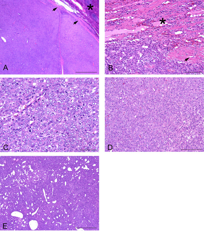
Primary splenic stromal tumors have rarely been reported in rodents. We report the case of a 90-week-old male WBN/Kob rat with a nodular demarcated mass in the spleen, which was kept as a non-treated animal in a long-term animal study. Histopathology revealed round to short spindle-shaped tumor cells arranged in a solid growth pattern. Invasive growth, anisokaryosis, and high mitotic activity (46 per 10 high-power fields [2.37 mm2]) were observed to be multifocal, but most tumor cells showed mild nuclear pleomorphism. The pattern of silver impregnation corresponded to that of the marginal zone of the red pulp. Immunohistochemistry revealed that the tumor cells were double positive for fascin and desmin and focally positive for Iba-1 and OX-6 expression. These characteristics were similar to those observed in fibroblastic reticular cells and dendritic cells in the marginal zone of the red pulp. These findings suggest that the malignant stromal cell tumor of the spleen in this case had characteristics of both fibroblastic reticular cells and dendritic cells.
Keywords: dendritic cell; fibroblastic reticular cell; rat; spleen; stromal cell tumor.
©2022 The Japanese Society of Toxicologic Pathology.
Figures


Similar articles
-
Monoclonal antibodies against mouse splenic stromal cells.Pathol Int. 1997 May;47(5):275-81. doi: 10.1111/j.1440-1827.1997.tb04493.x. Pathol Int. 1997. PMID: 9143021
-
Fibroblastic/cytokeratin-positive interstitial reticular cell tumor of the spleen with indolent behavior: a case report with review of the literature.Virchows Arch. 2023 Jun;482(6):1069-1077. doi: 10.1007/s00428-022-03463-9. Epub 2022 Nov 28. Virchows Arch. 2023. PMID: 36441242 Review.
-
Fibroblastic Reticulum Cell Tumor of Spleen: A Case Report.Int J Surg Pathol. 2014 Aug;22(5):447-50. doi: 10.1177/1066896913509009. Epub 2013 Nov 12. Int J Surg Pathol. 2014. PMID: 24220998
-
Heterogeneity of stromal cells in the human splenic white pulp. Fibroblastic reticulum cells, follicular dendritic cells and a third superficial stromal cell type.Immunology. 2014 Nov;143(3):462-77. doi: 10.1111/imm.12325. Immunology. 2014. PMID: 24890772 Free PMC article.
-
Human spleen microanatomy: why mice do not suffice.Immunology. 2015 Jul;145(3):334-46. doi: 10.1111/imm.12469. Immunology. 2015. PMID: 25827019 Free PMC article. Review.
References
-
- Willard-Mack CL, Elmore SA, Hall WC, Harleman J, Kuper CF, Losco P, Rehg JE, Rühl-Fehlert C, Ward JM, Weinstock D, Bradley A, Hosokawa S, Pearse G, Mahler BW, Herbert RA, and Keenan CM. Nonproliferative and proliferative lesions of the rat and mouse hematolymphoid system. Toxicol Pathol. 47: 665–783. 2019. - PMC - PubMed
-
- Dalia S, Shao H, Sagatys E, Cualing H, and Sokol L. Dendritic cell and histiocytic neoplasms: biology, diagnosis, and treatment. Cancer Contr. 21: 290–300. 2014. - PubMed
-
- Martel M, Sarli D, Colecchia M, Coppa J, Romito R, Schiavo M, Mazzaferro V, and Rosai J. Fibroblastic reticular cell tumor of the spleen: report of a case and review of the entity. Hum Pathol. 34: 954–957. 2003. - PubMed