

Polydopamine-modified poly(l-lactic acid) nanofiber scaffolds immobilized with an osteogenic growth peptide for bone tissue regeneration
- PMID: 35516986
- PMCID: PMC9063423
- DOI: 10.1039/c8ra08828d
Polydopamine-modified poly(l-lactic acid) nanofiber scaffolds immobilized with an osteogenic growth peptide for bone tissue regeneration
Abstract
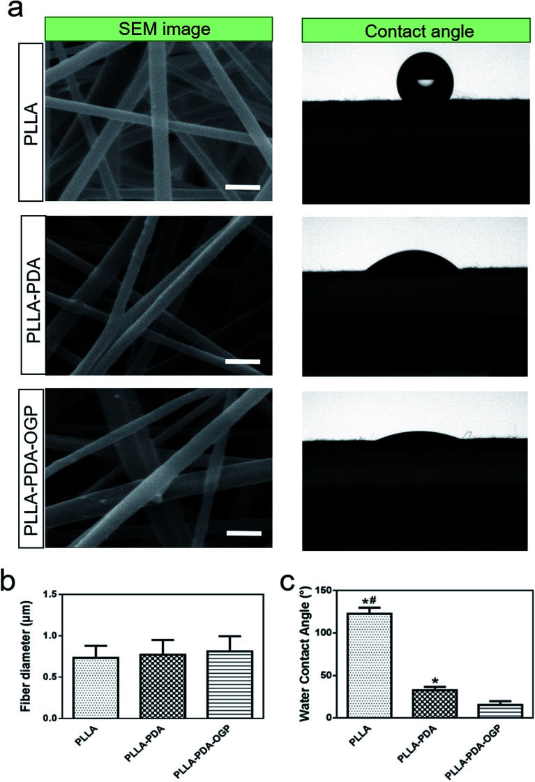
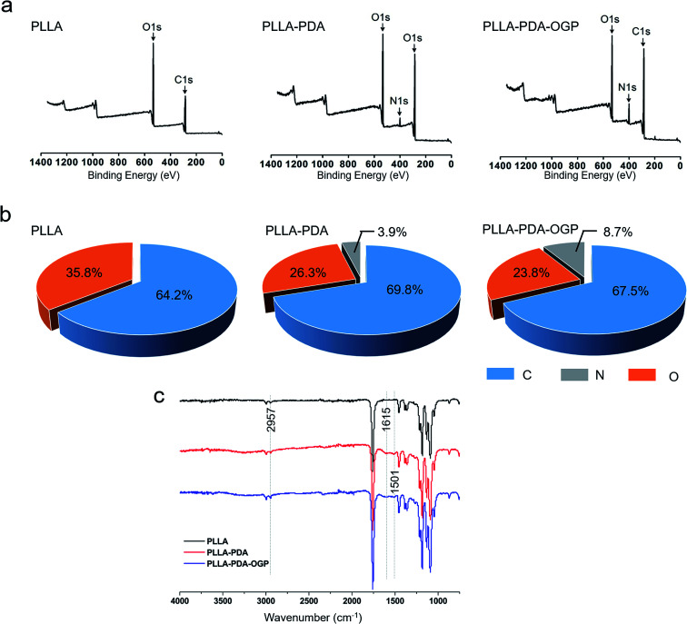
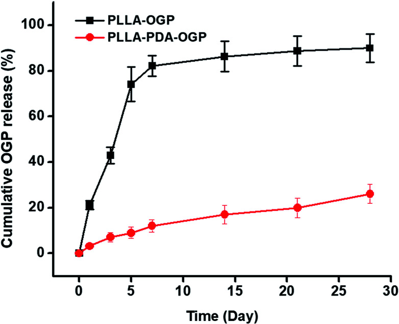
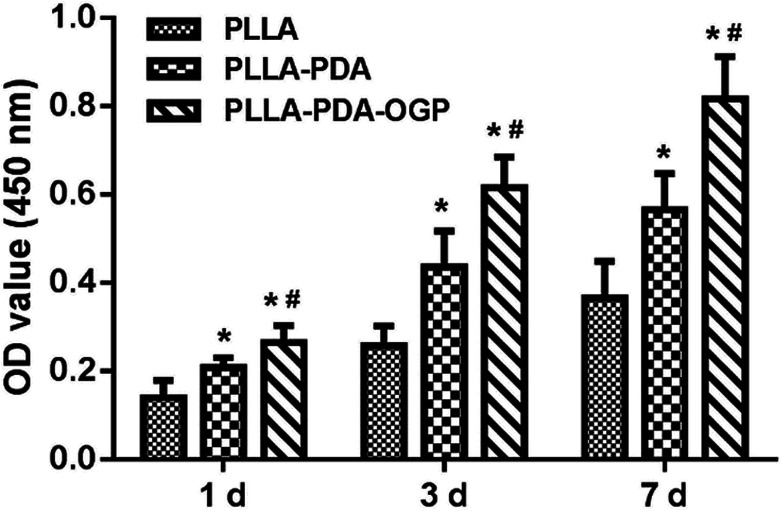
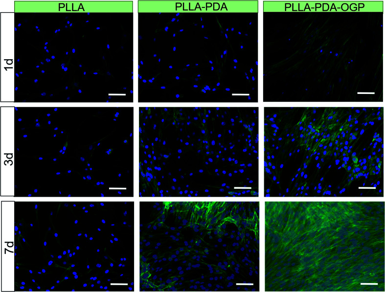
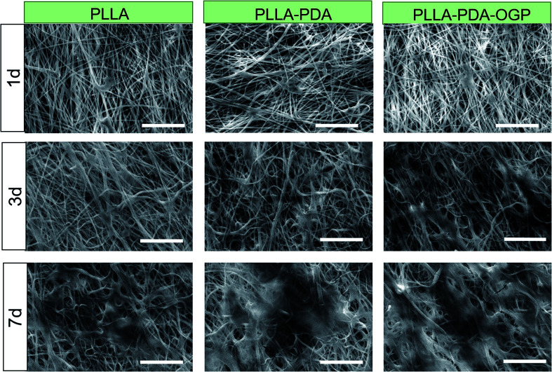
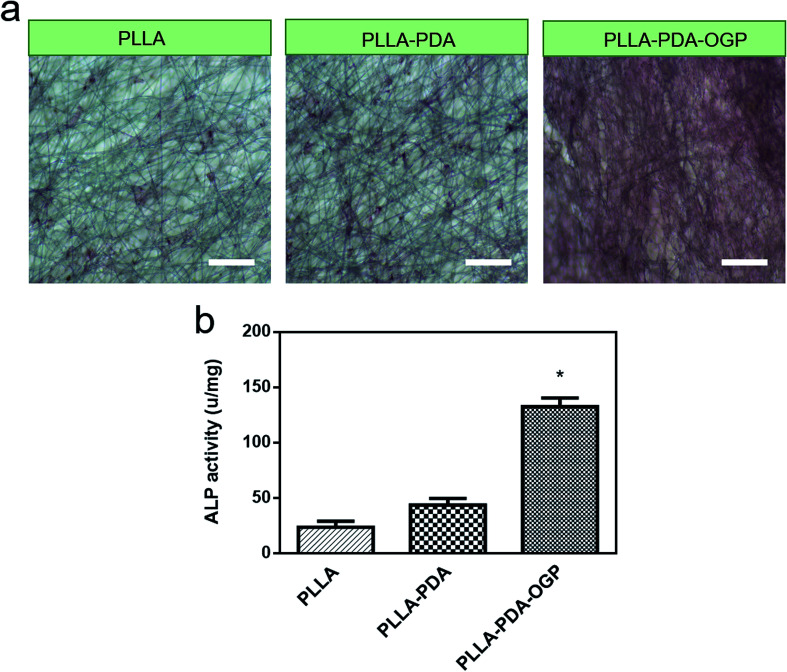
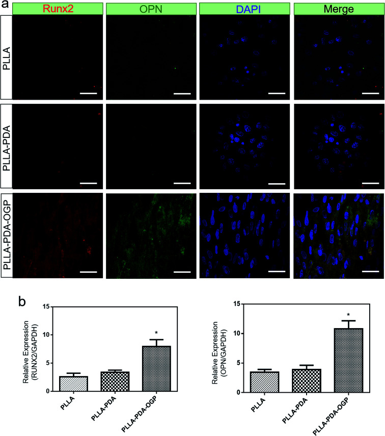
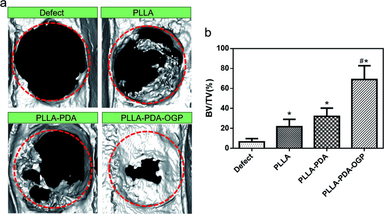
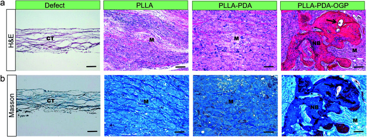
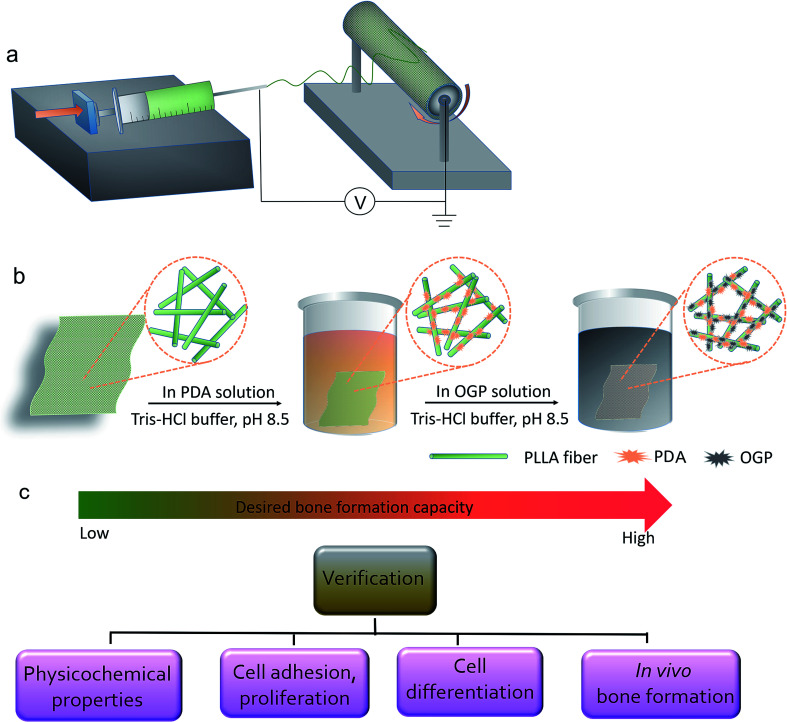
It is highly desirable for bone tissue engineering scaffolds to have significant osteogenic properties and capability to improve cell growth and thus enhance bone regeneration. In this study, a poly(l-lactic acid) (PLLA) nanofiber scaffold-immobilized osteogenic growth peptide (OGP) was prepared via polydopamine (PDA) coating. X-ray photoelectron spectroscopy (XPS), contact angle measurement, and scanning electron microscopy (SEM) were used to determine the OGP immobilization, hydrophilicity and surface roughness of the samples. The SEM and fluorescence images demonstrate that the PLLA nanofiber scaffolds immobilized with the OGP have excellent cytocompatibility in terms of cell adhesion and proliferation. The ALP activity and the Runx2 and OPN expression results indicated that the PLLA nanofiber scaffolds immobilized with OGP significantly enhanced the osteogenic differentiation and calcium mineralization of hMSCs in vitro. A rat model of critical skull bone defect was selected to evaluate the bone formation capacity of the scaffolds. Micro CT analysis and histological results demonstrated that the PLLA scaffolds immobilized with OGP significantly promoted bone regeneration in critical-sized bone defects. This study verifies that the PLLA scaffold-immobilized OGP has significant potential in bone tissue engineering.
This journal is © The Royal Society of Chemistry.
Conflict of interest statement
There are no conflicts to declare.
Figures
Similar articles
-
Evaluation of cell adhesion and osteoconductivity in bone substitutes modified by polydopamine.Front Bioeng Biotechnol. 2023 Jan 13;10:1057699. doi: 10.3389/fbioe.2022.1057699. eCollection 2022. Front Bioeng Biotechnol. 2023. PMID: 36727042 Free PMC article. Review.
-
Polydopamine-coated biomimetic bone scaffolds loaded with exosomes promote osteogenic differentiation of BMSC and bone regeneration.Regen Ther. 2023 Mar 30;23:25-36. doi: 10.1016/j.reth.2023.03.005. eCollection 2023 Jun. Regen Ther. 2023. PMID: 37063095 Free PMC article.
-
[Electrospun PLGA scaffold loaded with osteogenic growth peptide accelerates cranial bone repair in rats].Nan Fang Yi Ke Da Xue Xue Bao. 2021 Aug 20;41(8):1183-1190. doi: 10.12122/j.issn.1673-4254.2021.08.09. Nan Fang Yi Ke Da Xue Xue Bao. 2021. PMID: 34549709 Free PMC article. Chinese.
-
[Preparation and osteogenic properties of poly ( L-lactic acid)/lecithin porous scaffolds with open pore structure].Zhongguo Xiu Fu Chong Jian Wai Ke Za Zhi. 2018 Sep 15;32(9):1123-1130. doi: 10.7507/1002-1892.201804127. Zhongguo Xiu Fu Chong Jian Wai Ke Za Zhi. 2018. PMID: 30701727 Free PMC article. Chinese.
-
Polydopamine-assisted osteoinductive peptide immobilization of polymer scaffolds for enhanced bone regeneration by human adipose-derived stem cells.Biomacromolecules. 2013 Sep 9;14(9):3202-13. doi: 10.1021/bm4008343. Epub 2013 Aug 29. Biomacromolecules. 2013. PMID: 23941596
Cited by
-
Electrospinning: A New Frontier in Peptide Therapeutics.AAPS PharmSciTech. 2025 Feb 26;26(3):69. doi: 10.1208/s12249-025-03054-2. AAPS PharmSciTech. 2025. PMID: 40011310 Review.
-
3D Printed Gelatin/Sodium Alginate Hydrogel Scaffolds Doped with Nano-Attapulgite for Bone Tissue Repair.Int J Nanomedicine. 2021 Dec 30;16:8417-8432. doi: 10.2147/IJN.S339500. eCollection 2021. Int J Nanomedicine. 2021. PMID: 35002236 Free PMC article.
-
Sustained delivery of osteogenic growth peptide through injectable photoinitiated composite hydrogel for osteogenesis.Front Bioeng Biotechnol. 2023 Aug 8;11:1228250. doi: 10.3389/fbioe.2023.1228250. eCollection 2023. Front Bioeng Biotechnol. 2023. PMID: 37614629 Free PMC article.
-
Evaluation of cell adhesion and osteoconductivity in bone substitutes modified by polydopamine.Front Bioeng Biotechnol. 2023 Jan 13;10:1057699. doi: 10.3389/fbioe.2022.1057699. eCollection 2022. Front Bioeng Biotechnol. 2023. PMID: 36727042 Free PMC article. Review.
-
Liquid Crystal Modified Polylactic Acid Improves Cytocompatibility and M2 Polarization of Macrophages to Promote Osteogenesis.Front Bioeng Biotechnol. 2022 Jun 17;10:887970. doi: 10.3389/fbioe.2022.887970. eCollection 2022. Front Bioeng Biotechnol. 2022. PMID: 35782509 Free PMC article.
References
-
- Xu C. Liu H. Yang H. Yang L. A Green Biocompatible Fabrication of Highly Porous Functional Ceramics with High Strength and Controllable Pore Structures. J. Mater. Sci. Technol. 2016;32(8):729–732. doi: 10.1016/j.jmst.2016.07.002. - DOI
LinkOut - more resources
Full Text Sources
Research Materials
Miscellaneous